
Coeliac Disease: Diagnosis and Management
Key points
- Coeliac disease: an autoimmune condition in which dietary gluten triggers small bowel inflammation, villous atrophy and malabsorption in genetically susceptible people.
- Genetics: strongly associated with HLA-DQ2 and HLA-DQ8; their absence has a high negative predictive value, but their presence is common and non-diagnostic.
- Presentation: diarrhoea, steatorrhoea, bloating, weight loss and fatigue - but many present only with iron-deficiency anaemia or are asymptomatic.
- First-line test: IgA tissue transglutaminase (tTG) antibodies with a total IgA level, since IgA deficiency causes false negatives.
- Critical caveat: the patient must eat gluten in more than one meal a day for at least 6 weeks before testing, or serology and biopsy may be falsely negative.
- Confirmation: duodenal biopsy at endoscopy showing villous atrophy, crypt hyperplasia and intraepithelial lymphocytosis (Marsh classification).
- Associations: dermatitis herpetiformis, type 1 diabetes, autoimmune thyroid disease, Down syndrome and Turner syndrome.
- Management: lifelong strict gluten-free diet, correction of deficiencies, and pneumococcal vaccination because of functional hyposplenism.
Introduction
Coeliac disease is a chronic, immune-mediated enteropathy triggered by dietary gluten - the storage protein found in wheat, barley and rye - in genetically susceptible individuals.1 It is not an allergy or an intolerance but a true autoimmune condition.
It affects around 1% of the UK population, though a large proportion remain undiagnosed, partly because presentation is frequently subtle or extraintestinal rather than the classic picture of malabsorption. It can present at any age, with peaks in early childhood after gluten introduction and in the fourth to sixth decades.
Pathophysiology
Gluten is incompletely digested, leaving gliadin peptides that cross the intestinal epithelium. In the lamina propria, the enzyme tissue transglutaminase (tTG) deamidates these peptides, increasing their affinity for HLA-DQ2 or HLA-DQ8 molecules on antigen-presenting cells. Presentation to CD4+ T cells drives an inflammatory response with cytokine release and the production of antibodies against gliadin, tissue transglutaminase and endomysium.1
The resulting chronic inflammation produces the characteristic triad of histological changes: villous atrophy (loss of the absorptive surface), crypt hyperplasia (compensatory proliferation), and intraepithelial lymphocytosis. Because the damage is most severe proximally, where gluten concentration is highest, it preferentially affects the duodenum and proximal jejunum - the very sites where iron, folate and calcium are absorbed, explaining the classic deficiency pattern.
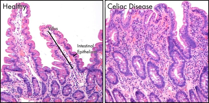
Around 95% of patients carry HLA-DQ2 and most of the remainder HLA-DQ8. However, roughly a third of the general population carries DQ2, so these alleles are neither sufficient nor useful for diagnosis - their value is a high negative predictive value, since their absence makes coeliac disease very unlikely.
Clinical features
Gastrointestinal features
- Chronic diarrhoea and steatorrhoea (pale, bulky, offensive stools that are difficult to flush)
- Bloating, abdominal distension and flatulence
- Abdominal pain and nausea
- Weight loss and, in children, failure to thrive and short stature
- Recurrent mouth ulcers
Extraintestinal and silent presentations
Many patients have no bowel symptoms at all, and coeliac disease is frequently uncovered during investigation of something else:
- Iron-deficiency anaemia - one of the commonest presentations, and unexplained iron deficiency should always prompt coeliac serology
- Folate deficiency (proximal small bowel) and, less commonly, B12 deficiency
- Fatigue and general malaise
- Dermatitis herpetiformis: an intensely itchy, blistering, symmetrical rash on the extensor surfaces (elbows, knees, buttocks), which is pathognomonic and treated with a gluten-free diet plus dapsone
- Osteoporosis or osteomalacia from calcium and vitamin D malabsorption
- Unexplained subfertility, recurrent miscarriage, or peripheral neuropathy and ataxia
- Deranged liver function tests
Associated conditions
Coeliac disease clusters with other autoimmune conditions, and NICE recommends offering testing to people with type 1 diabetes and autoimmune thyroid disease at diagnosis, as well as to first-degree relatives of affected people.2 It is also more common in Down syndrome, Turner syndrome, IgA deficiency and autoimmune liver disease.
Investigations
Serology
IgA tissue transglutaminase (tTG) antibodies are the first-line test, requested together with a total IgA level.2 This pairing matters because selective IgA deficiency is around 10 times more common in coeliac disease than in the general population and produces a falsely negative IgA-based test. If IgA is deficient, an IgG-based test (IgG tTG, IgG endomysial antibody or IgG deamidated gliadin peptide) is used instead.
IgA endomysial antibodies (EMA) are highly specific and used as a second-line or confirmatory test where tTG is equivocal. Anti-gliadin antibodies are no longer recommended.
Duodenal biopsy
Upper GI endoscopy with duodenal biopsy remains the diagnostic gold standard in adults, and should be arranged for anyone with positive serology.2 At least four biopsies are taken from the second part of the duodenum plus one or two from the duodenal bulb, because the changes can be patchy. Endoscopy may show scalloping of the duodenal folds, though a normal-looking mucosa does not exclude the diagnosis.
Histology is graded using the Marsh classification, which describes the progressive severity of mucosal damage.
| Stage | Histological findings |
|---|---|
| Marsh 0 | Normal mucosa |
| Marsh 1 | Increased intraepithelial lymphocytes, normal villi and crypts |
| Marsh 2 | Intraepithelial lymphocytosis with crypt hyperplasia, villi still normal |
| Marsh 3a | Partial villous atrophy |
| Marsh 3b | Subtotal villous atrophy |
| Marsh 3c | Total villous atrophy - a flat mucosa |
Other investigations
- Full blood count and blood film: anaemia, and features of hyposplenism such as Howell-Jolly bodies and target cells
- Haematinics: ferritin, folate and B12; and calcium, vitamin D and albumin
- Liver function tests and thyroid function tests
- DEXA scan to assess bone mineral density, given the risk of osteoporosis
- HLA-DQ2/DQ8 typing: used selectively to exclude the diagnosis, for instance in patients already established on a gluten-free diet who will not undertake a gluten challenge
Differential diagnosis
- Irritable bowel syndrome: overlapping symptoms of bloating and altered bowel habit, but with normal serology, calprotectin and biopsy - coeliac serology should be checked in all suspected IBS
- Non-coeliac gluten sensitivity: symptoms on gluten with negative serology and normal biopsy
- Lactose intolerance: may also occur secondarily to coeliac disease from loss of brush border lactase
- Inflammatory bowel disease: particularly Crohn's disease with small bowel involvement
- Other causes of villous atrophy: tropical sprue, giardiasis, small intestinal bacterial overgrowth, common variable immunodeficiency, and drugs such as olmesartan
- Pancreatic exocrine insufficiency: steatorrhoea with normal duodenal histology
Management
Gluten-free diet
The cornerstone of treatment is a lifelong, strict gluten-free diet, excluding all wheat, barley and rye. This allows mucosal healing, resolves symptoms, corrects malabsorption and reduces the long-term risks of osteoporosis and malignancy. Referral to a specialist dietitian is essential, both for practical education about hidden sources of gluten (sauces, processed foods, medications, cross-contamination) and to ensure nutritional adequacy.3
Oats are tolerated by the majority of patients but are frequently contaminated during milling, so only certified gluten-free oats should be used, and tolerance should be confirmed. Adherence is the single most important determinant of outcome and is often the greatest practical challenge for patients.
Correcting deficiencies and preventing complications
- Replace iron, folate, vitamin B12, calcium and vitamin D as indicated
- Bone protection: assess bone density and ensure adequate calcium and vitamin D intake; treat osteoporosis conventionally
- Vaccination: patients have functional hyposplenism, so pneumococcal vaccination is recommended with a booster every 5 years, alongside annual influenza and Haemophilus influenzae type b vaccination2
Monitoring
Patients are reviewed annually with assessment of symptoms, dietary adherence, weight, and blood tests including full blood count, haematinics, calcium and coeliac serology. Falling antibody titres provide a useful objective marker of dietary adherence. Persistent symptoms despite a strict diet warrant reassessment for inadvertent gluten exposure, an alternative or coexisting diagnosis, or the rare complications below.
Complications
- Nutritional deficiencies: iron, folate, B12, calcium, vitamin D and other fat-soluble vitamins
- Osteoporosis and osteomalacia, with an increased fracture risk
- Functional hyposplenism, increasing the risk of encapsulated organism infection
- Anaemia, often refractory to oral iron while gluten exposure continues
- Secondary lactose intolerance from brush border enzyme loss, usually reversible with mucosal healing
- Enteropathy-associated T-cell lymphoma (EATL): rare but serious, and should be suspected if symptoms, weight loss or abdominal pain recur after a period of good control on a gluten-free diet
- Small bowel adenocarcinoma and oesophageal carcinoma, both rare but with increased relative risk
- Refractory coeliac disease: persistent villous atrophy and symptoms despite 12 months of a strict gluten-free diet, requiring specialist management
- Subfertility and adverse pregnancy outcomes, which improve with dietary treatment
Red flags
Prognosis
With a strict, lifelong gluten-free diet the prognosis is excellent. Symptoms typically improve within weeks, serology normalises over 6-12 months, and mucosal healing follows over months to a couple of years, with most patients achieving a normal life expectancy and quality of life.1
The small excess risk of small bowel lymphoma and other malignancies falls with sustained dietary adherence, which is a strong argument for the strictness of the diet even in patients whose symptoms are mild. Outcomes are poorer in those diagnosed late, those with persistently poor adherence, and the small minority with refractory disease, in whom the risk of lymphoma is substantially higher.
References
- Lebwohl B, Sanders DS, Green PHR. Coeliac disease. Lancet. 2018. Available here
- NICE NG20. Coeliac disease: recognition, assessment and management. 2015 (updated 2020). Available here
- NICE Clinical Knowledge Summaries (CKS). Coeliac disease. 2023. Available here
- Ludvigsson JF et al. BSG guidelines on the diagnosis and management of adult coeliac disease. Gut. 2014. Available here
- GeneFood, CC BY 4.0, via Wikimedia Commons. Available here
- NHS. Coeliac disease. 2023. Available here
- Coeliac UK. Information for health professionals. Available here
This article is written for revision and education. It is not clinical guidance and must not be used to make decisions about the care of a patient. Always check current NICE guidance and local protocols.