
Thyroid Nodules and Goitre: Deciding Which Lumps Matter
Key points
- Thyroid nodules are very common: palpable in around 5% of women, but present on ultrasound in up to half of people over 60. Only about 5% are malignant.
- Start with the TSH: it determines the next investigation. A suppressed TSH means an isotope scan; a normal or raised TSH means an ultrasound.
- A hot nodule is almost never cancer: an autonomously functioning nodule that takes up isotope is benign in essentially all cases and is managed as thyrotoxicosis, not as a cancer risk.
- Ultrasound grading: the British Thyroid Association U score runs from U1 (normal) to U5 (malignant). Fine needle aspiration is indicated for U3 to U5.
- Cytology grading: the Thy score runs from Thy1 (non-diagnostic) to Thy5 (malignant), with Thy3 covering the indeterminate results that usually require diagnostic surgery.
- The key limitation of cytology: FNA cannot distinguish a follicular adenoma from a follicular carcinoma, because the diagnosis rests on capsular and vascular invasion seen only on the resected specimen.
- Red flags: rapid growth, a hard fixed lump, hoarseness, stridor, cervical lymphadenopathy, age under 20 or over 60, male sex and previous neck irradiation.
- Commonest cancer: papillary carcinoma, accounting for around 85%. It spreads via lymphatics and has an excellent prognosis.
Introduction
A goitre is any enlargement of the thyroid gland; a nodule is a discrete lesion within it. Both are extremely common, and the clinical task is almost always the same: identify the small minority that represent malignancy or that are causing compression, and avoid over-investigating the large majority that do not.
- Palpable nodules are found in around 5% of women and 1% of men
- On ultrasound, nodules are present in up to 50 to 60% of people over 60 - the phenomenon of the incidentaloma, found on scans done for something else entirely
- Around 5% of nodules are malignant, and the risk is broadly the same whether the nodule is solitary or one of many
Causes of goitre
| Diffuse goitre | Nodular goitre or discrete nodule |
|---|---|
| Physiological - puberty and pregnancy | Multinodular goitre - the commonest nodular cause |
| Iodine deficiency - the commonest cause worldwide | Colloid nodule and thyroid cyst |
| Graves' disease - smooth, with a bruit | Follicular adenoma |
| Hashimoto's thyroiditis - firm, sometimes bosselated | Toxic adenoma - a hot nodule |
| Subacute (de Quervain's) thyroiditis - painful and tender | Thyroid carcinoma - papillary, follicular, medullary or anaplastic |
| Drugs - lithium, amiodarone | Lymphoma - on a background of Hashimoto's |
| Dyshormonogenesis - including Pendred syndrome with deafness | Parathyroid adenoma or cyst mimicking a thyroid nodule |
Assessment
History
- How long has it been there and how fast is it growing? Rapid growth over weeks is concerning; a lump present unchanged for years usually is not - although a sudden increase may simply be haemorrhage into a benign cyst, which is painful and self-limiting.
- Compressive symptoms - dysphagia, dyspnoea, stridor, a choking sensation when lying flat, and hoarseness
- Hoarseness or voice change - which may indicate recurrent laryngeal nerve involvement and is an important red flag
- Thyroid status - symptoms of hyper- or hypothyroidism
- Pain - suggesting thyroiditis or haemorrhage into a cyst rather than malignancy
- Risk factors for malignancy - previous head or neck irradiation, especially in childhood; a family history of thyroid cancer, MEN 2, familial adenomatous polyposis or Cowden syndrome; and residence in an iodine-deficient area
- Age and sex - risk is higher under 20 and over 60, and higher in men
Examination
- Inspect from the front, then ask the patient to swallow - a thyroid swelling moves up on swallowing, and a thyroglossal cyst additionally moves on tongue protrusion
- Palpate from behind - assess size, symmetry, consistency (soft, firm or hard), whether a nodule is solitary or part of a multinodular gland, tenderness, and fixation to surrounding structures
- Examine the cervical lymph nodes - lymphadenopathy substantially raises the probability of malignancy
- Percuss for retrosternal extension, and check for tracheal deviation
- Pemberton's sign - ask the patient to raise both arms above the head for a minute. Facial plethora, distended neck veins and stridor indicate thoracic inlet obstruction by a retrosternal goitre.
- Listen for a bruit over the gland - a feature of Graves' disease
- Assess thyroid status, and examine for signs of Graves' disease
Investigations
Ultrasound and the U classification
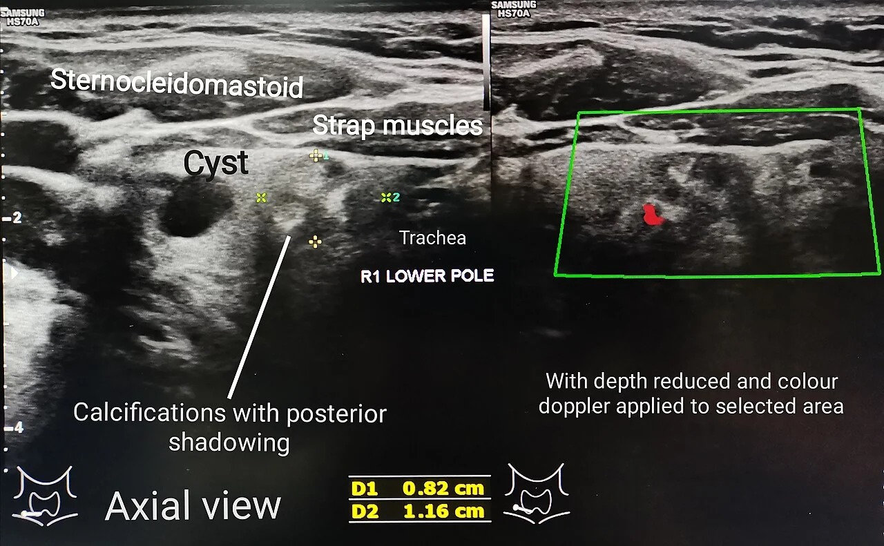
Ultrasound is the primary imaging test. The British Thyroid Association U classification grades the appearance and determines whether cytology is needed.
| Grade | Appearance | Action |
|---|---|---|
| U1 | Normal | No action |
| U2 | Benign - simple cyst, spongiform, halo, peripheral eggshell calcification | Reassure; no FNA required |
| U3 | Indeterminate - homogeneous hyperechoic solid nodule, mixed cystic and solid | FNA |
| U4 | Suspicious - solid hypoechoic, disrupted peripheral calcification, lobulated outline | FNA |
| U5 | Malignant - solid hypoechoic with microcalcification, taller than wide, irregular or infiltrative margin, extracapsular extension, associated abnormal lymph nodes | FNA |
Fine needle aspiration and the Thy classification
| Grade | Meaning | Management |
|---|---|---|
| Thy1 / Thy1c | Non-diagnostic (Thy1c indicates a cystic lesion) | Repeat FNA under ultrasound guidance |
| Thy2 / Thy2c | Non-neoplastic (benign) | Reassure and discharge or monitor; repeat if clinically suspicious |
| Thy3a | Atypia of undetermined significance | Repeat FNA or diagnostic hemithyroidectomy after multidisciplinary discussion |
| Thy3f | Follicular neoplasm suspected | Diagnostic hemithyroidectomy - cytology cannot resolve this |
| Thy4 | Suspicious of malignancy | Surgery |
| Thy5 | Diagnostic of malignancy | Definitive surgery |
Other investigations
- Calcitonin - if medullary thyroid carcinoma is suspected, particularly with a family history of MEN 2 or with diarrhoea and flushing. Not measured routinely in the UK.
- CT or MRI of the neck and thorax - for retrosternal extension, tracheal compression or suspected local invasion. Avoid iodinated contrast if radioiodine treatment is planned, since the iodine load blocks uptake for several months.
- Flexible nasendoscopy - to assess vocal cord function before thyroid surgery and in any patient with hoarseness
- Thyroglobulin is not a diagnostic test for nodules - it is used to monitor for recurrence after treatment of differentiated thyroid cancer
- Genetic testing for RET - in confirmed medullary carcinoma, and cascade testing of relatives
Thyroid cancer
| Type | Proportion | Features | Prognosis |
|---|---|---|---|
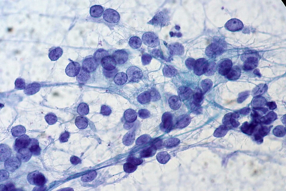
| Papillary carcinoma | Around 85% | Young adults, more common in women. Spreads via lymphatics to cervical nodes. Often multifocal. Histology shows Orphan Annie nuclei and psammoma bodies. Associated with previous radiation exposure. | Excellent - over 90% 10-year survival |
| Follicular carcinoma | Around 10% | Older patients, commoner in iodine-deficient areas. Spreads haematogenously to bone and lung. Diagnosed only on histology by capsular or vascular invasion. | Good, though worse than papillary |
| Medullary carcinoma | 3 to 5% | Arises from parafollicular C cells and secretes calcitonin. Around a quarter are familial, associated with MEN 2A and 2B (RET mutation). Histology shows amyloid stroma. Not iodine-avid, so radioiodine is ineffective. | Intermediate; depends on stage at diagnosis |
| Anaplastic carcinoma | Around 1% | Elderly patients with a rapidly enlarging, hard, fixed mass, often with stridor, hoarseness and dysphagia from local invasion | Very poor - median survival measured in months |
| Lymphoma | Under 5% | Arises on a background of Hashimoto's thyroiditis; presents as a rapidly enlarging goitre | Often good with chemotherapy and radiotherapy |
Treatment of differentiated thyroid cancer
- Surgery - hemithyroidectomy for small, low-risk unifocal tumours; total thyroidectomy for larger, multifocal or higher-risk disease, with central or lateral neck dissection if nodes are involved
- Radioiodine ablation after total thyroidectomy in intermediate and high-risk disease, to destroy residual thyroid tissue and micrometastases
- TSH suppression - levothyroxine at a dose that suppresses TSH, since TSH is a growth factor for differentiated thyroid tissue. The degree of suppression is balanced against the risks of atrial fibrillation and osteoporosis.
- Follow-up with thyroglobulin - a rising level after total thyroidectomy and ablation indicates recurrence. Thyroglobulin antibodies must be measured alongside, as they interfere with the assay.
- Medullary carcinoma is treated surgically with monitoring of calcitonin and CEA; it does not respond to radioiodine or TSH suppression, and relatives require RET genetic testing
- Anaplastic carcinoma is usually managed palliatively, with airway protection as the priority
Managing benign goitre and nodules
- Benign, asymptomatic and euthyroid - reassure and discharge, or arrange interval clinical review. Routine repeat ultrasound of a U2/Thy2 nodule is not required, and generates anxiety and further unnecessary testing.
- Treat the underlying cause - levothyroxine for hypothyroidism, antithyroid treatment for a toxic nodule or toxic multinodular goitre, and analgesia or steroids for subacute thyroiditis
- Levothyroxine to shrink a euthyroid goitre is not recommended - it is ineffective and exposes the patient to the risks of suppressed TSH
- Simple cysts - aspirate for symptom relief; they frequently recur, and persistent recurrence is an indication for surgery
- Radioiodine - effective for a toxic or large multinodular goitre, reducing volume by around 40% as well as treating hyperthyroidism
- Surgery - for compressive symptoms, retrosternal extension, suspected malignancy, cosmetic concern, or a toxic goitre where other treatments are unsuitable
- Consider percutaneous ethanol injection or radiofrequency ablation for selected benign nodules in specialist centres
Red flags
Prognosis
The overwhelming majority of thyroid nodules and goitres are benign and carry no threat to life. Most require reassurance and, at most, periodic clinical review - and a significant part of good practice here is not generating harm through over-investigation, since the incidental detection of tiny nodules on scans done for other reasons has driven a substantial rise in thyroid cancer diagnosis without a corresponding rise in mortality.
Where cancer is found, the outlook is generally excellent. Papillary carcinoma has a 10-year survival above 90%, and even with nodal metastases the prognosis remains good - which is why nodal disease influences the extent of surgery rather than the expectation of cure. Follicular carcinoma does slightly less well because of haematogenous spread. Medullary carcinoma depends heavily on stage at diagnosis, which is why cascade genetic testing of relatives with a RET mutation, and prophylactic thyroidectomy in childhood for those affected, changes outcomes so decisively.
Anaplastic carcinoma is the exception in every respect - median survival is measured in months, treatment is usually palliative, and the immediate clinical priority is the airway rather than the oncology.
Adverse prognostic factors in differentiated thyroid cancer are summarised by age (worse over 55), male sex, tumour size, extrathyroidal extension, distant metastases and unfavourable histological subtypes. For most patients, though, the honest message is that this is one of the more treatable malignancies in medicine, and that the follow-up - thyroglobulin monitoring and levothyroxine adjustment - matters more day to day than the cancer itself.
References
- British Thyroid Association. Guidelines for the management of thyroid cancer, third edition. Available here
- NICE NG145. Thyroid disease: assessment and management. 2019, updated 2023. Available here
- NICE NG12. Suspected cancer: recognition and referral. 2015, updated 2023. Available here
- NICE Clinical Knowledge Summaries. Neck lump. Available here
- Royal College of Pathologists. Guidance on the reporting of thyroid cytology specimens. Available here
- Haugen BR, Alexander EK, Bible KC et al. 2015 American Thyroid Association management guidelines for adult patients with thyroid nodules and differentiated thyroid cancer. Available here
- Cerevisae, CC BY-SA 4.0, via Wikimedia Commons. Available here
- Ed Uthman, CC BY 2.0, via Wikimedia Commons. Available here
This article is written for revision and education. It is not clinical guidance and must not be used to make decisions about the care of a patient. Always check current NICE guidance and local protocols.