
Superior Vena Cava Obstruction
Key points
- SVC obstruction: impaired venous return through the superior vena cava, from external compression, intraluminal thrombus or direct tumour invasion.
- Commonest cause: malignancy accounts for around 60-85% of cases, most often lung cancer - particularly small cell - and lymphoma.
- The rising cause: intravascular devices. Central lines, PICC lines, pacemaker leads and dialysis catheters now account for a substantial and growing minority.
- Presentation: facial and upper limb swelling, plethora, headache worse on bending forward or lying flat, and distended non-pulsatile neck and chest wall veins.
- Pemberton's sign: raising both arms above the head for one minute produces facial plethora, cyanosis and distress as venous return is further obstructed.
- Investigation: CT of the chest with contrast is the investigation of choice, defining the level, the cause and the collateral circulation.
- The key principle: obtain a tissue diagnosis before treatment wherever possible, because chemosensitive tumours are treated very differently from others.
- True emergency: stridor, laryngeal or cerebral oedema, or haemodynamic compromise. These need immediate endovascular stenting and airway assessment.
Introduction
Superior vena cava obstruction results from impaired venous drainage through the superior vena cava, causing venous congestion of the head, neck and upper limbs. It is often called SVC syndrome, reflecting the constellation of signs rather than a single pathological process.
The anatomy explains everything about it. The SVC is a thin-walled, low-pressure vessel carrying venous return from the entire upper body, and it runs in the right side of the superior mediastinum in a confined space bounded by the sternum, trachea, right main bronchus, aorta and pulmonary artery, surrounded by lymph nodes. It is therefore highly susceptible to compression by anything that enlarges in that space, and it has little capacity to resist.
Historically SVC obstruction was regarded as an oncological emergency requiring immediate radiotherapy. That view has been revised.2,5 In most patients the onset is gradual enough for collateral venous channels - through the azygos, internal mammary, lateral thoracic and vertebral venous systems - to develop, and there is time to establish a tissue diagnosis first. Establishing that diagnosis matters, because treatment differs enormously between a small cell lung cancer, a lymphoma and a catheter-related thrombosis.
Aetiology
Malignant causes
Malignancy accounts for approximately 60-85% of cases.
- Lung cancer - by far the commonest, responsible for around 70% of malignant cases. Small cell lung cancer is over-represented because it typically arises centrally and grows rapidly; non-small cell carcinoma, particularly squamous cell, also causes it.
- Non-Hodgkin lymphoma - the second commonest malignant cause, and the one where an urgent tissue diagnosis is most valuable given its chemosensitivity
- Metastatic disease to mediastinal nodes - most often from breast cancer
- Germ cell tumours and thymoma - important causes in younger patients with an anterior mediastinal mass
- Mesothelioma and other primary mediastinal tumours
Non-malignant causes
- Intravascular devices - central venous catheters, PICC lines, tunnelled dialysis catheters, pacemaker and ICD leads. These cause thrombosis and fibrotic stenosis, and now account for a substantial and increasing proportion of cases as device use has expanded. This is the cause most likely to be encountered in a non-oncological setting.
- Fibrosing mediastinitis - following histoplasmosis, tuberculosis, radiotherapy or IgG4-related disease
- Retrosternal goitre - a classic cause, and the setting in which Pemberton's sign was originally described
- Thoracic aortic aneurysm
- Benign mediastinal masses - dermoid cysts and bronchogenic cysts
- Post-radiotherapy fibrosis
- Constrictive pericarditis - producing similar features by a different mechanism
- Thrombophilia and thrombosis without an indwelling device, including Behcet disease
Clinical features
Symptoms reflect venous congestion above the level of obstruction, and characteristically worsen with anything that further increases venous pressure in the head - lying flat, bending forward, coughing or straining.
Symptoms
- Facial and periorbital swelling - often first noticed by the patient or family on waking, and frequently misattributed to allergy or renal disease
- Swelling of the neck and both arms
- Headache and a sensation of fullness or pressure in the head, classically worse on bending forward or lying flat
- Breathlessness - the commonest symptom, worse when supine, and partly from associated airway compression
- Cough
- Visual disturbance
- Hoarseness or stridor - indicating laryngeal oedema or airway compression, and a red flag
- Dysphagia - from oesophageal compression
- Confusion, drowsiness and reduced consciousness - from cerebral oedema, and a marker of severe disease
- Symptoms of the underlying cause - weight loss, haemoptysis, night sweats, lymphadenopathy
Examination
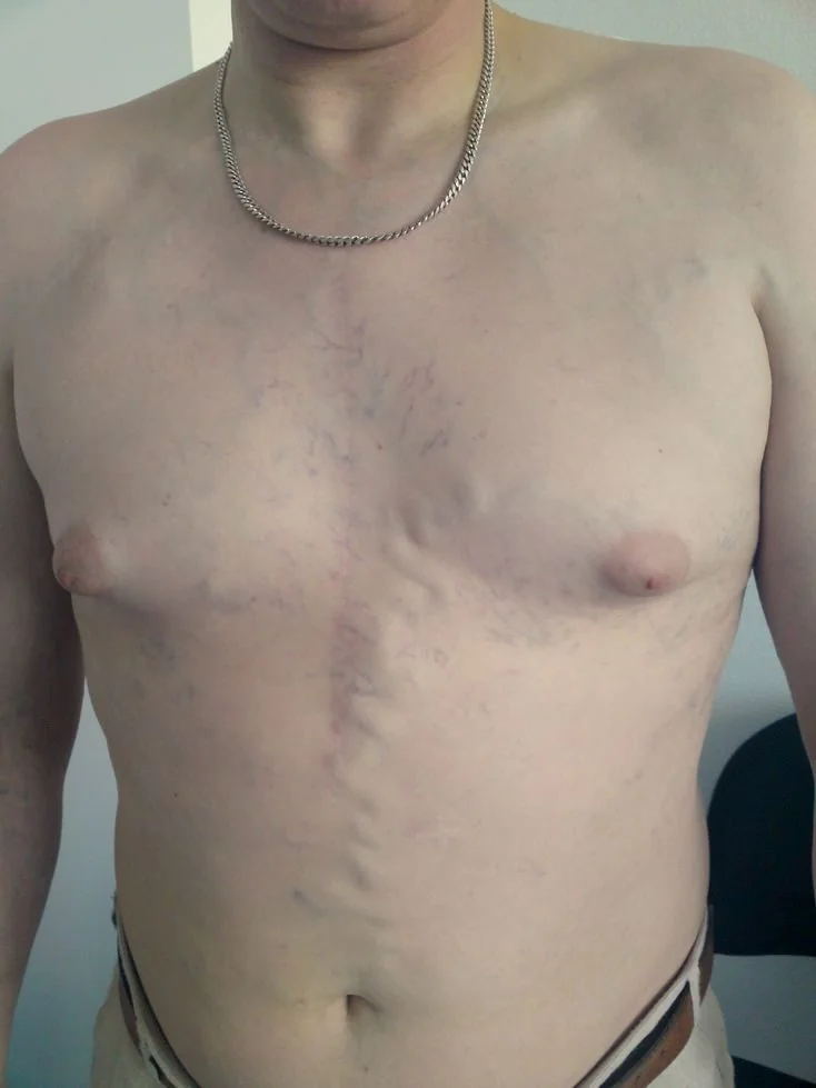
- Facial plethora and swelling, with periorbital and conjunctival oedema (chemosis)
- Distended neck veins that are non-pulsatile and do not vary with respiration - this is the key distinction from a raised JVP due to cardiac disease, where the waveform is pulsatile and varies
- Dilated collateral veins over the anterior chest wall and shoulders, with flow directed downwards. Their presence indicates the obstruction has been developing over weeks or longer.
- Bilateral upper limb oedema
- Cyanosis of the face and upper body
- Stridor - airway compromise
- Papilloedema - raised intracranial pressure from cerebral venous congestion
- Signs of the underlying cause - lymphadenopathy, a chest mass, clubbing, Horner syndrome, a goitre, or a central line or pacemaker in situ
Investigations
Imaging
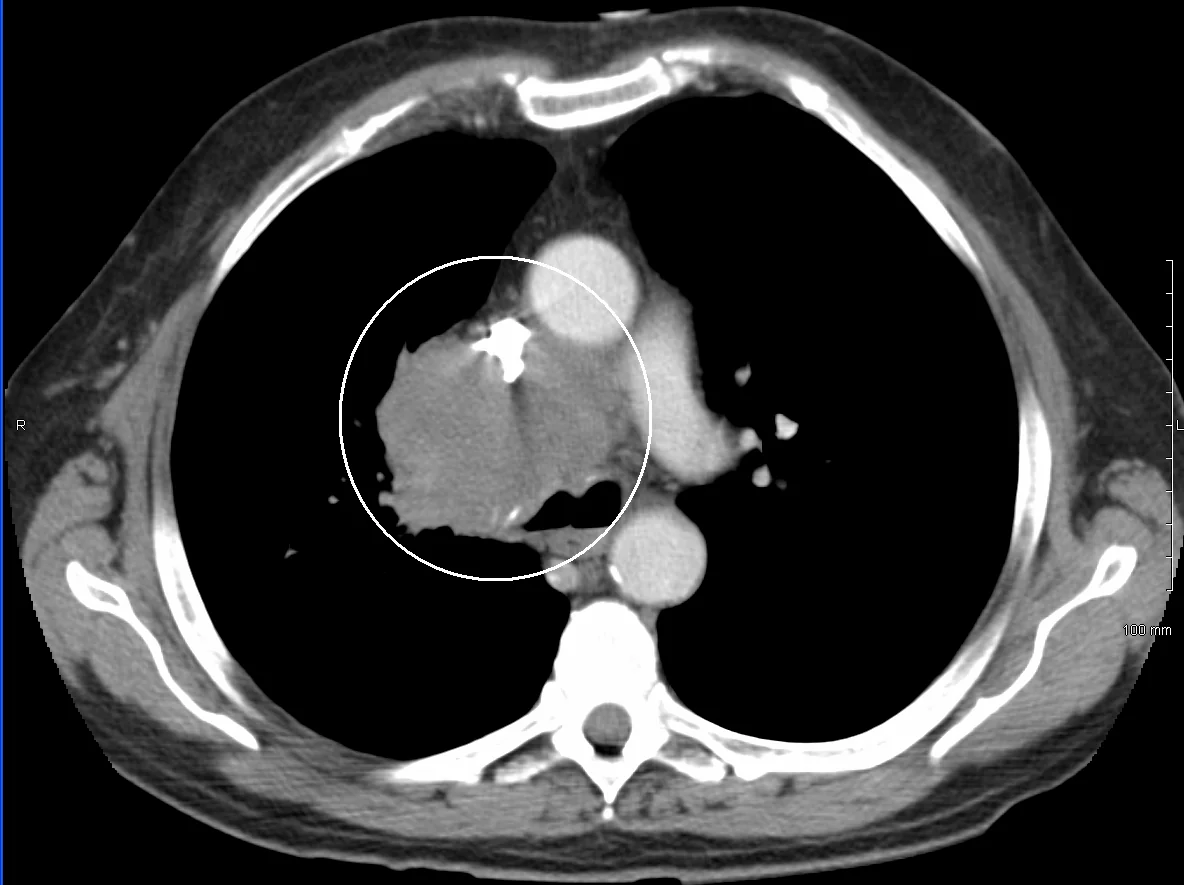
- Chest X-ray - abnormal in around 80% of cases, showing mediastinal widening, a right hilar or paratracheal mass, a pleural effusion or a lung lesion. A normal chest X-ray does not exclude the diagnosis.
- CT chest with intravenous contrast - the investigation of choice.1 It defines the level and length of obstruction, distinguishes external compression from intraluminal thrombus, demonstrates collateral circulation, identifies the underlying cause, and guides biopsy and stent planning. Contrast should be injected through a lower limb vein or with a protocol adapted for upper limb obstruction.
- CT or MR venography - where detailed venous anatomy is needed before intervention
- Ultrasound of the upper limb and neck veins - useful for detecting catheter-related thrombosis
- PET-CT - for staging once malignancy is confirmed
Establishing a tissue diagnosis
Other investigations
- FBC, U&Es, LFTs, calcium, LDH and clotting - LDH is often markedly raised in lymphoma, and hypercalcaemia may coexist in malignancy
- Tumour markers - AFP and beta-hCG where a germ cell tumour is suspected in a younger patient
- Thyroid function - if a retrosternal goitre is a possibility
- Blood cultures - if a line-related infection is suspected alongside thrombosis
- Thrombophilia screen - in unprovoked thrombosis without a device
Management
Immediate supportive measures
- Sit the patient upright - reduces hydrostatic pressure in the head and neck and relieves symptoms quickly
- Oxygen if hypoxic
- Assess the airway urgently if there is stridor, and involve anaesthetics and ENT early
- Avoid cannulating the upper limbs - infusions will not drain effectively and any drug given may pool. Use a femoral or lower limb vein instead.
- Analgesia and treatment of anxiety, which is often considerable
- Corticosteroids - dexamethasone is widely given, though the evidence base is weak and it is most justifiable where lymphoma or thymoma is suspected, or to reduce peritumoural oedema before radiotherapy. Avoid giving steroids before a lymphoma biopsy if at all possible, as they can render the tissue non-diagnostic.
- Diuretics - sometimes used for symptomatic relief, but of limited value and risk causing hypovolaemia
Endovascular stenting
Percutaneous SVC stenting has become the treatment of choice for symptomatic relief in most patients, particularly where symptoms are severe or where the tumour is unlikely to respond quickly to chemotherapy.2,3
- Rapid relief - symptoms typically improve within 24-72 hours, considerably faster than radiotherapy
- Does not require a tissue diagnosis first, so it can be used in the genuinely urgent patient while the diagnostic pathway continues
- Effective regardless of tumour type, and can be combined with subsequent chemotherapy or radiotherapy
- Complications - stent migration, thrombosis, infection, bleeding, and rarely SVC perforation
- Anticoagulation is usually given after stenting, though duration varies by centre and by whether thrombus was present
Treating the cause
| Cause | Treatment |
|---|---|
| Small cell lung cancer | Chemotherapy, often with a rapid response within days. Radiotherapy may be added. |
| Lymphoma | Chemotherapy - highly effective and potentially curative, which is why the tissue diagnosis matters so much |
| Germ cell tumour | Chemotherapy - also potentially curative even with bulky disease |
| Non-small cell lung cancer | Radiotherapy and/or stenting; systemic therapy according to stage and molecular profile |
| Catheter-related thrombosis | Anticoagulation.4 Remove the line if it is infected, no longer needed, or malpositioned; it may be retained if essential and the patient is anticoagulated. Consider catheter-directed thrombolysis in severe acute cases. |
| Retrosternal goitre | Surgical resection |
| Fibrosing mediastinitis | Stenting; treat any underlying infective or inflammatory cause |
| Benign stricture | Angioplasty with or without stenting |
Differential diagnosis
| Condition | Distinguishing features |
|---|---|
| Congestive cardiac failure | Pulsatile raised JVP varying with respiration, peripheral oedema affecting the legs, orthopnoea with pulmonary crackles, and no collateral chest wall veins |
| Cardiac tamponade | Pulsatile JVP, pulsus paradoxus, hypotension, muffled heart sounds, and effusion on echocardiography |
| Constrictive pericarditis | Kussmaul's sign, a pericardial knock, pericardial calcification on imaging |
| Nephrotic syndrome | Generalised oedema, heavy proteinuria, hypoalbuminaemia; facial swelling is typically periorbital and worst on waking |
| Angioedema | Rapid onset over minutes to hours, often with urticaria, lip and tongue involvement, and a trigger such as a drug or food |
| Cushing syndrome | Moon face with truncal obesity, striae and proximal weakness, developing over months |
| Bilateral upper limb DVT | Arm swelling without facial involvement or collateral chest wall veins |
The most useful single discriminator at the bedside is the character of the distended neck veins: non-pulsatile and fixed in SVC obstruction, pulsatile and respiration-varying in cardiac causes - together with the presence of dilated collateral veins over the chest wall, which are essentially specific for venous obstruction.
Complications
- Airway obstruction - from laryngeal oedema or direct tracheal compression by the causative mass. The commonest life-threatening complication.
- Cerebral oedema and raised intracranial pressure - causing headache, confusion, reduced consciousness, seizures and, rarely, coning
- Visual loss - from papilloedema and retinal venous congestion
- Extension of thrombus - into the brachiocephalic and jugular veins, and pulmonary embolism
- Pleural and pericardial effusion
- Aspiration - from oesophageal compression and impaired swallowing
- Stent complications - migration, occlusion, infection or perforation
- Bleeding - venous hypertension makes surgery and biopsy in the upper body more hazardous
Red flags
Prognosis
SVC obstruction itself is rarely the direct cause of death. Prognosis is determined almost entirely by the underlying condition, and this varies more widely than in almost any other presentation.
Benign causes carry an excellent outlook. Catheter-related obstruction usually resolves with anticoagulation, line management and stenting where needed, and a retrosternal goitre is cured by resection.
Lymphoma and germ cell tumours presenting with SVC obstruction remain potentially curable with chemotherapy despite the dramatic presentation, which is the strongest argument for obtaining tissue before treating.
Small cell lung cancer typically responds rapidly to chemotherapy with prompt symptomatic relief, but overall prognosis reflects the extensive-stage disease that SVC obstruction usually signifies, with median survival typically under a year.
Non-small cell lung cancer presenting with SVC obstruction indicates locally advanced or metastatic disease and carries a poor prognosis, with median survival of the order of six months. Stenting nonetheless provides worthwhile and rapid palliation, and quality of life rather than survival is the appropriate treatment goal in many of these patients.
Across all malignant causes, stenting achieves symptom relief in over 90% of patients, usually within days, with re-occlusion rates of around 10-15%. Given that many patients have limited life expectancy, this rapid and reliable palliation is often the most valuable intervention available.
References
- NICE NG122. Lung cancer: diagnosis and management. 2019, updated 2024. Available here
- Rowell NP, Gleeson FV. Steroids, radiotherapy, chemotherapy and stents for superior vena caval obstruction in carcinoma of the bronchus. Cochrane Database of Systematic Reviews. 2001. Available here
- UK Oncology Nursing Society and Royal College of Radiologists. Guidance on the management of oncological emergencies. Available here
- NICE NG158. Venous thromboembolic diseases: diagnosis, management and thrombophilia testing. 2020, updated 2023. Available here
- Wilson LD, Detterbeck FC, Yahalom J. Superior vena cava syndrome with malignant causes. New England Journal of Medicine. 2007. Available here
- NICE NG12. Suspected cancer: recognition and referral. 2015, updated 2023. Available here
- Macmillan Cancer Support. Superior vena cava obstruction. Available here
- Pemberton HS. Sign of submerged goitre. The Lancet. 1946. Available here
This article is written for revision and education. It is not clinical guidance and must not be used to make decisions about the care of a patient. Always check current NICE guidance and local protocols.