
Mitral Valve Disease: Stenosis, Regurgitation and Prolapse
Key points
- Mitral stenosis: almost always rheumatic. Obstruction to left ventricular filling loads the left atrium, not the ventricle.
- Stenosis signs: malar flush, tapping apex, loud first heart sound, opening snap, and a rumbling mid-diastolic murmur at the apex.
- Atrial fibrillation: very common in mitral stenosis, and the reason these patients need anticoagulation with warfarin rather than a DOAC.
- Mitral regurgitation: primary (valve disease) or secondary (a stretched annulus from a dilated ventricle). Volume overload dilates the ventricle.
- Regurgitation signs: displaced thrusting apex, a pansystolic murmur at the apex radiating to the axilla, a soft first heart sound and a third heart sound.
- Acute regurgitation: papillary muscle rupture 3-5 days after myocardial infarction, or endocarditis. Sudden pulmonary oedema with a short, soft murmur.
- Surgical thresholds: operate at an ejection fraction of 60% or below - a 'normal' figure that already represents impairment, because the ventricle empties into a low-pressure atrium.
- Mitral valve prolapse: the commonest valve abnormality, affecting 2-3% of people. Mid-systolic click with a late systolic murmur, usually benign.
Introduction
The mitral valve sits between the left atrium and the left ventricle, and unlike the aortic valve it is a complex apparatus rather than a simple set of leaflets: two leaflets, an annulus, chordae tendineae and two papillary muscles. Disease of any of these components produces mitral regurgitation, which is why the causes are so varied.
Three conditions dominate. Mitral stenosis obstructs ventricular filling and loads the left atrium. Mitral regurgitation allows backflow into the atrium and loads both chambers. Mitral valve prolapse is a leaflet abnormality that is usually harmless but can progress to significant regurgitation.
Mitral stenosis
Aetiology
Rheumatic heart disease accounts for over 90% of cases - there is no realistic second cause in exam terms. It follows group A streptococcal pharyngitis, often decades earlier, and causes commissural fusion, leaflet thickening and chordal shortening. Because rheumatic fever is now rare in the UK, mitral stenosis here is seen mostly in people who grew up overseas.1
Rare alternatives include severe mitral annular calcification, congenital stenosis, and infiltration by carcinoid, amyloid or the Libman-Sacks endocarditis of systemic lupus erythematosus.
Pathophysiology
The normal mitral valve area is 4-6 cm². As it narrows, a pressure gradient develops across the valve in diastole, and left atrial pressure rises. The consequences follow in sequence:
- Left atrial dilatation - producing atrial fibrillation, and stasis that predisposes to left atrial thrombus and systemic embolism
- Pulmonary venous congestion - breathlessness, orthopnoea and haemoptysis
- Pulmonary hypertension - initially reactive, later fixed
- Right ventricular failure - raised JVP, hepatomegaly, ascites and peripheral oedema
Anything that shortens diastole or increases flow demand causes abrupt decompensation, because there is less time to fill through a fixed narrow orifice. Tachycardia, the onset of atrial fibrillation, pregnancy, fever and exercise are the classic precipitants - which is why a patient can be stable for years and then present acutely when they develop AF.
Clinical features
- Exertional breathlessness, orthopnoea and paroxysmal nocturnal dyspnoea
- Palpitations from atrial fibrillation
- Haemoptysis - from rupture of congested bronchial veins
- Fatigue from a low cardiac output
- Hoarseness (Ortner syndrome) - the enlarged left atrium compresses the left recurrent laryngeal nerve
- Dysphagia - oesophageal compression by the enlarged atrium
- Systemic embolism, including stroke, sometimes as the presenting event
Examination
- Malar flush - dusky pink cheeks from a low cardiac output with peripheral vasoconstriction
- Pulse: often irregularly irregular from atrial fibrillation; low volume
- Apex: tapping and not displaced - a palpable first heart sound, since the ventricle is not enlarged
- Loud first heart sound, then an opening snap shortly after the second sound
- Murmur: low-pitched rumbling mid-diastolic murmur with pre-systolic accentuation, heard best at the apex, with the bell, in the left lateral position, in expiration
- Signs of pulmonary hypertension: a loud P2, right ventricular heave, raised JVP, tricuspid regurgitation and peripheral oedema
Investigations
- ECG - P mitrale (a broad, bifid P wave of left atrial enlargement) if in sinus rhythm; atrial fibrillation; right ventricular hypertrophy and right axis deviation with pulmonary hypertension
- Chest X-ray - left atrial enlargement giving a double right heart border, a splayed carina, and straightening of the left heart border; pulmonary congestion and Kerley B lines; a calcified valve
- Echocardiography - the definitive test. Measures valve area and gradient, and scores leaflet mobility, thickening, calcification and subvalvular disease to judge suitability for balloon valvuloplasty.
- Transoesophageal echocardiography - mandatory before balloon valvuloplasty to exclude left atrial thrombus
Severity: a valve area below 1.0 cm² is severe, and 1.0-1.5 cm² moderate.
Management
- Rate control - beta-blockers or digoxin. Slowing the heart lengthens diastole and improves filling, which is why rate control matters more here than in most conditions.
- Anticoagulation - for atrial fibrillation, previous embolism or left atrial thrombus
- Diuretics for pulmonary congestion
- Percutaneous balloon mitral valvuloplasty - the treatment of choice where the valve is pliable, not heavily calcified, without significant regurgitation, and with no left atrial thrombus
- Surgical valve replacement or repair - where the anatomy is unsuitable for valvuloplasty
Mitral regurgitation
Aetiology
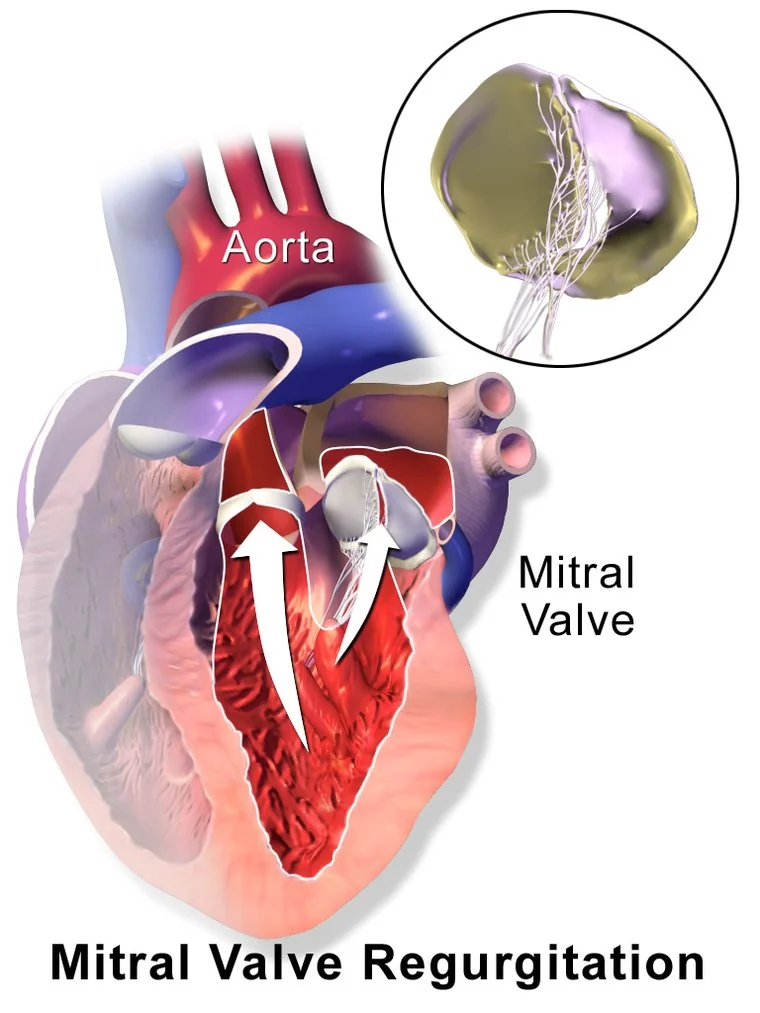
The critical distinction is between primary disease of the valve apparatus and secondary regurgitation where the valve is structurally normal but the ventricle has stretched the annulus. The distinction determines treatment: primary disease needs the valve fixed, secondary disease needs the ventricle treated.
| Primary (valve apparatus) | Secondary (functional) |
|---|---|
| Myxomatous degeneration and mitral valve prolapse | Dilated cardiomyopathy - annular stretching |
| Infective endocarditis - leaflet destruction | Ischaemic - papillary muscle displacement after infarction |
| Rheumatic heart disease | Atrial fibrillation with severe left atrial dilatation |
| Papillary muscle rupture after myocardial infarction | |
| Chordal rupture | |
| Connective tissue disease - Marfan, Ehlers-Danlos |
Acute versus chronic
Acute severe regurgitation gives a normal-sized, non-compliant left atrium a sudden large regurgitant volume. Left atrial and pulmonary venous pressures rise abruptly and the patient presents with sudden pulmonary oedema and cardiogenic shock. As in acute aortic regurgitation, the murmur is short and soft because pressures equalise quickly - so severity is easily underestimated.
The classic cause is papillary muscle rupture 3-5 days after a myocardial infarction, typically inferior, because the posteromedial papillary muscle has a single blood supply and is therefore vulnerable. It is a surgical emergency.
Chronic regurgitation allows the atrium and ventricle to dilate gradually, so patients remain asymptomatic for years before developing exertional breathlessness, fatigue and atrial fibrillation.
Examination
- Apex: displaced, diffuse and thrusting - a volume-loaded ventricle
- Murmur: pansystolic, blowing, loudest at the apex, radiating to the axilla, louder in expiration and in the left lateral position
- Soft first heart sound - the leaflets do not close properly
- Third heart sound from rapid ventricular filling of the volume-overloaded ventricle
- Loud P2 and right heart signs if pulmonary hypertension has developed
- Atrial fibrillation is common
Management
Secondary regurgitation is treated by treating the ventricle: full heart failure therapy with an ACE inhibitor or ARNI, beta-blocker, mineralocorticoid receptor antagonist and SGLT2 inhibitor, plus cardiac resynchronisation where indicated. Reverse remodelling reduces the regurgitation.
Primary regurgitation needs the valve addressed. Repair is preferred to replacement wherever feasible - it preserves the subvalvular apparatus, gives better ventricular function afterwards, avoids a prosthesis and avoids lifelong anticoagulation.
Transcatheter edge-to-edge repair clips the leaflets together via a catheter, and is used in selected patients who are poor surgical candidates - particularly in secondary regurgitation with heart failure, where trial results have been mixed and patient selection is critical.
Mitral valve prolapse
Displacement of one or both mitral leaflets into the left atrium during systole, caused by myxomatous degeneration. It is the commonest valve abnormality, present in 2-3% of the population, and in most people it is an incidental and harmless finding.3
It is associated with connective tissue disorders - Marfan syndrome and Ehlers-Danlos syndrome - so a young, tall patient with a click deserves a wider look.
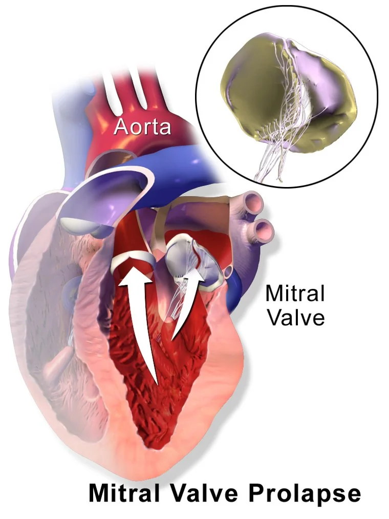
Most patients are asymptomatic. Some report atypical chest pain, palpitations or anxiety. The classic finding is a mid-systolic click, followed by a late systolic murmur if there is regurgitation.
Management is reassurance and periodic echocardiography. Complications are uncommon but include progressive mitral regurgitation, infective endocarditis, atrial and ventricular arrhythmias, and rarely sudden cardiac death - the last associated with bileaflet prolapse, marked leaflet redundancy and inferolateral T wave inversion.
Complications
- Atrial fibrillation - very common in both stenosis and regurgitation, from left atrial dilatation
- Systemic embolism and stroke - particularly mitral stenosis with atrial fibrillation and left atrial thrombus
- Pulmonary hypertension and right heart failure - the usual end point of untreated mitral stenosis
- Left ventricular dysfunction - in regurgitation, from chronic volume overload
- Infective endocarditis - any abnormal valve is a substrate
- Haemoptysis - from congested bronchial veins in mitral stenosis
- Ortner syndrome - hoarseness from recurrent laryngeal nerve compression
- Acute pulmonary oedema - abruptly, in acute regurgitation or with new atrial fibrillation in stenosis
Red flags
Prognosis
Untreated symptomatic mitral stenosis carries a poor outlook, with survival falling sharply once pulmonary hypertension or right heart failure develops. Balloon valvuloplasty in suitable anatomy produces excellent long-term results and can be repeated, which is why assessing valve morphology matters.
Chronic mitral regurgitation has a long asymptomatic phase, but once symptoms or ventricular dysfunction appear the prognosis without surgery is poor. Mitral repair carries a lower operative mortality and better long-term survival than replacement, and outcomes are best in high-volume centres - which is the practical argument for referring before the ventricle deteriorates rather than after.
Mitral valve prolapse without significant regurgitation has a normal life expectancy, and the main task is to reassure while arranging appropriate surveillance for the minority who progress.
References
- Vahanian A, Beyersdorf F, Praz F et al. ESC/EACTS Guidelines for the management of valvular heart disease. European Heart Journal. 2021. Available here
- Connolly SJ, Karthikeyan G, Ntsekhe M et al. Rivaroxaban in rheumatic heart disease-associated atrial fibrillation (INVICTUS). New England Journal of Medicine. 2022. Available here
- Freed LA, Levy D, Levine RA et al. Prevalence and clinical outcome of mitral-valve prolapse. New England Journal of Medicine. 1999. Available here
- NICE NG208. Heart valve disease presenting in adults: investigation and management. 2021. Available here
- NICE Clinical Knowledge Summaries. Heart murmurs. Available here
- Otto CM, Nishimura RA, Bonow RO et al. ACC/AHA Guideline for the management of patients with valvular heart disease. Circulation. 2021. Available here
- BNF. Warfarin sodium - indications and target INR. Available here
This article is written for revision and education. It is not clinical guidance and must not be used to make decisions about the care of a patient. Always check current NICE guidance and local protocols.