
Infective Endocarditis: Diagnosis and Management
Key points
- Infective endocarditis: infection of the endocardium, usually a heart valve, producing vegetations of organisms, platelets and fibrin.
- Think of it: in anyone with a fever and a new or changed murmur, and in any prosthetic valve patient with a fever.
- Commonest organism: Staphylococcus aureus overall, and in acute disease, intravenous drug use and early prosthetic valve infection.
- Blood cultures first: at least three sets from separate sites before antibiotics. Starting antibiotics first can make the diagnosis unprovable.
- Diagnosis: the modified Duke criteria - two major, one major with three minor, or five minor.
- Imaging: transthoracic echocardiography first, transoesophageal if it is negative with high suspicion, or if there is a prosthetic valve.
- Treatment: 4-6 weeks of intravenous antibiotics guided by the organism, managed by an endocarditis team.
- Surgery: for heart failure, uncontrolled infection such as an abscess, or to prevent embolism from a large vegetation.
Introduction
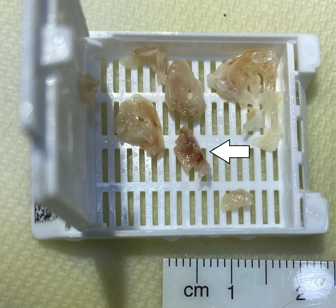
Infective endocarditis is infection of the endocardial surface of the heart, most often a valve. Organisms adhere to damaged endothelium and grow within a protective mesh of platelets and fibrin - a vegetation - which shields them from both the immune system and antibiotics. That is why treatment takes weeks of intravenous therapy rather than days of oral antibiotics.1
It is uncommon, with an incidence of around 3-10 per 100,000 per year, but it carries an in-hospital mortality of 15-30% and remains one of the diagnoses most often missed on first presentation. The reason is that its early features - fever, malaise, weight loss, aches - are indistinguishable from a hundred benign illnesses.
The epidemiology has shifted. Rheumatic heart disease in young people has largely been replaced by degenerative valve disease in older people, prosthetic valves, intracardiac devices, intravenous drug use and healthcare-associated bacteraemia. As a result Staphylococcus aureus has overtaken the oral streptococci as the commonest cause.
Aetiology
Two things must coincide: a surface organisms can stick to, and organisms in the bloodstream.
Turbulent flow across an abnormal valve damages the endothelium, and platelets and fibrin deposit on the injured surface to form a sterile thrombus. During a bacteraemia - from the mouth, gut, skin, or an intravenous line - organisms adhere to this thrombus, multiply within it, and the vegetation grows. Vegetations sit on the low-pressure side of the lesion: the atrial side of the mitral valve, the ventricular side of the aortic valve.
The organisms
| Organism | Typical setting |
|---|---|
| Staphylococcus aureus | The commonest cause overall. Acute, aggressive, rapidly destructive. Intravenous drug use, healthcare-associated bacteraemia, indwelling lines, prosthetic valves. |
| Viridans group streptococci (S. mitis, S. sanguinis) | Subacute, indolent. Oral flora, following dental work or with poor dentition. Native valves. |
| Enterococcus faecalis | Older patients, after genitourinary or gastrointestinal instrumentation. |
| Coagulase-negative staphylococci (S. epidermidis) | Early prosthetic valve endocarditis - contamination at the time of surgery. |
| Streptococcus gallolyticus (formerly S. bovis) | Associated with colorectal carcinoma - always arrange colonoscopy. |
| HACEK organisms | Haemophilus, Aggregatibacter, Cardiobacterium, Eikenella, Kingella. Fastidious slow-growing oral organisms; a classic cause of apparently culture-negative disease. |
| Coxiella burnetii, Bartonella, Brucella | Culture-negative endocarditis; need specific serology. Ask about farm animal and livestock exposure, homelessness and body lice. |
| Candida and other fungi | Intravenous drug use, immunosuppression, prolonged intravenous access, prosthetic valves. Large vegetations and a poor prognosis. |
Culture-negative endocarditis
Around 10% of cases have negative blood cultures. By far the commonest reason is antibiotics given before cultures were taken. The others are fastidious organisms (HACEK), intracellular organisms requiring serology (Coxiella, Bartonella), and fungi. This is the single strongest argument for taking cultures first.
Risk factors
Risk is best thought of as cardiac factors that provide the surface, and non-cardiac factors that provide the bacteraemia.
| Cardiac | Non-cardiac |
|---|---|
| Prosthetic valve - the highest-risk group | Intravenous drug use |
| Previous infective endocarditis | Indwelling central venous catheter or long line |
| Congenital heart disease, particularly unrepaired cyanotic disease or repairs with prosthetic material | Haemodialysis |
| Regurgitant valve lesions; bicuspid aortic valve | Poor dental hygiene and dental infection |
| Mitral valve prolapse with regurgitation | Immunosuppression, including HIV and diabetes |
| Hypertrophic cardiomyopathy | Recent surgery or instrumentation |
| Pacemaker or implantable defibrillator leads | Chronic skin conditions; tattooing and piercing |
Note that rheumatic heart disease remains an important risk factor globally and in people who grew up outside the UK, even though degenerative disease now dominates in the UK-born population.
Clinical features
Presentation ranges from a fulminant septic illness over days, typical of Staphylococcus aureus, to a vague malaise over weeks or months, typical of the viridans streptococci. The subacute form is the one that gets missed.
Constitutional
- Fever - present in around 90%, though it may be absent in the elderly, the immunosuppressed and those already given antibiotics
- Malaise, fatigue, anorexia and weight loss
- Night sweats and rigors
- Arthralgia and myalgia - so patients are sometimes investigated for a rheumatological illness
- Headache and confusion, particularly in older people
Cardiac
A new or changed murmur, usually regurgitant, from leaflet destruction or perforation. Symptoms of heart failure - breathlessness, orthopnoea, oedema - indicate significant valve destruction and are the commonest reason surgery becomes necessary.
Embolic and immunological
Fragments of vegetation break off and lodge distally, while circulating immune complexes deposit in small vessels. The classic peripheral stigmata arise from these two mechanisms, and knowing which is which makes them far easier to remember:
| Sign | Description | Mechanism |
|---|---|---|
| Osler nodes | Painful raised nodules on the finger and toe pulps | Immune complex deposition (Osler = Ouch) |
| Janeway lesions | Painless flat erythematous macules on palms and soles | Septic emboli |
| Roth spots | Retinal haemorrhages with pale centres | Immune complex vasculitis |
| Splinter haemorrhages | Linear red-brown streaks under the nails | Microemboli (also caused by trauma - check several nails) |
| Glomerulonephritis | Haematuria and proteinuria | Immune complex deposition |
| Splenomegaly | In subacute disease | Chronic immune stimulation and splenic infarcts |
| Clubbing | Late sign in longstanding subacute disease | Chronic infection |
Emboli to larger vessels produce stroke, splenic or renal infarction (flank or abdominal pain), limb ischaemia, and in right-sided disease septic pulmonary emboli with pleuritic pain, cough and cavitating lung nodules.
Clinical examination
- Observations - fever, tachycardia, and the features of sepsis
- Cardiovascular - listen carefully for a new or changed murmur, and compare with any previous documentation. Assess for heart failure: raised JVP, third heart sound, basal crackles, peripheral oedema.
- Hands - splinter haemorrhages, Osler nodes, clubbing, and needle marks
- Palms and soles - Janeway lesions
- Fundoscopy - Roth spots
- Abdomen - splenomegaly, and tenderness suggesting splenic infarction
- Neurological examination - focal deficits from embolic stroke or a mycotic aneurysm
- Mouth and teeth - dental caries and periodontal disease as the portal of entry
- Lines and devices - inspect every intravenous access site, and the pocket of any pacemaker or defibrillator
- Urinalysis - dipstick for blood and protein at the bedside
In a patient with a prosthetic valve, listen specifically for a muffled or absent click and any new murmur, both of which suggest dehiscence or obstruction.
Differential diagnosis
- Sepsis from another source - the commonest alternative, and the one that leads to antibiotics being started before cultures are taken
- Rheumatic fever - fever, arthritis and a murmur following streptococcal pharyngitis; use the Jones criteria
- Atrial myxoma - fever, weight loss, raised inflammatory markers, embolic events and a murmur that changes with posture; the great mimic
- Non-bacterial thrombotic (marantic) endocarditis - sterile vegetations in advanced malignancy or antiphospholipid syndrome
- Libman-Sacks endocarditis - sterile vegetations in systemic lupus erythematosus
- Vasculitis and connective tissue disease - fever, arthralgia, raised inflammatory markers, glomerulonephritis and a positive rheumatoid factor overlap closely
- Lymphoma and other occult malignancy - fever, weight loss and night sweats
- Tuberculosis and other chronic infection
Investigations
Blood cultures
Echocardiography
- Transthoracic echocardiography (TTE) first line in all suspected cases - looking for vegetations, new valvular regurgitation, abscess and prosthetic dehiscence
- Transoesophageal echocardiography (TOE) where TTE is negative but clinical suspicion remains high, where imaging is technically poor, and in all patients with a prosthetic valve or intracardiac device, since prosthetic material obscures transthoracic views
- A negative echocardiogram does not exclude the diagnosis - repeat after 5-7 days if suspicion persists
The modified Duke criteria
Diagnosis rests on the modified Duke criteria, which combine microbiology and imaging with clinical features.2 Definite endocarditis requires 2 major, 1 major plus 3 minor, or 5 minor criteria.
| Major criteria | Minor criteria |
|---|---|
| Positive blood cultures - a typical organism from two separate cultures, or persistently positive cultures with an organism consistent with endocarditis, or a single positive culture for Coxiella burnetii | Predisposition - a predisposing cardiac lesion, or intravenous drug use |
| Evidence of endocardial involvement - echocardiographic vegetation, abscess, or new dehiscence of a prosthetic valve; or new valvular regurgitation | Fever above 38°C |
| (2023 ESC update also accepts abnormal activity on PET-CT or cardiac CT around a prosthetic valve) | Vascular phenomena - arterial emboli, septic pulmonary infarcts, mycotic aneurysm, intracranial haemorrhage, conjunctival haemorrhage, Janeway lesions |
| Immunological phenomena - glomerulonephritis, Osler nodes, Roth spots, positive rheumatoid factor | |
| Microbiological evidence not meeting a major criterion |
Other investigations
- FBC - normocytic normochromic anaemia of chronic disease, neutrophilia
- CRP and ESR - raised, and used to track response to treatment
- U&Es - renal impairment from glomerulonephritis, sepsis or nephrotoxic antibiotics such as gentamicin and vancomycin
- LFTs, and rheumatoid factor (a minor criterion)
- Urinalysis and microscopy - haematuria, proteinuria and red cell casts
- ECG - repeat regularly. A new or lengthening PR interval suggests an aortic root abscess extending into the conducting system.
- Chest X-ray - pulmonary oedema, or cavitating nodules from septic emboli in right-sided disease
- CT head, chest, abdomen and pelvis - to identify embolic complications and, in the older patient, an occult source
- Serology for Coxiella, Bartonella and Brucella where cultures are negative
Management
Treatment is prolonged intravenous antibiotics, coordinated by a specialist endocarditis team including cardiology, microbiology and cardiac surgery. All patients should be discussed with such a team early, and any patient needing surgery should be managed in a centre where it is available.
Antibiotics
Empirical therapy is started after cultures, then narrowed once the organism and sensitivities are known. Regimens follow national and local protocols; the principles matter more than the exact drugs:
| Setting | Typical empirical therapy |
|---|---|
| Native valve, subacute, no penicillin allergy | Amoxicillin, with or without gentamicin |
| Native valve, acute or staphylococcal suspected | Flucloxacillin |
| Penicillin allergy, MRSA suspected, or prosthetic valve | Vancomycin plus gentamicin, with rifampicin added for prosthetic valves |
- Duration: usually 4 weeks for a native valve and 6 weeks for a prosthetic valve, counted from the first day of effective therapy or from the day of surgery
- Rifampicin is added for prosthetic material because of its activity against organisms embedded in biofilm
- Gentamicin and vancomycin need level monitoring, and both are nephrotoxic - track renal function closely
- Outpatient parenteral antibiotic therapy suits selected stable patients after an initial inpatient period
Surgery
Around half of patients need surgery. The three accepted indications are worth learning as a group:1
- Heart failure - from severe valvular regurgitation or obstruction. This is the commonest indication and the strongest.
- Uncontrolled infection - a perivalvular abscess, fistula, enlarging vegetation, persistent fever and positive cultures beyond 7-10 days of appropriate therapy, or infection with a fungus or a highly resistant organism
- Prevention of embolism - a large vegetation (typically above 10 mm) that has already embolised, or a very large vegetation above 15 mm, particularly on the mitral valve
Surgery is not delayed to complete an antibiotic course; where indicated it is performed while the patient is still on treatment. Infected pacemaker or defibrillator systems must be completely extracted, since antibiotics alone will not clear a colonised lead.
Supportive care and prevention
- Treat heart failure, arrhythmia and embolic complications
- Dental assessment and treatment of any source of infection
- Remove infected lines and address ongoing intravenous drug use with specialist support
- Anticoagulation is a difficult judgement - continue it for a mechanical valve, but stop it temporarily if there has been intracranial haemorrhage, on specialist advice
Complications
- Heart failure - from acute valvular regurgitation; the commonest cause of death and the leading indication for surgery
- Perivalvular abscess - most often at the aortic root, extending into the conducting system to cause first-degree, then complete, heart block. Watch the PR interval.
- Systemic embolism - stroke, splenic and renal infarction, limb ischaemia, and mesenteric ischaemia
- Mycotic aneurysm - infection weakening an arterial wall, classically intracranial, with a risk of catastrophic haemorrhage
- Septic pulmonary emboli and lung abscess - in right-sided disease
- Glomerulonephritis and acute kidney injury - from immune complex deposition, sepsis and nephrotoxic antibiotics
- Metastatic infection - vertebral osteomyelitis, discitis, septic arthritis and splenic abscess
- Valve dehiscence in prosthetic valve endocarditis
- Relapse or recurrence - relapse with the same organism usually reflects inadequate treatment; reinfection reflects continuing risk
Red flags
Prognosis
In-hospital mortality is 15-30%, and around 40% of patients die within five years. Outcome depends far more on the organism, the host and the complications than on how promptly antibiotics were started.1
Adverse prognostic features include Staphylococcus aureus or fungal infection, prosthetic valve involvement, heart failure, perivalvular abscess, stroke, older age, renal failure and diabetes. Viridans streptococcal native valve endocarditis in a younger patient, by contrast, usually responds well to antibiotics alone.
Around half of patients require surgery, and appropriately timed surgery improves survival rather than adding risk - delaying it in the hope that antibiotics will suffice is a recognised cause of avoidable death. Survivors need long-term follow-up: they have a substantially raised risk of a further episode, making dental care, prompt investigation of fever, and avoidance of unnecessary intravenous access lifelong priorities.
References
- Delgado V, Ajmone Marsan N, de Waha S et al. ESC Guidelines for the management of endocarditis. European Heart Journal. 2023. Available here
- Li JS, Sexton DJ, Mick N et al. Proposed modifications to the Duke criteria for the diagnosis of infective endocarditis. Clinical Infectious Diseases. 2000. Available here
- NICE CG64. Prophylaxis against infective endocarditis. 2008, updated 2016. Available here
- NICE Clinical Knowledge Summaries. Infective endocarditis. Available here
- British Society for Antimicrobial Chemotherapy. Guidelines for the diagnosis and antibiotic treatment of endocarditis in adults. Available here
- NICE NG208. Heart valve disease presenting in adults: investigation and management. 2021. Available here
- BNF. Flucloxacillin - indications and dosing. Available here
This article is written for revision and education. It is not clinical guidance and must not be used to make decisions about the care of a patient. Always check current NICE guidance and local protocols.