
Intestinal Obstruction and Ileus
Key points
- Mechanical obstruction: a physical blockage of the bowel lumen. Peristalsis continues against it, producing colic and high-pitched bowel sounds.
- Ileus: failure of propulsion without a physical obstruction, usually after surgery. The abdomen is distended but silent and painless.
- Four cardinal features: colicky abdominal pain, vomiting, distension and absolute constipation. Their order tells you how proximal the obstruction is.
- Commonest causes: adhesions then hernias in the small bowel; colorectal cancer then volvulus in the large bowel.
- The 3-6-9 rule: normal maximum diameters on abdominal radiograph are 3 cm small bowel, 6 cm colon and 9 cm caecum.
- Fluid losses: several litres are sequestered into the bowel lumen and wall, so patients are profoundly hypovolaemic with a hypochloraemic hypokalaemic metabolic alkalosis.
- Initial management: drip and suck - intravenous fluid and electrolyte replacement, nil by mouth and a nasogastric tube on free drainage - with CT to define the cause and level.
- When to operate: closed loop obstruction, strangulation or ischaemia, perforation, an irreducible hernia, an obstructing tumour, or failure of conservative management after 48 to 72 hours.
Introduction and definitions
Intestinal obstruction is one of the commonest reasons for emergency general surgical admission and accounts for a substantial share of emergency laparotomies in the UK. The first task at the bedside is not to name the cause but to place the patient into one of three categories, because they are managed differently.
| Mechanical obstruction | Paralytic ileus | Pseudo-obstruction | |
|---|---|---|---|
| Nature | Physical blockage of the lumen | Failure of peristalsis, no blockage | Failure of colonic motility, no blockage |
| Typical setting | Adhesions, hernia, tumour | First days after abdominal surgery, sepsis, electrolyte disturbance | Frail, immobile or medically unwell inpatients; Ogilvie syndrome |
| Pain | Colicky, prominent | Absent or a dull ache only | Usually mild |
| Bowel sounds | High-pitched and tinkling, later absent | Absent or reduced | Reduced |
| Radiology | Dilatation to a transition point, collapsed bowel beyond | Diffuse dilatation of small and large bowel with gas throughout to the rectum | Massive colonic dilatation, no transition point |
| Treatment | Drip and suck, then surgery if it does not resolve | Correct the cause, supportive care | Correct the cause, decompression, neostigmine |
Two further distinctions matter. Simple obstruction has an intact blood supply; strangulated obstruction does not, and is a surgical emergency. Partial obstruction still allows some flatus to pass and often settles; complete obstruction does not.
Causes
Small bowel obstruction
- Adhesions - the commonest cause in the UK, responsible for around 60% of cases. Almost any previous abdominal or pelvic operation can cause them, and they may present decades later.
- Hernias - the commonest cause worldwide and in patients who have never had surgery. Always examine the groins: an incarcerated femoral hernia in an older woman is the classic missed diagnosis.
- Malignancy - either a primary small bowel tumour, which is rare, or peritoneal disease from ovarian, colorectal or gastric cancer, which is not
- Crohn disease - inflammatory or fibrotic stricturing, often with a background of previous resections
- Intussusception - the commonest cause in infants; in adults it is rare and usually indicates a lead point such as a polyp or tumour
- Gallstone ileus - a large stone erodes through a cholecystoduodenal fistula and impacts at the ileocaecal valve
- Bezoar, foreign body or a swallowed object, and rarely radiation enteritis
Large bowel obstruction
- Colorectal carcinoma - around 60% of cases, and the reason every large bowel obstruction demands cancer staging
- Volvulus - about 20%, usually sigmoid, less commonly caecal
- Diverticular stricture - the sequel to repeated episodes of diverticulitis
- Faecal impaction, particularly in frail, immobile or opioid-treated patients
- Extrinsic compression from pelvic or retroperitoneal malignancy
- Hernia and anastomotic stricture, both less common than in the small bowel
Ileus
- Recent abdominal surgery - physiological for up to a few days, particularly after open surgery and bowel handling
- Electrolyte disturbance - hypokalaemia, hyponatraemia, hypomagnesaemia and hypercalcaemia
- Drugs - opioids, anticholinergics, tricyclic antidepressants
- Intra-abdominal inflammation - pancreatitis, peritonitis, an abscess, or retroperitoneal haemorrhage
- Systemic illness - sepsis, uraemia, diabetic ketoacidosis, hypothyroidism, spinal injury
Pathophysiology
Once the lumen is blocked, gas and fluid accumulate proximally. The gas is largely swallowed nitrogen, and the fluid comes from the several litres of gastrointestinal secretion produced daily. The bowel proximal to the obstruction dilates, and this dilatation drives the rest of the pathophysiology.
- Vigorous peristalsis against the obstruction produces colic. Later, as the bowel fatigues and becomes atonic, the pain becomes constant and bowel sounds disappear.
- Third-space fluid loss into the lumen and the oedematous bowel wall, compounded by vomiting, causes hypovolaemia. Losing gastric contents produces a hypochloraemic, hypokalaemic metabolic alkalosis.
- Bacterial overgrowth in stagnant contents leads to faeculent vomiting and translocation of bacteria across the bowel wall.
- Rising intraluminal pressure compresses first the mucosal capillaries, then the venous drainage, then the arterial supply, producing mucosal ischaemia, wall necrosis and eventually perforation.
Closed loop obstruction
A closed loop is a segment obstructed at two points, so that neither decompression proximally nor passage distally is possible. The two classic examples are a volvulus and a large bowel obstruction with a competent ileocaecal valve, where the valve prevents reflux of colonic contents into the ileum.
This is dangerous because pressure rises rapidly with no escape route, and ischaemia and perforation follow within hours rather than days. The caecum has the thinnest wall and largest radius, so by the law of Laplace it carries the highest wall tension and is where a closed-loop colon usually perforates. A caecal diameter above 9 cm, or above 12 cm in some units, mandates urgent decompression regardless of how well the patient looks.
Clinical features
Four cardinal features define obstruction, and the order in which they appear indicates the level.
| Feature | Proximal small bowel | Distal small bowel | Large bowel |
|---|---|---|---|
| Vomiting | Early, profuse, may be non-bilious if very proximal | Later, bilious then faeculent | Late or absent unless the ileocaecal valve is incompetent |
| Pain | Frequent colic every few minutes, upper abdominal | Colic every 5 to 10 minutes, periumbilical | Infrequent colic every 10 to 30 minutes, lower abdominal |
| Distension | Minimal | Moderate | Marked |
| Absolute constipation | Late | Intermediate | Early |
Absolute constipation means the passage of neither faeces nor flatus, and is the feature that distinguishes complete from partial obstruction. Patients may still open their bowels once or twice after the obstruction develops, emptying the bowel distal to the blockage, so a recent bowel motion does not exclude the diagnosis.
Examination
- Assess volume status first - tachycardia, hypotension, dry mucous membranes, reduced skin turgor and low urine output are the norm and are frequently underestimated
- Inspection - distension, laparotomy scars, visible peristalsis, and a distended abdomen that is tympanic to percussion
- Hernial orifices and the scrotum - examine every groin, standing if the patient can, and specifically feel for a tender lump below and lateral to the pubic tubercle
- Palpation - a soft, distended, non-tender abdomen suggests simple obstruction; localised tenderness or guarding suggests strangulation
- Auscultation - high-pitched tinkling sounds early, silence late. Bowel sounds are unreliable in isolation and should never be the deciding feature.
- Digital rectal examination - an empty, ballooned rectum supports obstruction; a rectal mass or hard impacted faeces gives the diagnosis directly
Investigations
Blood tests
- FBC - leucocytosis suggests strangulation or perforation; anaemia raises suspicion of a colorectal tumour
- U&Es - acute kidney injury and hypokalaemia are usual, and potassium must be corrected before it worsens the ileus
- Venous blood gas - the most useful single test. A metabolic alkalosis is expected from vomiting; a metabolic acidosis with a raised lactate suggests ischaemic bowel and should prompt immediate senior review.
- CRP, amylase, LFTs, clotting, and group and save before theatre
- Carcinoembryonic antigen is not a diagnostic test but may be sent as a baseline once a colorectal cancer is confirmed
Imaging
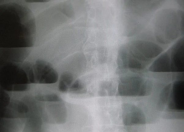
Abdominal radiograph is quick and retains a role here, unlike in undifferentiated abdominal pain. Learn the two distinctions:
- Small bowel - central loops, valvulae conniventes crossing the full width of the lumen, no gas in the colon or rectum, dilated above 3 cm
- Large bowel - peripheral loops, haustra that do not cross the full width, dilated above 6 cm, or above 9 cm at the caecum
- Ileus - dilatation of both small and large bowel with gas present all the way to the rectum, and no transition point
- Rigler sign (gas on both sides of the bowel wall) or free subdiaphragmatic gas indicates perforation
- A coffee bean or inverted U shadow arising from the pelvis suggests sigmoid volvulus
Contrast-enhanced CT of the abdomen and pelvis is the definitive investigation and should be obtained in essentially all adults with suspected obstruction. It identifies the level and the cause, demonstrates a transition point, distinguishes obstruction from ileus, and, crucially, assesses bowel viability. Signs of ischaemia include reduced or absent bowel wall enhancement, wall thickening, mesenteric fat stranding, pneumatosis intestinalis and portal venous gas.1
In suspected large bowel obstruction, a CT with rectal contrast or a water-soluble contrast enema can confirm a true mechanical obstruction and distinguish it from pseudo-obstruction, which is important because operating on Ogilvie syndrome is harmful.
Management
Initial resuscitation: drip and suck
- Nil by mouth
- Nasogastric tube on free drainage, with regular aspiration. This decompresses the stomach, relieves vomiting and reduces the risk of aspiration at induction of anaesthesia.
- Intravenous fluid resuscitation with a balanced crystalloid. Patients are typically several litres depleted, and the volume required is routinely underestimated.3
- Correct electrolytes, particularly potassium, which is nearly always low from vomiting and which perpetuates ileus if left uncorrected
- Urinary catheter and hourly urine output to guide resuscitation, aiming for at least 0.5 mL/kg/hour
- Analgesia and antiemetics, and venous thromboembolism prophylaxis
- Strict fluid balance chart, recording nasogastric losses and replacing them
- Serial abdominal examination and repeat bloods, because deterioration is detected clinically before it is detected radiologically
Adhesional small bowel obstruction
Most adhesional obstruction without strangulation settles on conservative management, and around 70 to 80% resolve within 48 to 72 hours. Conservative treatment should be abandoned in favour of surgery if there is any sign of strangulation, if the patient deteriorates, or if there is no resolution after roughly 72 hours, since prolonged temporising increases both morbidity and the eventual complexity of the operation.2
Surgery is by adhesiolysis, with resection of any non-viable bowel and primary anastomosis where the patient is stable and the bowel healthy. Laparoscopy is appropriate in selected cases with a single band adhesion and limited distension.
Large bowel obstruction
This rarely settles with conservative management, because the cause is usually a fixed lesion. The options depend on the cause, the site and the patient.
| Situation | Option | Considerations |
|---|---|---|
| Obstructing right-sided or transverse colon tumour | Right or extended right hemicolectomy with primary ileocolic anastomosis | The ileocolic anastomosis heals well even in the emergency setting, so a stoma is often avoidable |
| Obstructing left-sided or sigmoid tumour, unstable patient | Hartmann procedure - resection with an end colostomy and closure of the rectal stump | Avoids an anastomosis in unfavourable conditions. Reversal is possible later but many are never reversed. |
| Obstructing left-sided tumour, stable patient | Resection with primary anastomosis, with or without a defunctioning loop ileostomy | Requires a stable, well-resuscitated patient and appropriate expertise |
| Obstructing left-sided tumour, palliative or bridge to surgery | Self-expanding metal stent placed endoscopically | Relieves obstruction, allows staging and optimisation, and converts an emergency into an elective operation. Risks perforation and stent migration. |
| Unresectable or very frail patient | Defunctioning loop colostomy alone | Relieves the obstruction without the physiological insult of a resection |
Managing ileus
Treatment is supportive and directed at the cause. Correct electrolytes, minimise opioids by using multimodal and regional analgesia, exclude an intra-abdominal collection or anastomotic leak if it is prolonged or develops after an initial recovery, encourage early mobilisation, and use nasogastric drainage for symptomatic relief. Enhanced recovery programmes reduce ileus through early feeding, early mobilisation, avoidance of routine nasogastric tubes and opioid-sparing analgesia.4
Acute colonic pseudo-obstruction (Ogilvie syndrome)
This is massive colonic dilatation without a mechanical cause, typically in an elderly or unwell inpatient after trauma, orthopaedic surgery, or with sepsis or electrolyte disturbance. Mechanical obstruction must be excluded on CT or contrast enema before treatment.
- Correct electrolytes, stop opioids and anticholinergics, treat sepsis and mobilise the patient
- Nasogastric decompression and, where tolerated, a flatus tube
- Intravenous neostigmine if conservative measures fail and the caecum continues to dilate. It is highly effective, but must be given with cardiac monitoring and atropine available because it can cause profound bradycardia. It is contraindicated in mechanical obstruction, bradycardia and active bronchospasm.
- Colonoscopic decompression if neostigmine fails or is contraindicated
- Surgery only for perforation or ischaemia, as it carries high mortality in this group
Complications
- Bowel ischaemia, infarction and perforation - the feared endpoint, with faecal peritonitis and septic shock
- Hypovolaemic shock and acute kidney injury from unreplaced third-space losses
- Aspiration pneumonitis from vomiting, particularly at induction of anaesthesia, which is why the nasogastric tube matters
- Electrolyte disturbance - hypokalaemia, hypochloraemic alkalosis and hypomagnesaemia
- Short bowel syndrome after extensive small bowel resection, with lifelong nutritional consequences
- Anastomotic leak, stoma complications and wound dehiscence after surgery
- Recurrent adhesional obstruction - each operation generates further adhesions, so the risk compounds
- Abdominal compartment syndrome in massive distension
Red flags
Prognosis
Simple obstruction managed promptly has a good outcome, and most adhesional small bowel obstruction resolves without an operation. Recurrence is the main long-term issue: after one episode of adhesional obstruction, a substantial minority have another within five years, and each subsequent operation increases the adhesive burden.
Outcome is far worse once ischaemia has occurred. Mortality after resection of infarcted bowel is high, and the risk rises steeply with age, frailty and delay to theatre. Emergency surgery for obstructing colorectal cancer carries a worse oncological outcome than elective resection, which is part of the rationale for bowel cancer screening and for stenting as a bridge to elective surgery.
The practical lesson is that the decisions determining outcome are made in the first 24 hours: resuscitating adequately, obtaining a CT, recognising a closed loop or strangulation, and not allowing a patient to drift on a conservative pathway they are not responding to.
References
- Royal College of Radiologists. iRefer: making the best use of clinical radiology. Available here
- Ten Broek RPG, Krielen P, Di Saverio S et al. Bologna guidelines for diagnosis and management of adhesive small bowel obstruction: 2017 update. World Journal of Emergency Surgery. 2018. Available here
- NICE CG174. Intravenous fluid therapy in adults in hospital. 2013, updated 2017. Available here
- Association of Coloproctology of Great Britain and Ireland. Guidelines for the management of colorectal cancer. Available here
This article is written for revision and education. It is not clinical guidance and must not be used to make decisions about the care of a patient. Always check current NICE guidance and local protocols.