
Kawasaki Disease
Key points
- Definition: an acute self-limiting vasculitis of medium-sized muscular arteries, of unknown cause, with a particular predilection for the coronary arteries.
- Why it matters: it is the commonest cause of acquired heart disease in children in the UK and other high-income countries.
- Who gets it: children under 5 in 80% of cases, with a peak in the second year of life, and a higher incidence in children of East Asian ancestry.
- Diagnosis: clinical - fever for 5 days or more plus at least 4 of conjunctivitis, rash, cervical lymphadenopathy, mucosal changes and extremity changes. There is no confirmatory test.
- The clinical signature: a strikingly irritable young child with unremitting fever that does not respond to antibiotics or antipyretics.
- Treatment: intravenous immunoglobulin 2 g/kg as a single infusion, given within 10 days of fever onset, plus aspirin.
- The aspirin exception: Kawasaki disease is one of the very few indications for aspirin in a child under 16, and this is regularly examined.
- Outcome: untreated, about a quarter develop coronary artery aneurysms. Treated within 10 days, fewer than 5% do.
Introduction
Kawasaki disease is an acute febrile vasculitis of childhood, first described by Tomisaku Kawasaki in 1967. It is self-limiting - the fever will eventually settle whatever you do - but the vasculitis can permanently damage the coronary arteries in the process, and that damage is largely preventable if treatment is given in time.
This is what makes it an important and frequently examined diagnosis. There is no diagnostic test, the individual features are all shared with commoner illnesses, and the therapeutic window closes at around 10 days. A child with 6 days of fever who has been treated for tonsillitis, then for a presumed viral exanthem, and is now on their third antibiotic, is the classic missed case.
Incidence in the UK is roughly 8-9 per 100,000 children under 5. In Japan it is more than thirty times higher, and children of Japanese and Korean ancestry living outside Asia retain an elevated risk - which points to genetic susceptibility rather than environmental exposure alone.1
Aetiology and pathophysiology
The cause remains unknown. The epidemiology - seasonal peaks, geographical clustering, an age distribution that spares young infants with maternal antibody and older children who are presumably already immune - strongly suggests a widespread infectious trigger that causes disease only in genetically susceptible children. Polymorphisms in ITPKC, a negative regulator of T-cell activation, and in CASP3 are the best-established susceptibility loci. Kawasaki disease is not contagious, and siblings do not need isolating.
Pathologically the arteritis proceeds in three overlapping stages:
- Necrotising arteritis in the first 2 weeks - neutrophilic destruction of the arterial wall from lumen outwards, which is what produces saccular aneurysms
- Subacute and chronic vasculitis - a lymphocytic, plasma cell and eosinophilic infiltrate that may persist for months or years and produces fusiform aneurysms
- Luminal myofibroblastic proliferation - progressive intimal thickening that can cause late stenosis and ischaemia even after an aneurysm appears to have regressed
Two consequences follow. First, aneurysms form during a defined window in the first fortnight, which is why treatment timing matters so much. Second, the arterial wall never truly returns to normal, so a child with significant coronary involvement needs lifelong cardiology follow-up even if the vessel calibre looks normal on later imaging.
Clinical features and diagnostic criteria
Diagnosis requires fever for 5 days or more, plus at least 4 of the 5 principal clinical features.1,2 The fever is typically high, remittent, and conspicuously unresponsive to antipyretics and antibiotics.
| Feature | Detail |
|---|---|
| Bilateral conjunctival injection | Bulbar, non-purulent, with sparing of the limbus - a clear ring around the iris. Painless. |
| Mucous membrane changes | Cracked, red, fissured and bleeding lips; strawberry tongue; diffuse erythema of the oropharynx. No ulcers or exudate. |
| Polymorphous rash | Any morphology except vesicular or bullous - maculopapular, morbilliform, urticarial or erythroderma. Early desquamation in the nappy area is characteristic. |
| Extremity changes | Acutely, erythema and firm oedema of the palms and soles. In weeks 2-3, periungual desquamation peeling from the fingertips. |
| Cervical lymphadenopathy | At least one node of 1.5 cm or more, usually unilateral - the least frequently present of the five criteria |
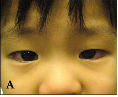
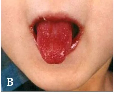
Other features that support the diagnosis
- Extreme irritability - disproportionate, inconsolable and very characteristic; possibly related to an aseptic meningitis
- Erythema and induration at the site of a previous BCG scar - highly suggestive where present, and specific enough to be worth looking for deliberately
- Arthralgia or arthritis, typically of large joints
- Sterile pyuria from urethritis - a common source of confusion with urinary tract infection
- Gastrointestinal upset: vomiting, diarrhoea, abdominal pain, and occasionally hydrops of the gallbladder
- Anterior uveitis on slit lamp examination
- Cardiac involvement: myocarditis, pericardial effusion, tachycardia out of proportion to fever, and a gallop rhythm
The three phases
| Phase | Timing | Features |
|---|---|---|
| Acute febrile | Days 1-14 | Fever, the five principal criteria, irritability, myocarditis. This is the window in which treatment prevents aneurysms. |
| Subacute | Weeks 2-4 | Fever settles, periungual desquamation appears, platelet count rises steeply. Coronary aneurysms form and the risk of sudden death is highest. |
| Convalescent | Weeks 4-8 | Clinical recovery with normalisation of inflammatory markers. Beau's lines may appear on the nails. |
Differential diagnosis
| Condition | Points against Kawasaki disease |
|---|---|
| Scarlet fever | Sandpaper rash sparing the palms and soles, rapid response to penicillin, positive throat swab, no conjunctivitis |
| Measles | Koplik spots, prominent cough and coryza, rash spreading downwards from behind the ears, exudative rather than limbal-sparing conjunctivitis |
| Adenovirus | Exudative conjunctivitis and pharyngitis, positive respiratory PCR, less marked inflammatory response |
| Staphylococcal scalded skin syndrome | Tender skin, positive Nikolsky sign, sheet-like peeling, mucosae spared |
| Toxic shock syndrome | Hypotension and multi-organ dysfunction early, with a source of staphylococcal or streptococcal infection |
| Stevens-Johnson syndrome | Recent drug exposure, target lesions, mucosal ulceration and skin detachment |
| Systemic juvenile idiopathic arthritis | Quotidian spiking fever for weeks, salmon-pink evanescent rash, arthritis, hepatosplenomegaly |
| PIMS-TS / MIS-C | Older children, prominent gastrointestinal symptoms and shock, temporally linked to SARS-CoV-2, markedly raised ferritin and troponin |
Investigations
No test confirms or excludes Kawasaki disease. Investigations serve three purposes: to support the diagnosis in incomplete cases, to exclude mimics, and to assess the coronary arteries.
Blood and urine
- FBC - normocytic normochromic anaemia and neutrophilia in the acute phase; a marked thrombocytosis after day 7 is characteristic, with platelet counts often above 450 and sometimes above 1,000 x10^9/L
- CRP and ESR - markedly raised, and their absence should make you question the diagnosis
- LFTs - raised transaminases and a low albumin, which correlates with severity
- U&Es - hyponatraemia is common
- Urinalysis - sterile pyuria with no organisms on culture
- Troponin and BNP where myocarditis is suspected
- Exclusion tests: throat swab and ASO titre, blood cultures, and viral PCR for measles, adenovirus and EBV
Cardiac assessment
- Echocardiography at diagnosis, repeated at 1-2 weeks and again at 6-8 weeks. Coronary dimensions are reported as Z-scores corrected for body surface area, since absolute diameters are meaningless in a growing child.
- ECG - may show a prolonged PR interval, non-specific ST and T wave changes, or arrhythmia in myocarditis
- More frequent imaging, and cardiac CT or MR angiography, where aneurysms are identified or the child is unwell
- A normal early echocardiogram does not exclude the diagnosis or remove the need for treatment - aneurysms typically appear in the second or third week
Management
Intravenous immunoglobulin
IVIG 2 g/kg as a single infusion over 10-12 hours is the definitive treatment. Given within 10 days of the onset of fever - and ideally within 7 - it reduces the incidence of coronary artery aneurysms from around 25% to under 5%.1,3
- Treat on clinical suspicion. Do not delay waiting for an echocardiogram or for further criteria to appear.
- IVIG is still given after day 10 if the child remains febrile or has persistently raised inflammatory markers or coronary abnormalities, though the benefit is smaller
- About 10-20% of children have IVIG-resistant disease, defined as persistent or recrudescent fever more than 36 hours after the infusion. These children are given a second dose of IVIG, usually with corticosteroids.
- Corticosteroids are added to first-line treatment in children assessed as high risk, and are used in refractory disease. Infliximab, ciclosporin and anakinra are third-line options managed in specialist centres.
Aspirin
- High dose (anti-inflammatory): 7.5-12.5 mg/kg four times daily, continued until the child has been afebrile for 48 hours5
- Low dose (antiplatelet): 2-5 mg/kg once daily thereafter, continued for 6-8 weeks and stopped if the 6-8 week echocardiogram is normal
- Indefinite low-dose aspirin if coronary abnormalities persist, with the addition of warfarin or low molecular weight heparin for giant aneurysms, where the thrombotic risk is high
- Kawasaki disease is one of the few situations in which aspirin is deliberately prescribed to a child under 16, accepting a small Reye's syndrome risk because the cardiac benefit is far greater
Supportive care and follow-up
- Admit for observation, fluids and analgesia; these children are miserable and often not drinking
- Daily clinical review for recrudescent fever in the 48 hours after IVIG
- Cardiology follow-up determined by the maximum coronary Z-score, ranging from discharge after a normal 6-8 week echocardiogram to lifelong surveillance with imaging and functional testing for those with giant aneurysms
- Long-term cardiovascular risk factor advice for children with persistent coronary abnormalities - no smoking, activity, lipid monitoring
- Advise parents that recurrence occurs in around 2-3% and that they should seek review early for any prolonged fever in the future
Complications
- Coronary artery aneurysms - the defining complication. Giant aneurysms, 8 mm or more in diameter or with a Z-score of 10 or above, carry the worst prognosis.
- Coronary thrombosis and myocardial infarction - the leading cause of death, occurring most often in the subacute phase or years later in a giant aneurysm. Infarction in a child may present only as pallor, vomiting, abdominal pain or shock rather than chest pain.
- Coronary stenosis from late myointimal proliferation, causing ischaemia in adolescence or adulthood
- Myocarditis, pericardial effusion and mitral regurgitation in the acute phase
- Arrhythmia and sudden cardiac death
- Kawasaki disease shock syndrome - hypotension with myocardial dysfunction and capillary leak, more often IVIG-resistant
- Macrophage activation syndrome - suspect if the child deteriorates with falling platelets, falling ESR, very high ferritin and hypofibrinogenaemia
- Peripheral arterial aneurysms - axillary, iliac or renal, which is why a systemic vasculitis rather than a cardiac disease is the right way to think about it
Red flags
Prognosis
With timely IVIG, mortality in the UK is well under 0.5%, and the great majority of children make a complete recovery with no cardiac sequelae. Around half to two-thirds of small and medium aneurysms regress angiographically within 1-2 years, though the vessel wall remains abnormal histologically.
Giant aneurysms behave differently. They rarely regress, they thrombose, and they require lifelong anticoagulation, surveillance and sometimes coronary intervention or bypass grafting in adolescence or adult life. Whether children who had Kawasaki disease without aneurysms carry any excess long-term cardiovascular risk remains an open question, and current guidance is to manage conventional risk factors attentively.1
The single determinant of outcome that a clinician controls is time to treatment. That is why the practical message of this topic is not really about vasculitis at all: it is that a young child with fever lasting 5 days or more and no explanation should have Kawasaki disease actively considered and inflammatory markers checked, rather than another course of antibiotics.
References
- McCrindle BW, Rowley AH, Newburger JW et al. Diagnosis, treatment, and long-term management of Kawasaki disease: a scientific statement for health professionals from the American Heart Association. Circulation. 2017. Available here
- NICE Clinical Knowledge Summaries. Kawasaki disease. Available here
- Eleftheriou D, Levin M, Shingadia D et al. Management of Kawasaki disease. Archives of Disease in Childhood. 2014. Available here
- NICE NG143. Fever in under 5s: assessment and initial management. 2019, updated 2021. Available here
- BNF for Children. Aspirin. Available here
- UKHSA. Contraindications and special considerations: the Green Book, chapter 6. Available here
- RCPCH. Paediatric multisystem inflammatory syndrome temporally associated with COVID-19 (PIMS-TS): guidance. Available here
This article is written for revision and education. It is not clinical guidance and must not be used to make decisions about the care of a patient. Always check current NICE guidance and local protocols.