
Conjunctivitis: Bacterial, Viral and Allergic
Key points
- Definition: inflammation of the conjunctiva, producing diffuse redness, discharge and grittiness with normal visual acuity and a normal pupil.
- The safety rule: conjunctivitis does not reduce vision, does not cause true photophobia and does not cause ciliary flush. If any of those are present, the diagnosis is something else.
- Discharge: purulent and sticky suggests bacterial, watery suggests viral, stringy and itchy suggests allergic.
- Commonest cause: adenovirus, which is highly contagious, often bilateral in sequence, and accompanied by a tender preauricular lymph node.
- Bacterial treatment: most cases are self-limiting within 5-7 days. Topical chloramphenicol shortens symptoms modestly and is reserved for severe or non-resolving disease.
- Allergic treatment: allergen avoidance, cool compresses, then a topical antihistamine or mast cell stabiliser such as sodium cromoglicate.
- Never miss: ophthalmia neonatorum, gonococcal hyperacute conjunctivitis, and a red eye in a contact lens wearer, which is keratitis until proved otherwise.
- Advice: no school or work exclusion is required for simple infective conjunctivitis, but hand hygiene, separate towels and no shared eye make-up are.
Introduction
Conjunctivitis is inflammation of the conjunctiva, the thin vascular membrane that lines the inner eyelids and covers the sclera as far as the limbus. It is the commonest cause of a red eye in every setting, and around one in eight of the population will consult about it at some point.1
Almost all of it is benign and self-limiting. The clinical importance of the topic lies in two directions: recognising the small number of presentations that are dangerous, and resisting the strong reflex to prescribe an antibiotic for something that will settle on its own. UK antimicrobial stewardship guidance identifies acute infective conjunctivitis as one of the conditions where topical antibiotics are most consistently over-prescribed.2
The conjunctiva has no role in refraction and lies entirely outside the visual axis. This single anatomical fact generates the safety rule that structures the whole assessment: conjunctivitis does not reduce visual acuity. A red eye with a genuine drop in acuity has something else going on, and looking no further than the conjunctiva is how corneal ulcers and uveitis are missed.
Aetiology and classification
Conjunctivitis is conventionally divided into infective and allergic, with a smaller group of irritant and toxic causes that are easy to forget and easy to treat once identified.
Viral
- Adenovirus accounts for around 75% of infective conjunctivitis in adults. Serotypes 8, 19 and 37 cause epidemic keratoconjunctivitis, which is more severe and can leave subepithelial corneal infiltrates that blur vision for weeks or months.
- Herpes simplex virus causes a unilateral follicular conjunctivitis, often with vesicles on the lid margin, and may accompany a dendritic corneal ulcer
- Varicella zoster in herpes zoster ophthalmicus
- Enterovirus and coxsackievirus cause acute haemorrhagic conjunctivitis, with subconjunctival haemorrhages, seen in outbreaks
- Molluscum contagiosum on the lid margin can shed viral particles and cause a chronic follicular conjunctivitis that only resolves when the lesion is removed
Bacterial
- Staphylococcus aureus, Streptococcus pneumoniae and Haemophilus influenzae cause most simple acute bacterial conjunctivitis. H. influenzae is disproportionately common in children and is associated with concurrent otitis media - the conjunctivitis-otitis syndrome.
- Moraxella catarrhalis typically causes a more indolent angular conjunctivitis
- Neisseria gonorrhoeae causes hyperacute conjunctivitis with copious, rapidly reaccumulating purulent discharge, marked lid swelling and a genuine risk of corneal perforation within 48 hours. This is a sight-threatening emergency.
- Chlamydia trachomatis serovars D-K cause a chronic follicular conjunctivitis in sexually active adults, often lasting weeks and unresponsive to standard drops
- Chlamydia trachomatis serovars A-C cause trachoma, the world's leading infectious cause of blindness, through repeated reinfection, conjunctival scarring, entropion and trichiasis
Allergic
- Seasonal allergic conjunctivitis (hay fever eyes) - IgE-mediated, tied to pollen seasons, almost always bilateral
- Perennial allergic conjunctivitis - house dust mite and animal dander, symptoms year-round
- Vernal keratoconjunctivitis - a severe, chronic form in atopic boys and young men, with giant cobblestone papillae on the upper tarsal conjunctiva and a risk of corneal shield ulcers and permanent visual loss
- Atopic keratoconjunctivitis - the adult counterpart, associated with atopic dermatitis, and carrying a risk of corneal scarring, cataract and keratoconus
- Giant papillary conjunctivitis - a mechanical and immunological reaction to contact lenses, sutures or an ocular prosthesis
Risk factors
- Contact with an affected person - adenovirus survives on surfaces for weeks and spreads readily in schools, households and eye clinics
- Young age, both for viral and for Haemophilus conjunctivitis
- Atopy - eczema, asthma and allergic rhinitis for the allergic forms
- Contact lens wear, particularly for giant papillary conjunctivitis and as a risk factor for the far more serious keratitis
- Recent unprotected sexual intercourse or a new partner, for chlamydial and gonococcal conjunctivitis
- Blepharitis, dry eye and previous ocular surface disease
- Immunosuppression, which prolongs and worsens viral disease
- Crowding, poor sanitation and limited access to water, for trachoma
Clinical features
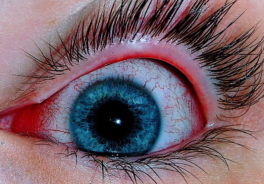
Every form of conjunctivitis shares a core picture: diffuse conjunctival injection greatest in the fornices and least at the limbus, a gritty or foreign body sensation, some form of discharge, and normal vision. The subtype is then distinguished largely by the character of the discharge and the accompanying symptoms.
| Feature | Bacterial | Viral | Allergic |
|---|---|---|---|
| Defining symptom | Sticky discharge | Watery discharge, often with a preceding cold | Itch |
| Discharge | Purulent, yellow-green, reaccumulates through the day | Watery, clear | Stringy, ropy mucus |
| Lids on waking | Stuck together | Crusted but usually openable | Puffy rather than stuck |
| Laterality | Often unilateral then spreads | Starts unilateral, second eye in 1-2 days | Bilateral from the outset |
| Conjunctival reaction | Papillae | Follicles (inferior fornix) | Papillae, chemosis |
| Preauricular node | Absent | Tender node present | Absent |
| Duration | 5-7 days | 1-3 weeks, sometimes longer | As long as the allergen exposure |
| Systemic clues | Otitis media in children | Sore throat, coryza, fever | Rhinitis, eczema, asthma |
Papillae versus follicles
These are examined by everting the upper lid and are worth understanding rather than memorising. Papillae are raised, flat-topped, cobblestoned elevations each with a central vessel, produced by a fibrovascular response, and are seen in bacterial and allergic disease. Follicles are pale, round, avascular elevations most obvious in the inferior fornix, representing lymphoid aggregates, and are seen in viral and chlamydial disease. A follicular conjunctivitis that has lasted more than three weeks in a sexually active adult is chlamydial until proved otherwise.
Features that exclude simple conjunctivitis
Examination
- Visual acuity in each eye with glasses or a pinhole, before instilling anything. This is the single most important measurement and must be documented.
- Pattern and distribution of injection, comparing fornices with limbus
- Character and volume of discharge, and whether it reaccumulates within minutes of being wiped away
- Lid margins for crusting, collarettes, telangiectasia and blocked meibomian orifices, and the lid skin for vesicles
- Upper lid eversion for papillae, follicles, giant papillae or a subtarsal foreign body
- Fluorescein staining for corneal involvement - punctate erosions, a dendrite, or an infiltrate
- Preauricular lymph nodes, palpated in front of the tragus - tenderness strongly suggests viral or chlamydial disease
- Pupil reactions and, where possible, slit lamp assessment of the anterior chamber
Differential diagnosis
- Anterior uveitis - photophobia, aching pain, ciliary flush, a small or irregular pupil and reduced vision
- Microbial keratitis - severe pain, a white corneal infiltrate and contact lens wear
- Herpes simplex keratitis - a fluorescein-staining dendrite, reduced corneal sensation, unilateral
- Acute angle closure glaucoma - severe pain, vomiting, haloes and a fixed mid-dilated pupil, occasionally mistaken for conjunctivitis because the eye is red
- Episcleritis and scleritis - sectoral or violaceous redness rather than diffuse injection, and no discharge
- Subconjunctival haemorrhage - a solid block of blood rather than injected vessels
- Blepharitis, meibomian gland dysfunction and dry eye - chronic, bilateral, worse in the morning or at the end of the day respectively
- Preseptal or orbital cellulitis - lid swelling with fever, and in the orbital form proptosis and restricted eye movements
- Nasolacrimal duct obstruction in infants - chronic watering and stickiness with a white, quiet eye
Investigations
Conjunctivitis is a clinical diagnosis and routine swabbing is not recommended. Investigation is reserved for the presentations that behave unusually.3
- Charcoal swab for bacterial culture - in hyperacute, severe or treatment-resistant cases, and in all neonates
- Nucleic acid amplification test for Chlamydia trachomatis and Neisseria gonorrhoeae - in any chronic follicular conjunctivitis in a sexually active adult, and in every neonate with conjunctivitis. Send a conjunctival sample and refer to sexual health for full screening and partner notification.
- Viral PCR - available for adenovirus and herpes simplex, mostly used to control outbreaks or where the diagnosis alters treatment
- Gram stain and urgent microscopy - in suspected gonococcal conjunctivitis, where Gram-negative intracellular diplococci allow treatment to start immediately
- Corneal scrape - if there is any corneal infiltrate, arranged by ophthalmology
- Allergy testing - rarely needed; skin prick testing or specific IgE only if allergen identification would change management
Management
Infective conjunctivitis
Around 65% of cases of acute infective conjunctivitis resolve within 2-5 days without any treatment, and topical antibiotics shorten symptoms by roughly a day.2 NICE therefore recommends self-care as the default, with a delayed or backup prescription as an option for those who want one.
- Self-care first - clean the lids with cooled boiled water and cotton wool, use lubricating drops for comfort, and avoid contact lenses until 24 hours after symptoms resolve
- Chloramphenicol 0.5% drops two-hourly for 48 hours then four times daily, or 1% ointment four times daily, for 5-7 days, if treatment is chosen4
- Fusidic acid 1% gel twice daily is an alternative, and is preferred in pregnancy
- Review if not improving after a week, and reconsider the diagnosis rather than changing antibiotic
- Chlamydial conjunctivitis needs systemic treatment - oral doxycycline 100 mg twice daily for 7 days, or azithromycin 1 g as a single dose - together with sexual health referral and partner notification. Topical treatment alone is inadequate.
- Gonococcal conjunctivitis needs admission, intramuscular ceftriaxone, hourly saline irrigation and same-day ophthalmology review
Viral conjunctivitis
There is no effective antiviral for adenoviral conjunctivitis, and antibiotics do nothing. Treatment is symptomatic: cool compresses, preservative-free lubricants and simple analgesia. Explain that the eye may get worse before it gets better over the first three to five days, and that symptoms can persist for two to three weeks. Topical steroids are sometimes used by ophthalmologists for the subepithelial infiltrates of epidemic keratoconjunctivitis, but they prolong viral shedding and are never started in primary care.
Allergic conjunctivitis
- Allergen avoidance where feasible, plus cool compresses and preservative-free lubricants to dilute and wash out allergen
- Advise against rubbing - it degranulates mast cells directly and worsens symptoms, and chronic rubbing is implicated in keratoconus
- Topical antihistamine such as olopatadine or ketotifen, which have both antihistamine and mast cell stabilising activity, for rapid relief
- Mast cell stabiliser such as sodium cromoglicate 2% four times daily, started two weeks before the pollen season, for prophylaxis - it takes days to work and is not a rescue treatment
- Oral antihistamine if there is concurrent rhinitis
- Refer for topical steroid or ciclosporin if there is vernal or atopic keratoconjunctivitis, corneal involvement or failure of the above
Complications
- Subepithelial corneal infiltrates after adenoviral epidemic keratoconjunctivitis, causing glare and blurring for weeks to months
- Corneal perforation in untreated gonococcal conjunctivitis - the organism can penetrate intact corneal epithelium, which is unusual among bacteria
- Punctate epithelial keratitis and, in severe vernal disease, a corneal shield ulcer which can scar the visual axis
- Conjunctival scarring, symblepharon and entropion in trachoma, adenoviral membranous conjunctivitis and Stevens-Johnson syndrome
- Chronic dry eye from loss of goblet cells and damage to accessory lacrimal glands after severe or repeated inflammation
- Delayed diagnosis of a serious alternative - the commonest and most examinable complication of all, when uveitis or keratitis is treated for weeks as conjunctivitis
Ophthalmia neonatorum
Conjunctivitis in the first 28 days of life is given its own name because the differential and the stakes are different. It is a notifiable disease in the UK and every case warrants a specific microbiological diagnosis rather than empirical drops.5
| Onset | Likely cause | Features and management |
|---|---|---|
| First 24-48 hours | Chemical or Neisseria gonorrhoeae | Gonococcus gives hyperacute purulent discharge and marked lid oedema; admit, systemic cefotaxime, urgent ophthalmology |
| Day 5-14 | Chlamydia trachomatis | Mucopurulent discharge, often mild; oral erythromycin for 14 days, and treat the mother and her partners |
| Day 5 onwards | Staphylococci, streptococci, Haemophilus | Topical antibiotic after swabs |
| Any time in the first month | Herpes simplex | Vesicles, keratitis; systemic aciclovir, and assess for disseminated neonatal HSV |
Prognosis
Simple bacterial conjunctivitis resolves in 5-7 days, with or without antibiotics, and leaves no sequelae. Adenoviral conjunctivitis takes one to three weeks and can leave corneal infiltrates that resolve slowly but almost always completely. Allergic conjunctivitis persists as long as exposure continues and recurs predictably each season, but does not damage the eye in its seasonal and perennial forms.
The forms with a genuinely guarded prognosis are the ones that involve the cornea: vernal and atopic keratoconjunctivitis, gonococcal infection, and trachoma. Trachoma remains the leading infectious cause of blindness worldwide, and the World Health Organization's SAFE strategy - surgery for trichiasis, antibiotics, facial cleanliness and environmental improvement - has driven elimination in a growing number of countries.6
For the great majority of patients, the useful message is that the eye will settle on its own, that an antibiotic changes little, and that they should return if vision drops, pain develops or symptoms have not improved after a week. That last safety net is the part that matters, because it is what catches the cases that were never conjunctivitis.
References
- NICE Clinical Knowledge Summaries. Conjunctivitis - infective. Available here
- NICE and Public Health England. Managing common infections: guidance for primary care. Available here
- NICE Clinical Knowledge Summaries. Conjunctivitis - allergic. Available here
- BNF. Chloramphenicol - eye preparations. Available here
- UK Health Security Agency. Notifiable diseases and causative organisms: how to report. Available here
- World Health Organization. Trachoma fact sheet. Available here
This article is written for revision and education. It is not clinical guidance and must not be used to make decisions about the care of a patient. Always check current NICE guidance and local protocols.