
Placental Abruption
Key points
- Definition: premature separation of a normally sited placenta from the uterine wall before delivery of the fetus.
- Frequency: complicates around 0.6-1% of pregnancies and accounts for a disproportionate share of perinatal mortality.
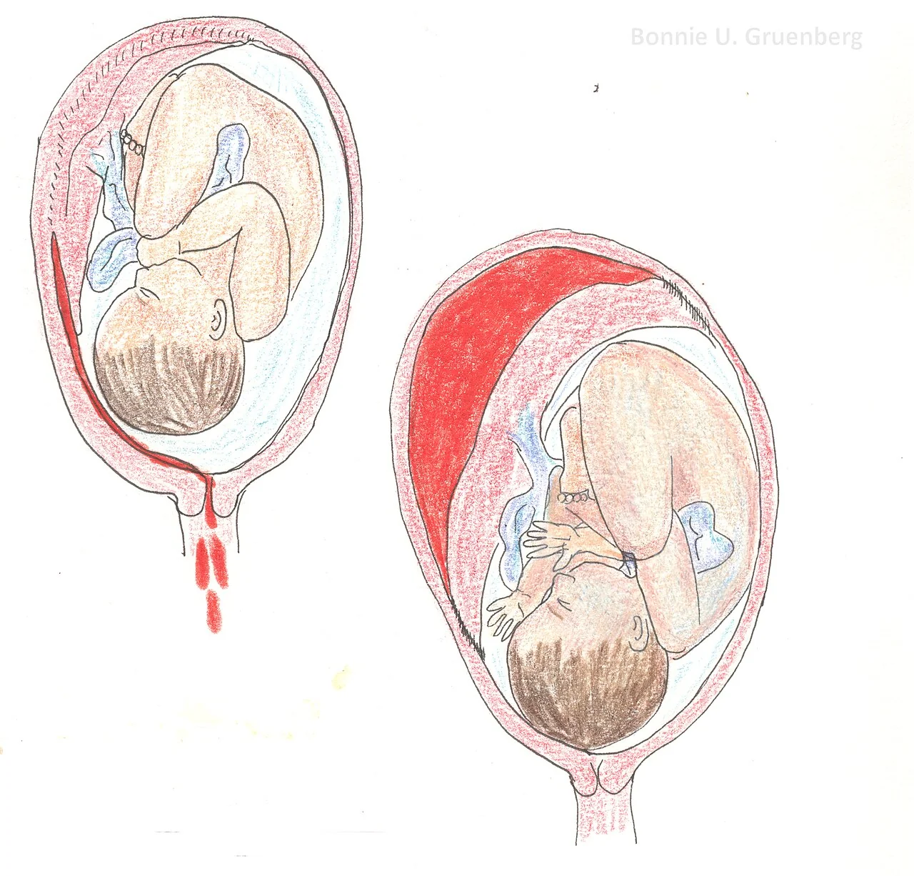
- Classification: revealed, concealed or mixed, depending on whether blood tracks out through the cervix or is retained behind the placenta.
- Presentation: sudden continuous severe abdominal or back pain with a tense, tender, woody uterus; the vaginal bleeding may be dark, scanty or absent.
- Diagnosis: clinical - a normal ultrasound does not exclude abruption because fresh clot is isoechoic with placental tissue.
- Trap: shock out of proportion to the visible bleeding, because the loss is concealed behind the placenta.
- Coagulopathy: abruption is the leading obstetric cause of disseminated intravascular coagulation; check fibrinogen, which should be above 2 g/L in pregnancy.
- Management: resuscitate then deliver; category 1 caesarean if the fetus is alive and compromised, vaginal birth if the fetus has died and the mother is stable.
Introduction
Placental abruption is the premature separation of a normally sited placenta from the uterine wall before the birth of the baby. It complicates around 0.6-1% of pregnancies but accounts for a disproportionate share of perinatal deaths, because the fetus loses its gas exchange surface abruptly and without warning.1
It is the diagnosis in obstetrics most likely to be underestimated at the bedside, for one reason: the volume of blood visible per vaginam may bear no relationship at all to the volume lost. A woman may have two litres of blood behind her placenta and a single stained pad. Any assessment that anchors on the visible loss will therefore under-resuscitate her, and the fetus will be the first to suffer.
Pathophysiology and classification
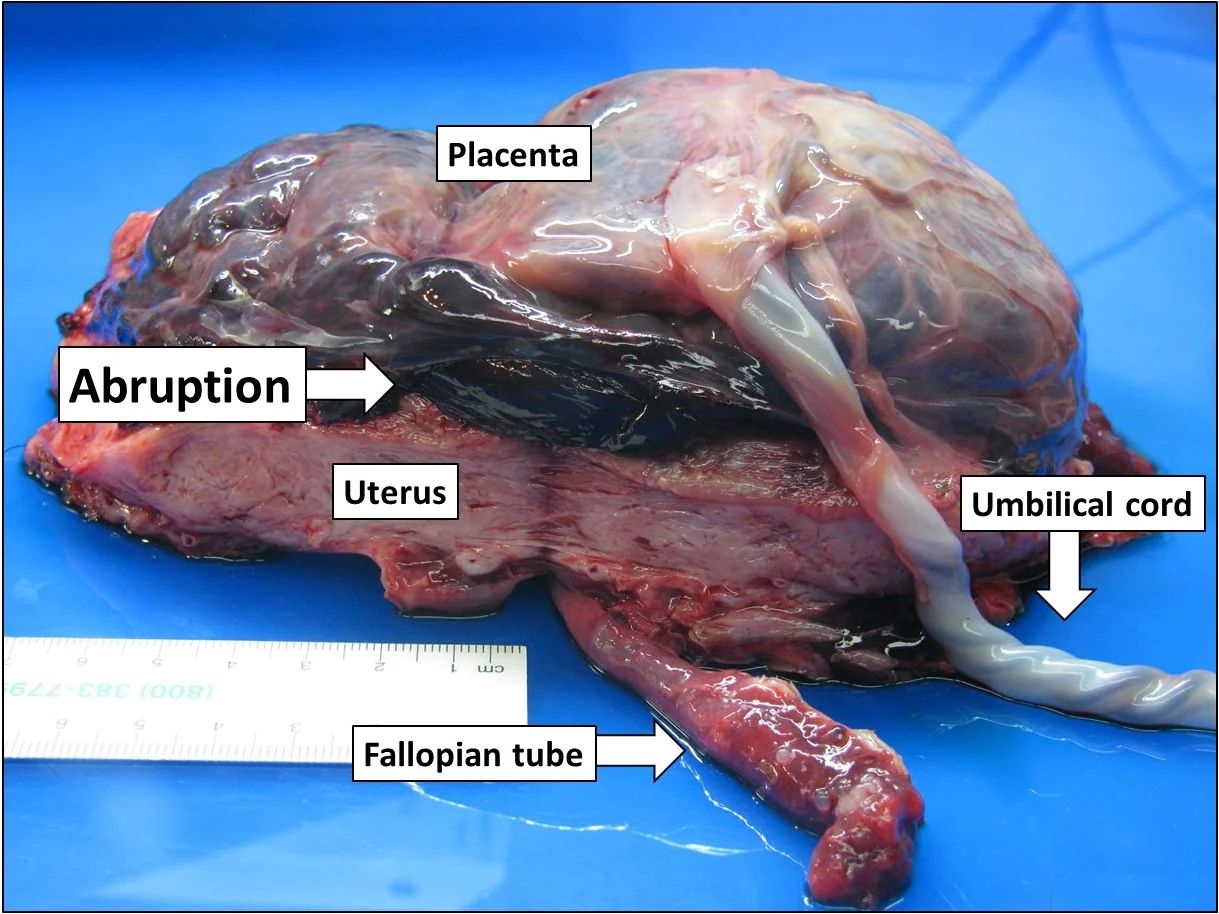
Abruption begins with rupture of a maternal decidual vessel. Blood at arterial pressure enters the decidua basalis and splits it, forming a haematoma that strips further placenta from the uterine wall. The process is self-propagating: each increment of separation shears more vessels and enlarges the haematoma. Because the exchange surface is lost immediately and irreversibly, fetal compromise is proportional to the area separated, and loss of more than about half the placental surface is usually fatal to the fetus.
Two rather different processes lead to the same endpoint. Chronic abruption arises from the same defective spiral artery remodelling that underlies pre-eclampsia and fetal growth restriction; these vessels are thin-walled, atherotic and prone to rupture, and the woman often has a history of small-for-dates growth or hypertension. Acute abruption is mechanical, following trauma or a sudden change in uterine volume, and occurs in a previously normal pregnancy.
| Type | What happens | Clinical consequence |
|---|---|---|
| Revealed | Blood tracks between the membranes and the uterine wall and escapes through the cervix | Visible bleeding roughly reflects the loss; less pain and less coagulopathy |
| Concealed | Blood is retained behind the placenta with no external loss | Severe pain, a rapidly enlarging tense uterus, profound shock with no visible bleeding, and a high risk of DIC |
| Mixed | Some blood escapes and some is retained | The commonest pattern; the visible loss always underestimates the total |
Risk factors
- Previous abruption - the strongest predictor, with a recurrence risk of around 4-12%, rising to roughly 25% after two previous abruptions
- Pre-eclampsia and chronic hypertension - shared pathology of defective placentation
- Fetal growth restriction - a marker of the same placental disease
- Smoking, cocaine and amphetamine use - cocaine causes intense vasoconstriction and is a classic cause of abruption in a young woman
- Abdominal trauma - road traffic collisions, falls, and domestic abuse, which must be asked about directly and in private
- Sudden uterine decompression - after rupture of membranes in polyhydramnios, or after delivery of the first twin
- Preterm prelabour rupture of membranes and chorioamnionitis
- Multiple pregnancy, multiparity and advanced maternal age
- Thrombophilia, including antiphospholipid syndrome
Clinical features
The classic presentation is sudden onset, continuous, severe abdominal pain, in contrast to the intermittent pain of labour. Where the placenta is posterior the pain is felt in the back and may be mistaken for musculoskeletal pain or renal colic. Vaginal bleeding, when present, is characteristically dark rather than bright red, because the blood has been retained for a period before escaping.
- Continuous severe abdominal or back pain, of sudden onset
- Dark vaginal bleeding, which may be scanty or entirely absent
- Reduced or absent fetal movements
- Frequent low-amplitude contractions, or a uterus that does not relax between contractions
- Maternal shock disproportionate to the visible loss
- Symptoms of associated pre-eclampsia: headache, visual disturbance, epigastric pain
Examination
The uterus is tense, tender and woody hard, and fetal parts are difficult or impossible to palpate. The symphysis-fundal height may be larger than expected because of the retroplacental haematoma, and may increase on serial measurement, which is a sinister sign. Observations show tachycardia and, later, hypotension. The cardiotocograph is frequently abnormal, with reduced variability, late decelerations, tachysystole, or in severe cases a bradycardia or a sinusoidal pattern indicating fetal anaemia.
Differential diagnosis
- Placenta praevia - painless bright red bleeding with a soft non-tender uterus and a high presenting part
- Uterine rupture - severe pain with loss of contractions, cardiovascular collapse, fetal bradycardia and easily palpable fetal parts, almost always in a scarred uterus
- Labour - intermittent pain with relaxation between contractions and cervical change
- Chorioamnionitis - uterine tenderness with fever, maternal and fetal tachycardia and offensive liquor
- Acute abdomen unrelated to pregnancy - appendicitis, pancreatitis, renal colic, ovarian torsion
- HELLP syndrome - epigastric pain with abnormal liver enzymes and thrombocytopenia; may coexist with abruption
Investigations
Abruption is a clinical diagnosis. Investigations serve to assess severity, detect coagulopathy, exclude praevia and monitor the fetus, not to confirm the diagnosis.
| Investigation | Interpretation |
|---|---|
| Full blood count | Baseline haemoglobin, which lags behind acute loss; platelets fall in DIC |
| Coagulation screen and fibrinogen | The single most important laboratory test; fibrinogen below 2 g/L indicates significant coagulopathy in pregnancy |
| Crossmatch 4 units | Anticipate transfusion; a group and save alone is insufficient in major abruption |
| Urea, electrolytes and creatinine | Acute kidney injury from hypovolaemia and from associated pre-eclampsia |
| Kleihauer test | In RhD negative women after 20 weeks, to size the fetomaternal haemorrhage and the anti-D dose |
| Ultrasound | Excludes placenta praevia and assesses the fetus; a retroplacental clot is seen in only a minority of cases |
| Cardiotocography | Detects fetal compromise and uterine irritability, and is often the earliest objective abnormality |
Management
Resuscitation
- Call for senior obstetric, anaesthetic, midwifery, haematology and neonatal help; activate the major obstetric haemorrhage protocol
- ABCDE with high-flow oxygen, left lateral tilt to relieve aortocaval compression, and two large-bore cannulae
- Take FBC, coagulation, fibrinogen, U&E, LFTs, crossmatch and Kleihauer as the lines are sited
- Transfuse red cells early, using O negative if needed, and give fresh frozen plasma, cryoprecipitate or fibrinogen concentrate and platelets guided by results
- Catheterise and monitor hourly urine output; aim for at least 30 mL per hour
- Continuous cardiotocography once maternal resuscitation is under way
- Anti-D within 72 hours for RhD negative women, at a dose guided by the Kleihauer
Deciding on delivery
| Situation | Approach |
|---|---|
| Fetus alive and viable with an abnormal CTG, or maternal instability | Category 1 caesarean section as soon as the mother is being adequately resuscitated |
| Fetus alive, reassuring CTG, at or beyond 37 weeks | Deliver, usually by induction with amniotomy, with continuous monitoring and immediate access to theatre |
| Fetus alive, reassuring CTG, preterm and bleeding settled | Admit, give antenatal corticosteroids, magnesium sulfate for neuroprotection if under 30-34 weeks, and monitor closely; tocolysis is contraindicated if bleeding continues |
| Fetus has died and the mother is stable | Vaginal birth is preferred, with amniotomy and oxytocin; correct the coagulopathy in parallel |
| Fetus has died and the mother is unstable or bleeding heavily | Caesarean section may still be needed to control haemorrhage |
Vaginal birth after fetal death is usually rapid because the uterus is already highly irritable, and it avoids a laparotomy in a coagulopathic patient. Regional anaesthesia is contraindicated where there is coagulopathy or ongoing haemorrhage, because of the risk of spinal haematoma and because a sympathetic block worsens hypotension in a hypovolaemic woman.1,2
The third stage
Postpartum haemorrhage should be actively anticipated in every abruption. Give active management of the third stage, have uterotonics drawn up, and continue an oxytocin infusion after delivery. Blood that has infiltrated the myometrium produces a Couvelaire uterus, bruised and purple, which contracts poorly and bleeds; combined with a consumptive coagulopathy this is one of the classic routes to peripartum hysterectomy.
Complications
| Group | Complications |
|---|---|
| Maternal haemodynamic | Hypovolaemic shock, acute kidney injury, acute tubular necrosis, multi-organ failure, maternal death |
| Haematological | Disseminated intravascular coagulation, massive transfusion and its complications |
| Obstetric | Couvelaire uterus with uterine atony, postpartum haemorrhage, emergency hysterectomy |
| Endocrine | Sheehan's syndrome - anterior pituitary necrosis after prolonged hypotension, presenting with failure of lactation and amenorrhoea |
| Fetal and neonatal | Hypoxic-ischaemic encephalopathy, fetal anaemia, stillbirth, iatrogenic preterm birth, fetal growth restriction in chronic abruption |
Red flags
Prognosis and future pregnancies
Perinatal mortality in significant abruption remains high, driven by acute hypoxia and by preterm birth. Maternal outcome is generally good where haemorrhage and coagulopathy are recognised and treated early, and poor where the visible blood loss was used as the measure of severity.
Recurrence risk is substantial, at roughly 4-12% after one abruption and around 25% after two, so a subsequent pregnancy is managed as high risk. That means smoking cessation and cessation of any stimulant use, aspirin 75-150 mg from 12 weeks where there are risk factors for placental disease, serial growth scans with umbilical artery Doppler, and a documented plan for early presentation and for the timing of birth. Women should be told what symptoms to act on, and told to attend rather than telephone.1
A debrief should be offered after any major abruption. These events are frightening, frequently involve a general anaesthetic and an unplanned separation from the baby, and are a recognised trigger for post-traumatic stress. Explaining what happened, and why it is unlikely to be anything the woman did, is part of the clinical work rather than an optional courtesy.4
References
- RCOG Green-top Guideline No. 63. Antepartum haemorrhage. Available here
- RCOG Green-top Guideline No. 47. Blood transfusion in obstetrics. Available here
- NICE NG201. Antenatal care. 2021. Available here
- MBRRACE-UK. Saving Lives, Improving Mothers' Care. Available here
This article is written for revision and education. It is not clinical guidance and must not be used to make decisions about the care of a patient. Always check current NICE guidance and local protocols.