
Vasa Praevia
Key points
- Definition: fetal vessels running through the membranes over or within 2 cm of the internal cervical os, unsupported by placental tissue or umbilical cord.
- Types: type I from a velamentous cord insertion; type II from vessels bridging a bilobed or succenturiate placenta.
- Frequency: approximately 1 in 1,200 to 1 in 5,000 pregnancies, and considerably commoner after IVF.
- The blood is fetal: total fetoplacental blood volume at term is only around 250 mL, so a loss that looks trivial on a pad is exsanguinating.
- Presentation: painless fresh red bleeding at the moment the membranes rupture, followed within minutes by fetal bradycardia or a sinusoidal CTG.
- Diagnosis: transvaginal ultrasound with colour Doppler showing vessels crossing the os that do not move when the woman or the probe does.
- Outcome: perinatal mortality is around 60% when undiagnosed, but survival exceeds 95% when it is diagnosed antenatally and delivery is planned.
- Management: corticosteroids at around 32 weeks and elective caesarean section at 34-36 weeks, before the membranes can rupture.
Introduction
Vasa praevia is the presence of fetal blood vessels running through the fetal membranes over the internal cervical os, or within 2 cm of it, unsupported by either the umbilical cord or placental tissue. It affects somewhere between 1 in 1,200 and 1 in 5,000 pregnancies.1
It is a rare condition that receives disproportionate attention for a good reason: the outcome is almost entirely determined by whether the diagnosis was made before labour. Undiagnosed, perinatal mortality is around 60%, and the deaths occur within minutes of membrane rupture. Diagnosed antenatally, with planned caesarean birth before the membranes rupture, survival is well over 95%. There are few conditions in obstetrics where a single ultrasound finding makes so much difference.
The mechanism of death is exsanguination, not asphyxia, and this is the point that makes the whole topic make sense. The vessels are fetal. When the membranes rupture across them, the fetus bleeds out into the vagina. Total fetoplacental blood volume at term is around 250 mL, and the fetal circulation cannot tolerate the loss of even a modest fraction of that. A volume of blood that would be an unremarkable maternal bleed is fatal to the fetus.
Anatomy and classification
Normally the umbilical cord inserts into the centre of the placental disc and its vessels are protected along their whole length: by Wharton's jelly within the cord, and by chorionic plate and placental tissue once they branch. Vasa praevia arises whenever a length of fetal vessel runs through bare membrane instead, and that bare segment happens to lie across the cervix.
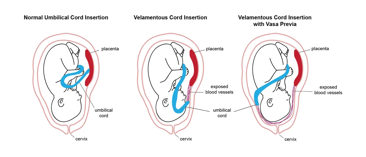
| Type | Anatomy |
|---|---|
| Type I | Velamentous cord insertion: the cord inserts into the membranes rather than the placental disc, and the vessels run unprotected across the membranes to reach it |
| Type II | Vessels bridging between the lobes of a bilobed placenta, or between the main placenta and a succenturiate (accessory) lobe |
| Type III | Vessels running along the placental margin within the membranes, with an otherwise normal cord insertion; a more recently described variant |
Risk factors
- Velamentous cord insertion - the underlying abnormality in type I disease
- Bilobed or succenturiate lobed placenta - the underlying abnormality in type II disease
- A low-lying placenta or placenta praevia in the second trimester, even if it subsequently resolves; the placental tissue over the os regresses but the vessels supplying it may remain in the membranes
- In vitro fertilisation - risk rises to as high as 1 in 200-300, for reasons that are not fully understood but probably relate to abnormal implantation
- Multiple pregnancy, in which velamentous insertion is far more common
- Multiparity and increasing maternal age, to a modest degree
The point about resolving low-lying placenta is worth dwelling on because it is counter-intuitive. Trophotropism means the placenta grows preferentially towards a well-vascularised fundus and atrophies over the poorly vascularised lower segment. The chorion regresses, but a vessel that was already running to that part of the placenta can be left behind, stranded in bare membrane over the cervix.
Clinical presentation
The classical presentation is the triad of rupture of membranes, painless fresh red vaginal bleeding, and immediate fetal compromise. The bleeding characteristically starts at the exact moment the waters break, whether spontaneously or after amniotomy, and the cardiotocograph deteriorates within minutes.
- Sudden painless vaginal bleeding coinciding with rupture of the membranes
- Fetal bradycardia, or a sinusoidal cardiotocograph pattern indicating severe fetal anaemia
- A mother who remains entirely well, with normal observations, despite an apparently significant bleed - because it is not her blood
- Occasionally, pulsatile vessels felt through the membranes on vaginal examination before rupture
- Rarely, vessels seen on direct visualisation of the membranes at speculum examination or amnioscopy
Differential diagnosis
| Condition | Whose blood | Pain | Maternal state | Fetal state |
|---|---|---|---|---|
| Vasa praevia | Fetal | Painless, at rupture of membranes | Well, normal observations | Rapid, profound compromise |
| Placenta praevia | Maternal | Painless | Compromised if the bleed is large | Normal until the mother decompensates |
| Placental abruption | Maternal | Severe, continuous | Shocked, often out of proportion to visible loss | Compromised, often severely |
| Uterine rupture | Maternal | Severe, with loss of contractions | Collapsed | Bradycardia |
| Bloody show | Maternal | Contractions | Well | Normal |
Vasa praevia must also be distinguished on ultrasound from funic presentation, in which a loop of free umbilical cord lies over the cervix. The distinction is made by asking the woman to change position or by applying gentle transducer pressure: a free cord loop moves, whereas vessels fixed within the membranes do not.
Diagnosis
Antenatal diagnosis is made with transvaginal ultrasound and colour Doppler. Linear vascular structures are seen crossing the internal os within the membranes; pulsed wave Doppler confirms a fetal arterial waveform, with a rate matching the fetal heart rate rather than the maternal pulse. The vessels remain fixed in position when the woman moves or when transducer pressure is applied, which is the finding that excludes funic presentation.
The UK does not currently operate a universal screening programme for vasa praevia, and RCOG does not recommend routine screening of the whole population, because the condition is rare, the false positive rate of screening is appreciable and many second-trimester findings resolve. In practice, targeted assessment is offered where risk factors are present: a low-lying placenta at the anomaly scan, a bilobed or succenturiate placenta, a velamentous cord insertion, an IVF pregnancy, or a multiple pregnancy.1,3
Management
When diagnosed antenatally
- Confirm the diagnosis on a repeat transvaginal scan in the third trimester, since a proportion of second-trimester findings resolve
- Counsel the woman fully, including the reason she must attend immediately if she has any bleeding, contractions or rupture of membranes
- Consider admission from around 30-34 weeks where the woman lives far from the hospital or has any additional risk factor for preterm birth, so that she is already in a unit with theatre access if she labours
- Antenatal corticosteroids at around 32 weeks, timed so they are on board before any planned or unplanned birth
- Plan elective caesarean section between 34+0 and 36+0 weeks, before the onset of labour and before spontaneous rupture of membranes
- Ensure the neonatal team is present at delivery, with O negative blood immediately available
- Deliver in a unit with a neonatal unit capable of caring for a late preterm and potentially severely anaemic baby
The timing of birth is a deliberate compromise. Delivering earlier reduces the chance of labour starting first but increases neonatal respiratory morbidity; delivering later reduces prematurity but risks the very event the whole plan exists to prevent. The RCOG range of 34-36 weeks reflects modelling suggesting this window minimises overall perinatal mortality.1
When it presents undiagnosed
- Category 1 caesarean section without delay - every minute of continued bleeding is fetal blood volume lost
- Do not wait for confirmatory tests of any kind
- Alert the neonatal team before delivery so that resuscitation equipment and emergency O negative blood are ready in the room
- Immediate cord clamping is appropriate here, in contrast to normal practice, because the priority is to stop further loss and give volume directly
- Neonatal resuscitation is volume resuscitation: the baby is exsanguinated, not primarily asphyxiated, so early transfusion is the intervention that works
- Send the placenta and membranes for histological examination to confirm the diagnosis, which also informs counselling for future pregnancies
At caesarean section
Where vasa praevia is known, the surgeon should be aware that the vessels lie in the lower segment and can be incised during hysterotomy. The uterine incision is planned to avoid them where possible, and the membranes are delivered intact if feasible. A senior obstetrician should perform or directly supervise the operation.
Red flags
The last of these is the single most useful discriminator in an exam and at the bedside. In every other cause of antepartum haemorrhage the mother is losing the blood, so maternal compromise precedes or accompanies fetal compromise. In vasa praevia the fetus is losing the blood, so the pattern is reversed.1
Complications and prognosis
Maternal complications relate to caesarean birth rather than to the condition itself, since the mother does not lose blood: the risks are those of surgery, and of preterm caesarean with an unformed lower segment. The fetal and neonatal complications are the substance of the condition.
- Fetal exsanguination and intrapartum death
- Severe neonatal anaemia requiring immediate transfusion
- Hypoxic-ischaemic encephalopathy in survivors of significant blood loss
- Complications of late preterm birth: respiratory distress, feeding difficulty, jaundice, hypoglycaemia
- Compression of the unsupported vessels by the presenting part during labour, causing intermittent fetal bradycardia even without rupture
The prognostic figures bear repeating because they are the reason the condition is taught at all. Undiagnosed vasa praevia carries a perinatal mortality of approximately 60%, and many survivors have suffered significant hypoxic-ischaemic injury. With antenatal diagnosis and planned caesarean birth, survival is reported at 97-99% with low rates of neurological morbidity.1,2
Vasa praevia is not thought to recur in a predictable fashion, but the underlying risk factors may. A woman who had a velamentous insertion or an accessory lobe should have careful assessment of placental morphology and cord insertion in a subsequent pregnancy. Where a baby has died, a full review, examination of the placenta and a follow-up appointment to explain what happened are essential, not least because parents in this situation frequently and wrongly believe that something they did precipitated the bleed.4
References
- RCOG Green-top Guideline No. 27b. Vasa praevia: diagnosis and management. 2018. Available here
- Oyelese Y, Catanzarite V, Prefumo F et al. Vasa previa: the impact of prenatal diagnosis on outcomes. Obstet Gynecol. 2004. Available here
- UK National Screening Committee. Screening for vasa praevia in pregnancy. Available here
- RCOG Green-top Guideline No. 55. Late intrauterine fetal death and stillbirth. Available here
This article is written for revision and education. It is not clinical guidance and must not be used to make decisions about the care of a patient. Always check current NICE guidance and local protocols.