
Venous Thromboembolism in Pregnancy
Key points
- Importance: thromboembolism remains a leading direct cause of maternal death in the UK, and most deaths are considered preventable.
- Risk: pregnancy raises VTE risk four- to sixfold; the puerperium raises it around twentyfold, with the greatest risk in the first three weeks after birth.
- Side: around 85-90% of pregnancy-associated DVTs are left-sided and iliofemoral, because the right common iliac artery compresses the left common iliac vein.
- D-dimer: is physiologically raised in pregnancy and must not be used to exclude VTE.
- Imaging: compression duplex ultrasound for suspected DVT; chest X-ray then V/Q scan or CTPA for suspected PE.
- Treat first: start treatment-dose low molecular weight heparin on clinical suspicion, before imaging, unless anticoagulation is contraindicated.
- Duration: continue for the remainder of pregnancy and for at least 6 weeks postnatally, with a minimum total of 3 months.
- Contraindicated drugs: warfarin is teratogenic in the first trimester and direct oral anticoagulants are avoided in pregnancy and breastfeeding.
Introduction
Venous thromboembolism is one of the leading direct causes of maternal death in the UK, and successive MBRRACE-UK reports have concluded that the majority of these deaths involve elements of substandard care: risk assessments not performed, prophylaxis not prescribed or prescribed at too low a dose, and symptoms attributed to normal pregnancy.1
Absolute risk is low, at roughly 1-2 events per 1,000 pregnancies, but relative risk is high: four- to sixfold antenatally and around twentyfold in the six weeks after birth, concentrated in the first three weeks. Pulmonary embolism is the presentation that kills, and it frequently follows a deep vein thrombosis that was never reported because the woman assumed leg swelling was normal in pregnancy.
The two clinical rules that follow are simple. Every pregnant woman gets a documented risk assessment at booking, on every admission, and after birth. And every pregnant woman with symptoms suggestive of thrombosis is investigated properly, because the consequences of over-investigating are trivial and the consequences of missing it are not.
Why pregnancy is prothrombotic
Pregnancy satisfies all three components of Virchow's triad, which is unusual and explains why the risk is so consistently elevated.
| Component | Changes in pregnancy |
|---|---|
| Hypercoagulability | Rise in factors VII, VIII, X, von Willebrand factor and fibrinogen; fall in protein S; acquired activated protein C resistance; rise in plasminogen activator inhibitors 1 and 2, reducing fibrinolysis |
| Venous stasis | Progesterone-mediated venodilatation from the first trimester; mechanical compression of the inferior vena cava and left common iliac vein by the gravid uterus; reduced mobility |
| Endothelial injury | Vascular damage at delivery, particularly with caesarean section, instrumental delivery or prolonged labour |
These changes are teleologically sensible: they exist to limit haemorrhage at placental separation, which was historically the dominant cause of maternal death. They begin in the first trimester, so prophylaxis in a high-risk woman starts early rather than in the third trimester, and they do not resolve immediately at delivery, which is why the puerperium is the highest-risk period of all.
Risk factors and risk assessment
RCOG Green-top Guideline 37a provides a scored risk assessment which is completed at booking, on every antenatal admission, intrapartum and postnatally.3 Risk is dynamic, and a woman who scored zero at booking may score four after a caesarean section complicated by postpartum haemorrhage.
| Points | Risk factors |
|---|---|
| 4 (or high risk) | Previous VTE (except a single event related to major surgery); antenatal high-risk thrombophilia such as antithrombin deficiency or antiphospholipid syndrome |
| 3 | Known low-risk thrombophilia with a family history; medical comorbidity such as active cancer, heart failure, active inflammatory bowel disease, nephrotic syndrome or sickle cell disease; any surgical procedure in pregnancy; ovarian hyperstimulation syndrome (first trimester) |
| 2 | BMI 40 or above; caesarean section in labour; postpartum haemorrhage over 1 litre requiring transfusion; preterm birth or stillbirth in this pregnancy |
| 1 | Age over 35; BMI 30 or above; parity 3 or more; smoking; gross varicose veins; current pre-eclampsia; multiple pregnancy; assisted conception; elective caesarean section; prolonged labour over 24 hours; mid-cavity or rotational operative delivery; immobility or dehydration; current systemic infection; long-distance travel |
- Antenatal total of 4 or more - prophylactic low molecular weight heparin from the first trimester
- Antenatal total of 3 - prophylactic low molecular weight heparin from 28 weeks
- Postnatal total of 2 or more - prophylactic low molecular weight heparin for at least 10 days
- Women admitted to hospital antenatally, and those with hyperemesis or ovarian hyperstimulation syndrome, should be considered for prophylaxis for the duration of the admission
- Prophylactic doses are weight-banded, so use a recent measured weight rather than the booking weight
Clinical features
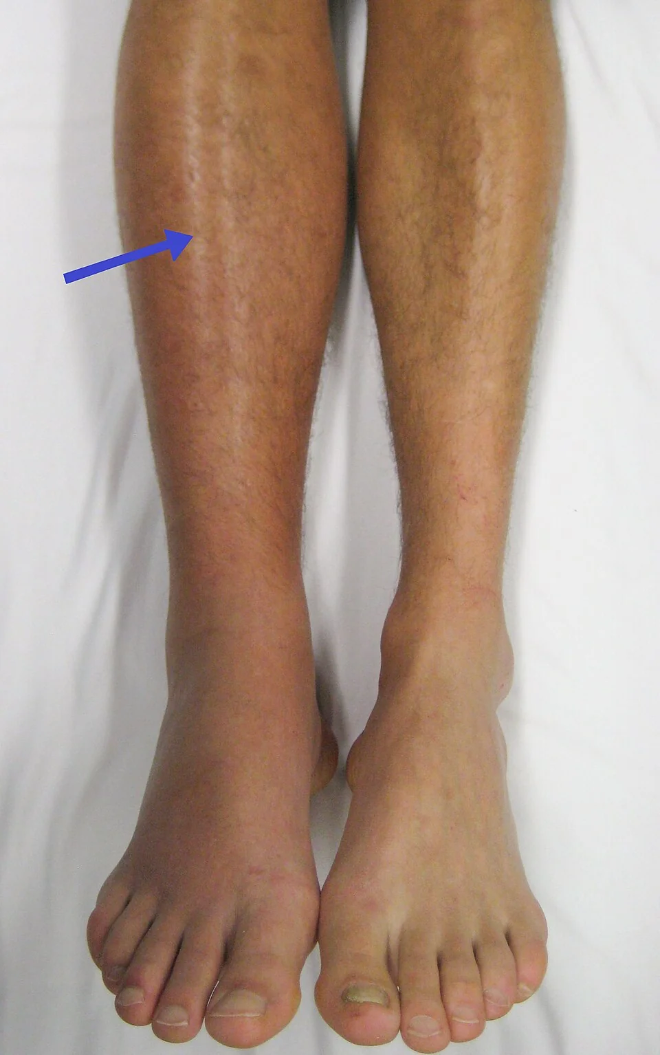
Deep vein thrombosis
- Unilateral leg pain, swelling, warmth and erythema, most often on the left
- Calf circumference measured 10 cm below the tibial tuberosity, with a difference of more than 2 cm between legs considered significant
- Tenderness along the deep venous system rather than just the calf muscle
- Lower abdominal, buttock or back pain, which suggests iliac vein involvement and is easily attributed to musculoskeletal pain of pregnancy
- Low-grade fever and a mildly raised white cell count, which can be mistaken for infection
Pulmonary embolism
- Dyspnoea, which is the commonest symptom and also the most easily dismissed
- Pleuritic chest pain, cough and haemoptysis
- Tachycardia, tachypnoea and hypoxia
- Syncope, hypotension or cardiac arrest in massive PE
- Signs of a coexisting DVT, which are present in a substantial minority and shortcut the investigation
Investigation
Suspected deep vein thrombosis
Compression duplex ultrasound of the whole leg is the investigation of choice. If it is negative and clinical suspicion is low, anticoagulation can be stopped. If it is negative but suspicion remains high, anticoagulation is continued and the scan repeated on days 3 and 7. Where iliac vein thrombosis is suspected because of back or buttock pain and whole-leg swelling, magnetic resonance venography or pulsed Doppler of the iliac vein should be considered, since a standard leg scan may not visualise it.
Suspected pulmonary embolism
- Baseline tests: ECG, chest X-ray, full blood count, urea and electrolytes, liver function and coagulation screen before starting anticoagulation
- ECG most often shows sinus tachycardia; right axis deviation, right bundle branch block, T wave inversion in the anterior leads and the classical S1Q3T3 pattern are all uncommon
- Chest X-ray is normal in more than half of cases; its main value is identifying pneumonia, pneumothorax or another explanation, and it determines the choice of definitive imaging
- Bilateral leg compression duplex if there are any leg symptoms, since a positive result confirms VTE and avoids irradiating the chest at all
- V/Q scan where the chest X-ray is normal and there is no coexisting lung disease
- CTPA where the chest X-ray is abnormal, or where V/Q is unavailable
| Consideration | V/Q scan | CTPA |
|---|---|---|
| Fetal radiation dose | Higher, giving a small increase in childhood cancer risk of roughly 1 in 280,000 | Lower fetal dose |
| Maternal breast dose | Low | High; a relative increase in lifetime breast cancer risk of the order of 13%, and breast tissue is proliferating in pregnancy |
| Diagnostic yield | Good when the chest X-ray is normal; indeterminate results are common with lung pathology | Also identifies alternative diagnoses; better with abnormal lung fields |
| Practical point | Preferred with a normal chest X-ray and no lung disease | Preferred with an abnormal chest X-ray |
Both tests deliver radiation doses well below the threshold associated with fetal harm, and the risk of an undiagnosed pulmonary embolism vastly exceeds the radiation risk. Women should be given this information and their choice documented, but the discussion must not become a reason for delay.2
Treatment
Treatment-dose low molecular weight heparin should be started immediately when VTE is clinically suspected, and continued until the diagnosis is excluded by objective testing. It does not cross the placenta, is not teratogenic, and is safe in breastfeeding.
- Dose by the woman's booking or early pregnancy weight, using the manufacturer's treatment regimen; either once or twice daily dosing is acceptable, though twice daily is often preferred around delivery because the anticoagulant effect can be more easily timed
- Routine anti-Xa monitoring is not required except at extremes of body weight, in renal impairment or in recurrent VTE
- Routine platelet monitoring is not required with low molecular weight heparin unless there has been prior unfractionated heparin exposure, since heparin-induced thrombocytopenia is rare
- Continue for the remainder of the pregnancy and for at least 6 weeks postnatally, with a minimum total duration of 3 months
- Graduated elastic compression stockings for the affected leg reduce symptoms, and early mobilisation is encouraged
- Arrange thrombophilia testing and haematology review after the acute episode, not during it, since pregnancy and acute thrombosis both distort the results
Massive pulmonary embolism
Haemodynamic collapse from pulmonary embolism is managed by an immediate multidisciplinary team including senior obstetric, anaesthetic, intensive care and cardiothoracic input. Options are intravenous unfractionated heparin, systemic thrombolysis, percutaneous catheter thrombectomy or surgical embolectomy. Thrombolysis is not contraindicated by pregnancy: the mortality of untreated massive PE is far greater than the bleeding risk, although it should not be given in the immediate postpartum period if avoidable. In cardiac arrest, perimortem caesarean section within four minutes improves both maternal and fetal survival by relieving aortocaval compression.
Anticoagulation around delivery
- Advise the woman to stop injecting heparin as soon as she thinks she is in labour, or 24 hours before a planned induction or caesarean section if on treatment dose
- Regional anaesthesia may be sited 12 hours after a prophylactic dose and 24 hours after a treatment dose
- An epidural catheter should not be removed within 12 hours of the last dose, and the next dose should be at least 4 hours after removal
- Restart low molecular weight heparin at least 4 hours after removal of the epidural catheter, and no sooner than 4 hours after a spinal, provided there is no ongoing bleeding
- Consider converting to unfractionated heparin near term in very high-risk women, because of its shorter half-life and reversibility
Complications and prognosis
- Death from pulmonary embolism, which remains among the commonest direct causes of maternal mortality in the UK
- Post-thrombotic syndrome - chronic leg pain, swelling, pigmentation and ulceration, particularly after iliofemoral thrombosis, affecting a substantial minority
- Recurrent VTE, both in this pregnancy and in subsequent ones, which is why previous VTE mandates antenatal prophylaxis
- Chronic thromboembolic pulmonary hypertension, a rare late consequence
- Bleeding from anticoagulation, particularly around delivery
With prompt diagnosis and adequate anticoagulation, the prognosis of pregnancy-associated VTE is good and most women recover fully. A woman who has had a pregnancy-associated VTE requires prophylactic low molecular weight heparin throughout any subsequent pregnancy and for six weeks after it, starting in the first trimester. She should be advised to avoid combined hormonal contraception, and referred for haematology review to determine whether long-term anticoagulation is warranted.2
Red flags
References
- MBRRACE-UK. Saving Lives, Improving Mothers' Care. Available here
- RCOG Green-top Guideline No. 37b. Thromboembolic disease in pregnancy and the puerperium: acute management. Available here
- RCOG Green-top Guideline No. 37a. Reducing the risk of venous thromboembolism during pregnancy and the puerperium. Available here
- James Heilman, MD, CC BY-SA 3.0, via Wikimedia Commons. Available here
This article is written for revision and education. It is not clinical guidance and must not be used to make decisions about the care of a patient. Always check current NICE guidance and local protocols.