
Spinal Cord Injury: Diagnosis and Management
Key points
- Spinal cord injury (SCI): damage to the spinal cord causing loss of motor, sensory and autonomic function below the level of the lesion, from trauma or non-traumatic causes.
- Complete versus incomplete: complete injury means no motor or sensory function preserved in the lowest sacral segments (S4-S5); any sacral sparing means the injury is incomplete and carries a better prognosis.
- Immediate priority: ABCDE with full spinal immobilisation, preventing secondary injury from movement, hypoxia and hypotension.
- Spinal shock: transient flaccidity and areflexia immediately after injury, which resolves over days to weeks to reveal the true upper motor neurone picture - it is not the same as neurogenic shock.
- Neurogenic shock: hypotension with bradycardia from loss of sympathetic outflow in lesions above T6 - distinguish from hypovolaemic shock, where the patient is tachycardic.
- Central cord syndrome: the commonest incomplete syndrome - hyperextension injury in an older patient with cervical spondylosis, causing weakness worse in the arms than the legs.
- Autonomic dysreflexia: a life-threatening hypertensive emergency in lesions at or above T6, usually triggered by a blocked catheter or bowel impaction.
- Investigation: CT for bony injury in trauma; MRI to assess the cord, ligaments and any compressive lesion.
Introduction
Spinal cord injury (SCI) is damage to the cord producing loss of motor, sensory and autonomic function below the level of the lesion. Most traumatic injuries in the UK follow falls (particularly in older adults) and road traffic collisions, with sport and assault accounting for a smaller proportion; non-traumatic causes include tumour, infection, ischaemia and inflammation.1
The central principle of acute management is preventing secondary injury. The primary injury - the mechanical damage at the moment of impact - cannot be undone, but a great deal of additional cord damage occurs afterwards from oedema, ischaemia, hypoxia and hypotension, and from further mechanical movement of an unstable spine. Almost everything done in the first hours is aimed at limiting that second wave.
The examinable core is the anatomy: knowing which tracts run where lets you predict the pattern of deficit from the mechanism, which is exactly what the incomplete cord syndromes test.
Aetiology
- Falls - the commonest cause in the UK, particularly low-energy falls in older adults with pre-existing cervical spondylosis, who can sustain significant cord injury without fracture
- Road traffic collisions - typically higher-energy injuries in younger patients
- Sports injuries - diving into shallow water, rugby, horse riding
- Violence - stab and gunshot wounds
- Non-traumatic - metastatic or primary tumour, epidural abscess, disc prolapse, transverse myelitis, spinal cord infarction, and haematoma (see Spinal cord compression)
Classification
Complete versus incomplete
The most important distinction is whether the injury is complete or incomplete, because it drives prognosis. A complete injury means no motor or sensory function is preserved in the lowest sacral segments (S4-S5) - which is why examining perianal sensation and voluntary anal contraction is a non-negotiable part of the assessment. Any preserved sacral function ('sacral sparing') means the injury is incomplete and there is meaningful potential for recovery.
The ASIA (American Spinal Injury Association) Impairment Scale grades severity from A (complete) through B, C and D (progressively more preserved motor function) to E (normal), and is the standard framework for documenting and tracking cord injury.2
Incomplete cord syndromes
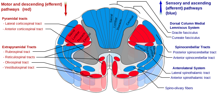
| Syndrome | Mechanism | Clinical picture |
|---|---|---|
| Central cord syndrome | Hyperextension injury, typically in an older patient with pre-existing cervical spondylosis | The commonest incomplete syndrome. Weakness worse in the upper limbs than the lower limbs (because arm fibres run more medially in the corticospinal tract), with variable sensory loss and bladder dysfunction |
| Anterior cord syndrome | Anterior spinal artery infarction, or flexion injury compressing the anterior cord | Loss of motor function and of pain and temperature below the lesion, with preserved proprioception and vibration (dorsal columns spared). Poor prognosis |
| Brown-Sequard syndrome | Cord hemisection - classically penetrating trauma | Ipsilateral loss of motor function, proprioception and vibration; contralateral loss of pain and temperature (which crosses within a few segments of entry). Best prognosis of the incomplete syndromes |
| Posterior cord syndrome | Posterior spinal artery infarction, B12 deficiency, trauma to the dorsal cord | Loss of proprioception and vibration with preserved motor, pain and temperature - producing marked sensory ataxia |
| Cauda equina syndrome | Compression below L1-L2 affecting the nerve roots rather than the cord | Lower motor neurone signs: flaccid weakness, absent reflexes, saddle anaesthesia, and early bladder and bowel dysfunction with reduced anal tone |
Clinical features
- Weakness or paralysis below the level of injury - tetraplegia with cervical lesions, paraplegia with thoracic or lumbar lesions
- Sensory loss with a definable level on the trunk
- Bladder and bowel dysfunction - urinary retention initially, later a reflex or flaccid bladder depending on the level
- Neck or back pain, and local tenderness or deformity
- Priapism - an uncommon but well-recognised sign of acute cord injury
- Respiratory compromise - lesions above C3-C5 ('C3, 4, 5 keeps the diaphragm alive') threaten diaphragmatic function and may require immediate ventilation; lower cervical and high thoracic lesions impair intercostal function and cough
Spinal shock versus neurogenic shock
Clinical examination
- ABCDE first, with full spinal immobilisation maintained throughout until the spine is cleared
- Motor examination of key myotomes in all four limbs, graded formally
- Sensory examination of key dermatomes to light touch and pinprick, working upwards to identify a level
- Perianal sensation, voluntary anal contraction and anal tone (with consent and a chaperone) - determines complete versus incomplete injury and is essential, not optional
- Reflexes, including bulbocavernosus reflex, whose return marks the end of spinal shock
- Log-roll with adequate staff to inspect and palpate the whole spine for tenderness, step or deformity
- Respiratory assessment - respiratory rate, effort, vital capacity, and paradoxical abdominal movement in high lesions
- Cardiovascular assessment - blood pressure and heart rate, looking specifically for the hypotension-with-bradycardia pattern of neurogenic shock
- Full trauma survey for associated injuries, which are common and can distract from or mask the cord injury
Investigations
- CT of the whole spine - the first-line imaging investigation in significant trauma, defining fractures, dislocation and bony canal compromise; usually performed as part of a trauma CT
- MRI spine - the investigation of choice for assessing the cord itself, ligamentous injury, disc herniation, haematoma and cord oedema or haemorrhage; essential where there is neurological deficit, and where the deficit is unexplained by the CT findings
- Plain radiographs - largely superseded by CT in major trauma, though still used in some lower-risk settings
- Clinical decision rules - the Canadian C-spine rule and NEXUS criteria help identify which alert, stable patients can have their cervical spine cleared clinically without imaging
- Bloods and ABG - baseline, plus assessment of ventilation in high lesions
- Bladder scan - to detect retention
Management
Immediate
- ABCDE with spinal immobilisation - and early consideration of intubation in high cervical lesions, before respiratory failure becomes an emergency
- Avoid hypoxia and hypotension - both worsen cord ischaemia and secondary injury. Mean arterial pressure is typically supported (often targeting around 85-90 mmHg for the first days) with fluids and vasopressors as needed
- Manage neurogenic shock - vasopressors are usually needed since fluids alone do not correct the loss of vascular tone; atropine or pacing for symptomatic bradycardia
- Urinary catheterisation - retention is near-universal acutely
- Early referral to a specialist spinal injuries centre
- Log-roll and pressure area care from the outset - patients with sensory loss develop pressure ulcers extremely quickly, sometimes within hours on a hard trauma board
- VTE prophylaxis - the risk of venous thromboembolism after spinal cord injury is very high
Definitive and ongoing management
- Surgical decompression and stabilisation - for unstable injuries or ongoing cord compression; early surgery (generally within 24 hours) is associated with better neurological outcomes in appropriate patients3
- Bracing or halo immobilisation - for selected stable injuries managed non-operatively
- Bladder management - intermittent self-catheterisation is the preferred long-term approach where feasible, reducing infection risk compared with indwelling catheters
- Bowel management - a structured, regular bowel programme is essential and has a large impact on quality of life
- Spasticity management - baclofen, tizanidine, botulinum toxin, and physiotherapy
- Neuropathic pain - gabapentin, pregabalin or amitriptyline
- Comprehensive rehabilitation in a specialist spinal injuries unit - physiotherapy, occupational therapy, wheelchair assessment, home adaptation, vocational rehabilitation
- Psychological support - depression and adjustment difficulty are common and should be actively addressed
- Sexual health and fertility counselling - important and frequently neglected aspects of long-term care
Autonomic dysreflexia
Complications
- Respiratory failure, pneumonia and atelectasis - the leading causes of early mortality, particularly in cervical injuries
- Autonomic dysreflexia in lesions at or above T6
- Pressure ulcers - largely preventable, and a major cause of morbidity and hospital readmission
- Venous thromboembolism
- Recurrent urinary tract infection and, over time, renal impairment
- Neuropathic pain and spasticity
- Heterotopic ossification and contractures
- Osteoporosis below the level of injury, with fragility fractures
- Depression, anxiety and adjustment disorder
- Sexual dysfunction and fertility concerns
- Postural hypotension, particularly early in rehabilitation
Red flags
Prognosis
Prognosis depends principally on the level and completeness of the injury. Sacral sparing - any preserved perianal sensation or voluntary anal contraction - indicates an incomplete injury and substantially better prospects for recovery, which is why that part of the examination matters so much. Most neurological recovery occurs in the first 6-12 months, though functional gains from rehabilitation continue well beyond this.
Among the incomplete syndromes, Brown-Sequard has the best prognosis and anterior cord syndrome the worst; central cord syndrome often shows useful recovery, though hand function frequently remains the most persistently affected. Life expectancy after spinal cord injury has improved considerably with specialist care but remains reduced, with respiratory complications, urinary sepsis and pressure ulcer-related infection being the principal causes of late mortality - most of which are, at least in part, preventable with good long-term care.
Long-term follow-up in a specialist spinal injuries service is what turns those preventable causes of late mortality into avoided ones. Lifelong surveillance covers renal tract imaging and function, since neurogenic bladder remains the principal threat to the kidneys; skin integrity, with education so that patients and carers perform regular pressure relief and skin checks; and cardiovascular risk, which is elevated because of reduced physical activity and altered autonomic control.
Equally important is the wider rehabilitation goal of participation rather than simply survival. Vocational rehabilitation, wheelchair skills, adapted driving, sport, sexual health and fertility services all contribute to long-term outcome, and outcomes measured only in neurological terms consistently underestimate what a well-supported patient can achieve.
References
- NICE NG41. Spinal injury: assessment and initial management. 2016. Available here
- American Spinal Injury Association. International Standards for Neurological Classification of Spinal Cord Injury (ISNCSCI). Available here
- Fehlings MG, Vaccaro A, Wilson JR et al. Early versus delayed decompression for traumatic cervical spinal cord injury (STASCIS). PLoS One. 2012. Available here
- Krassioukov A, Warburton DE, Teasell R, Eng JJ. A systematic review of the management of autonomic dysreflexia after spinal cord injury. Archives of Physical Medicine and Rehabilitation. 2009. Available here
This article is written for revision and education. It is not clinical guidance and must not be used to make decisions about the care of a patient. Always check current NICE guidance and local protocols.