
Cauda Equina Syndrome
Key points
- Definition: compression of the cauda equina - the bundle of lumbosacral nerve roots (L2-S5) below the level the spinal cord itself ends, around L1/L2 in adults.
- Why it matters: a surgical emergency - delayed decompression can cause irreversible bladder, bowel and sexual dysfunction and permanent lower limb paralysis.
- Commonest cause: central lumbar disc prolapse, particularly at L4/5 or L5/S1.
- Cardinal feature: bladder dysfunction - loss of urge to void, altered urinary sensation, or painless retention with overflow incontinence. This is the single most important symptom to elicit and document.
- Staging: suspected CES (red flag symptoms, exam and bladder function may be normal) through to CES with retention - a later, more urgent stage with a worse prognosis.
- Examination caveat: a normal PR exam and normal perianal sensation do NOT exclude cauda equina syndrome and must never be used to avoid urgent imaging if the history is suggestive.
- Investigation: emergency, same-day whole-spine MRI - out of hours if necessary. This cannot wait for routine imaging.
- Management: urgent discussion with the on-call spinal/neurosurgical team and surgical decompression, with outcomes best when this occurs within 24-48 hours of symptom onset.
Introduction
The cauda equina (Latin for 'horse's tail') is the bundle of lumbar, sacral and coccygeal nerve roots (L2-S5) that continues down the spinal canal after the spinal cord itself terminates at the conus medullaris, around the L1/L2 vertebral level in adults. Because the vertebral column grows faster than the spinal cord during development, these nerve roots must travel a long distance within the canal before exiting at their respective neural foramina, forming a loose collection of roots that resembles a horse's tail and gives the syndrome its name.
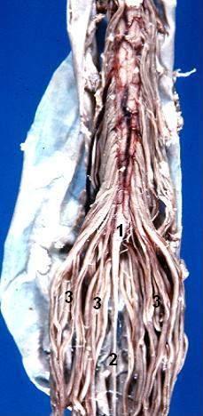
Cauda equina syndrome (CES) is compression of this nerve root bundle within the spinal canal, and it is a surgical emergency. Unlike compression of the spinal cord itself, which produces upper motor neurone signs, compression here affects peripheral nerve roots and so produces a lower motor neurone pattern of deficit. Left untreated, or treated too late, it causes permanent bladder, bowel and sexual dysfunction and permanent lower limb weakness - outcomes that are life-changing and, once established, largely irreversible.
CES is rare in absolute terms, but it is one of the most heavily examined presentations in the whole curriculum, and for good reason: it is time-critical, the diagnosis rests on a specific set of history and examination findings that every student must be able to elicit without prompting, and a missed or delayed diagnosis is one of the leading causes of medico-legal litigation against the NHS in orthopaedics and spinal surgery. Every patient presenting with back pain must be screened for it.
Causes
Anything that reduces the calibre of the spinal canal at the lumbosacral level, or that occupies space within it, can compress the cauda equina.
- Central lumbar disc prolapse - by far the most common cause, particularly a large central or centrolateral disc herniation at L4/5 or L5/S1, the two levels that carry the greatest mechanical load
- Lumbar spinal stenosis - degenerative narrowing of the canal, usually a more gradual onset than an acute disc prolapse
- Spinal tumour - primary (e.g. ependymoma, schwannoma, meningioma) or, more commonly, metastatic disease and vertebral collapse from bone metastases (breast, prostate, lung, renal and myeloma being the classic primaries)
- Spinal trauma or fracture with retropulsion of bone into the canal, compromising its diameter
- Spinal epidural abscess - risk factors include intravenous drug use, diabetes, immunosuppression, recent spinal instrumentation or epidural/spinal anaesthesia, and a bacteraemic source such as skin or line infection
- Spinal epidural haematoma - most often associated with anticoagulation or a coagulopathy, and a recognised complication of epidural or spinal anaesthesia and lumbar puncture
- Ankylosing spondylitis - a rare but recognised late association, sometimes with an arachnoiditis-related cauda equina syndrome
- Iatrogenic causes - a late complication of lumbar spinal surgery, including graft or implant migration, epidural haematoma, or recurrent disc herniation
Clinical features
The clinical picture reflects compression of multiple lumbosacral nerve roots simultaneously, so the deficit is typically bilateral, though it can be markedly asymmetric, and it follows a lower motor neurone pattern throughout since these are peripheral nerve roots rather than the spinal cord itself.
Pain and lower limb signs
Bilateral leg pain or sciatica is common, often severe, and may have been preceded by a longer history of unilateral back pain and sciatica that has now become bilateral - a change in pattern that should always be asked about specifically. Lower limb neurological signs follow a lower motor neurone distribution: weakness that can range from mild to a flaccid paraparesis, reduced tone, and reduced or absent knee and ankle reflexes. Sensory loss affects the legs in a root or patchy distribution and can vary considerably in severity between patients and even between the two legs.
Saddle anaesthesia
Saddle anaesthesia is numbness or altered sensation over the perianal, perineal and inner thigh region - the area of skin that would be in contact with a saddle. It reflects compression of the lower sacral roots (S3-S5) and is one of the defining features of the syndrome. It may be described by the patient as numbness, or noticed only when specifically asked about, for example difficulty feeling toilet paper when wiping, or numbness noticed during sex or when sitting.
Bladder dysfunction
Bladder dysfunction is the single most important feature to elicit and document, because it is the feature most closely tied to prognosis and to the staging of the syndrome. It must be asked about explicitly and in detail, since patients often do not volunteer it unprompted, either through embarrassment or because they have not connected it to their back pain.
- Loss of the normal urge or desire to void
- Altered urinary sensation - not feeling the bladder fill or empty normally
- Difficulty initiating micturition, a poor or interrupted stream, or having to strain to pass urine
- Painless urinary retention, often with overflow incontinence - urine leaking involuntarily from an overfull, insensate bladder without the patient feeling the urge to pass it
Bowel and sexual dysfunction
Bowel involvement mirrors the bladder picture: loss of the sensation of rectal fullness, faecal incontinence, and reduced anal tone on examination. Sexual dysfunction includes erectile dysfunction in men and loss of genital sensation in either sex, and should be asked about as part of the same systematic enquiry, however sensitive the questions may feel.
| System | Features |
|---|---|
| Motor | Bilateral (may be asymmetric) lower motor neurone weakness of the legs, ranging from mild to flaccid paraparesis; reduced tone |
| Sensory | Bilateral leg pain/sciatica and sensory loss; saddle anaesthesia over the perianal, perineal and inner thigh region |
| Reflexes | Reduced or absent knee and ankle reflexes; reduced or absent anal reflex and tone |
| Bladder | Loss of urge to void, altered urinary sensation, poor stream/straining, painless retention with overflow incontinence |
| Bowel | Loss of sensation of rectal fullness, faecal incontinence, reduced anal tone on PR |
| Sexual | Erectile dysfunction, loss of genital sensation, anorgasmia |
A spectrum, not a single moment
It is important to understand CES as a spectrum rather than an all-or-nothing diagnosis, because this staging is genuinely used in UK spinal referral pathways to communicate urgency and prognosis between the referring team and the on-call spinal service. A patient can present with red flag symptoms and a completely normal neurological and bladder examination - this is still an emergency requiring the same urgent MRI.
| Stage | Features | Significance |
|---|---|---|
| CES suspected (CESS) | Bilateral radicular pain and/or other red flag symptoms, but no objective bladder, bowel or perianal sensory disturbance on examination | Still requires emergency same-day MRI - a normal examination does not exclude evolving compression |
| CES incomplete (CESI) | Altered urinary sensation, loss of desire to void, or a poor/interrupted stream, with or without altered perianal sensation, but without painless retention or overflow incontinence | Genuine cauda equina compression with bladder involvement - urgent decompression is indicated |
| CES with retention (CESR) | Painless urinary retention with overflow incontinence, and typically established saddle anaesthesia and reduced anal tone | The most advanced and urgent stage - a much worse prognosis for recovery of bladder, bowel and sexual function even with prompt surgery |
Red flag screening questions
Every patient presenting with new or worsening lower back pain and leg pain should be screened with a specific, structured set of questions. These should be asked directly and explicitly - not inferred from a general systems enquiry - because patients frequently will not volunteer bladder, bowel or sexual symptoms unless asked. This list is exactly what should be used, and documented, in an OSCE history or a real clinical encounter.
A positive answer to any of these questions, in the context of back pain, mandates the same pathway: urgent examination including a PR exam, and same-day MRI. The answers, and the fact the questions were asked, must be documented explicitly - both because they change immediate management and because this documentation is precisely what is scrutinised in the medico-legal claims that follow a missed diagnosis.
Clinical examination
Examination has three components: a full neurological assessment of both lower limbs, assessment of perianal sensation, and a digital rectal examination.
- Full lower limb neurological examination - tone, power in all major muscle groups (including hip flexion/extension, knee flexion/extension, ankle dorsiflexion/plantarflexion and great toe extension), sensation to light touch and pinprick in all dermatomes, knee and ankle reflexes, and plantar responses
- Perianal sensation - light touch tested in the S2-S5 dermatomes around the anus, perineum and inner thighs, comparing side to side
- Digital rectal examination (PR) - assessing resting anal tone and the ability to voluntarily contract the sphincter around the examining finger
- Post-void bladder scan - an ultrasound measurement of post-void residual volume, used as an objective marker of bladder dysfunction; a significant residual volume supports the diagnosis, particularly when the history of urinary symptoms is ambiguous
- Gait, where safe to assess, and a general abdominal examination to feel for a palpably distended bladder
Investigations
Emergency, same-day whole-spine MRI is the investigation of choice to confirm the diagnosis and to identify the level and cause of compression, and it should be requested whenever cauda equina syndrome is genuinely suspected on history, regardless of examination findings.1 Whole-spine imaging is used because the level of compression cannot always be predicted from the clinical picture alone, and because it identifies alternative or additional pathology such as more proximal cord compression.
This scan cannot wait for the next routine or elective imaging slot. If suspicion is genuine, it must be arranged out of hours if necessary, with a radiologist and, where appropriate, the on-call spinal or neurosurgical team informed directly rather than the request simply being left in a queue. Delay in obtaining imaging is one of the most consistently identified failure points in cases that go on to litigation.
- Whole-spine MRI - the definitive investigation; contraindications (e.g. certain pacemakers, some metalwork) should prompt urgent discussion with radiology and the spinal team about an alternative rather than simply deferring the scan
- Post-void bladder scan - quantifies the post-void residual volume as an objective, reproducible marker of bladder dysfunction
- Bloods - FBC and CRP if infection or epidural abscess is suspected, clotting screen and INR if the patient is anticoagulated, and group and save/crossmatch if urgent surgery is anticipated
- Blood cultures if sepsis or an epidural abscess is a possibility
Management
Any patient with suspected cauda equina syndrome requires immediate discussion with the on-call spinal or neurosurgical team - this is not a referral that waits for the next working day or the next ward round. The discussion should happen as soon as red flag symptoms are identified, in parallel with arranging the emergency MRI, not after the scan result is back.
Definitive treatment is emergency surgical decompression, most commonly a discectomy for a prolapsed disc, aimed at relieving pressure on the nerve roots before the damage becomes permanent. Where the cause is different, treatment is directed at it in parallel: antibiotics and surgical drainage for an epidural abscess, and reversal of anticoagulation with surgical evacuation for an epidural haematoma. Radiotherapy or surgery may be used for malignant cord/cauda equina compression depending on the tumour type, extent of disease and patient fitness, in discussion with oncology.
Supportive measures run alongside definitive treatment: urinary catheterisation for anyone in retention, analgesia, and involvement of the wider multidisciplinary team (physiotherapy, continence services, and clinical psychology) from an early stage, since recovery - where it occurs - is often gradual and rehabilitation needs are considerable.
- Urgent same-day discussion with the on-call spinal/neurosurgical team, in parallel with imaging
- Emergency surgical decompression - most commonly discectomy for a central disc prolapse
- Urinary catheterisation if the patient is in retention
- Antibiotics and surgical drainage for an epidural abscess
- Reversal of anticoagulation and surgical evacuation for an epidural haematoma
- Analgesia and thromboprophylaxis review while awaiting surgery
- Early involvement of physiotherapy, continence services and psychological support
Complications
Even with prompt recognition and surgery, cauda equina syndrome can leave permanent deficits, and the later the stage at presentation (particularly established retention), the worse the expected outcome.
- Permanent bladder dysfunction - may require long-term intermittent self-catheterisation or an indwelling catheter
- Permanent bowel dysfunction - ongoing incontinence or the need for structured bowel management programmes
- Permanent saddle anaesthesia
- Sexual dysfunction - erectile dysfunction and loss of genital sensation, which may not recover
- Persistent lower limb weakness and sensory loss, affecting mobility and independence
- Chronic pain
- Substantial psychological and quality-of-life impact - depression, loss of employment, relationship difficulties and loss of independence are all well recognised
Differential diagnosis
- Conus medullaris syndrome - compression higher up, at the termination of the spinal cord itself (around T12-L2). Because both the cord and the exiting nerve roots can be affected, it can present with a mixed picture of upper and lower motor neurone signs (for example brisk reflexes with a positive Babinski sign alongside weakness), unlike cauda equina syndrome, which is a pure lower motor neurone picture throughout
- Mechanical back pain or sciatica without red flag features - the overwhelming majority of back pain presentations, managed conservatively; distinguished by the absence of bladder, bowel, saddle or bilateral neurological features
- Multiple sclerosis or other causes of myelopathy - can cause bladder dysfunction and leg weakness, but typically with upper motor neurone signs, a relapsing-remitting or progressive course, and other neurological features outside the lumbosacral distribution
- Peripheral neuropathy - can cause bilateral leg sensory loss and weakness, but usually a length-dependent, symmetrical, distal pattern evolving over a longer timescale, without back pain or bladder/bowel involvement
- Guillain-Barre syndrome - ascending, symmetrical weakness with areflexia; usually not associated with back pain or a sensory level, and bladder involvement is less prominent early on
Red flags
References
- NICE Clinical Knowledge Summaries. Cauda equina syndrome. Available here
- NICE NG59. Low back pain and sciatica in over 16s: assessment and management. 2016, updated 2020. Available here
- British Association of Spine Surgeons (BASS). Suspected Cauda Equina Syndrome - Standards of Care and referral pathway. Available here
- Getting It Right First Time (GIRFT). Spinal Surgery: GIRFT Programme National Specialty Report. 2019. Available here
- Society of British Neurological Surgeons and British Association of Spine Surgeons. Standards of care for investigation and management of Cauda Equina Syndrome. 2018. Available here
- Todd NV. Cauda equina syndrome: the timing of surgery probably does influence outcome. British Journal of Neurosurgery. 2005. Available here
- Gleave JR, Macfarlane R. Cauda equina syndrome: what is the relationship between timing of surgery and outcome? British Journal of Neurosurgery. 2002. Available here
- Fraser S, Roberts L, Murphy E. Cauda equina syndrome: a literature review of its definition and clinical presentation. Archives of Physical Medicine and Rehabilitation. 2009. Available here
- NHS Resolution. Learning from claims - cauda equina syndrome. Available here
- NICE NG75. Metastatic spinal cord compression in adults: diagnosis and management. Available here
This article is written for revision and education. It is not clinical guidance and must not be used to make decisions about the care of a patient. Always check current NICE guidance and local protocols.