
Leukaemia
Key points
- Leukaemia: malignant clonal proliferation of white cell precursors within the bone marrow, classified along two axes: acute vs chronic (how mature the cells are and how fast the disease evolves) and myeloid vs lymphoid (which lineage is affected).
- The four classical types: acute myeloid leukaemia (AML), acute lymphoblastic leukaemia (ALL), chronic myeloid leukaemia (CML) and chronic lymphocytic leukaemia (CLL) - each with a distinct age distribution, presentation and prognosis.
- Acute leukaemia is a same-day emergency: AML and ALL present with bone marrow failure (anaemia, infection, bleeding) from a marrow packed with blasts, and can deteriorate within hours - neutropenic sepsis and tumour lysis syndrome must be anticipated from diagnosis.
- AML: the commonest acute leukaemia in adults. Acute promyelocytic leukaemia (APL), a distinct subtype with t(15;17), causes life-threatening DIC and is treated with ATRA, a genuine medical emergency requiring immediate treatment on suspicion.
- ALL: the commonest cancer in childhood, with excellent cure rates (~90%) in children but a worse prognosis in adults. CNS involvement is common, so CNS-directed prophylaxis is part of every regimen.
- CLL: the commonest leukaemia overall in the West, typically an incidental finding in older adults; associated with warm autoimmune haemolytic anaemia and hypogammaglobulinaemia. Many patients need only "watch and wait".
- CML: defined by the Philadelphia chromosome (BCR-ABL1); covered in detail alongside the other myeloproliferative disorders, given its shared biology with that group.
- Diagnosis: FBC and blood film, bone marrow aspirate/trephine, immunophenotyping (flow cytometry) and cytogenetics/molecular testing together define the subtype and drive both treatment and prognosis.
Introduction
Leukaemia is malignant clonal proliferation arising from a haematopoietic precursor cell within the bone marrow, which then typically spills over into the peripheral blood. The four classical types are distinguished along two axes: whether the disease is acute (immature blast cells, rapid onset, life-threatening within weeks if untreated) or chronic (more mature cells, indolent course over years), and whether the affected lineage is myeloid or lymphoid.1
| Myeloid | Lymphoid | |
|---|---|---|
| Acute | AML - commonest acute leukaemia in adults | ALL - commonest cancer in childhood |
| Chronic | CML - defined by the Philadelphia chromosome | CLL - commonest leukaemia overall, elderly |
CML shares its biology and treatment principles with the other myeloproliferative neoplasms and is covered in full there; this article focuses on AML, ALL and CLL, with CML included for completeness of the classification.
Acute myeloid leukaemia (AML)
AML is the commonest acute leukaemia in adults, with incidence rising steeply with age (median age at diagnosis around 68). It arises either de novo, or secondary to a prior myelodysplastic syndrome or previous cytotoxic chemotherapy/radiotherapy (which carries a worse prognosis).
Clinical features
Presentation reflects bone marrow failure, as leukaemic blasts crowd out normal haematopoiesis:
- Anaemia - fatigue, dyspnoea, pallor
- Neutropenia - infections, sometimes severe, despite a high total white cell count (the circulating cells are non-functional blasts)
- Thrombocytopenia - bruising, petechiae, mucosal bleeding
- Gum hypertrophy and skin infiltration (leukaemia cutis) - more typical of monocytic subtypes
- Bone pain, hepatosplenomegaly and lymphadenopathy in some patients
- Signs of hyperviscosity/leukostasis (visual disturbance, confusion, respiratory distress) with very high blast counts - a medical emergency
Investigations
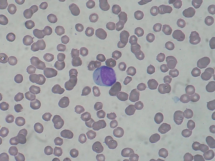
- Blood film - circulating myeloblasts, sometimes with Auer rods (needle-like cytoplasmic inclusions of fused primary granules) - these confirm myeloid lineage and, in practice, point strongly to AML, though they can also be seen in high-grade myelodysplastic syndrome with excess blasts
- Bone marrow aspirate - ≥20% blasts defines AML (below this, consider MDS)
- Immunophenotyping (flow cytometry), cytogenetics and molecular testing (e.g. FLT3, NPM1, CEBPA mutations) - essential for subtyping and prognosis, and increasingly guide targeted therapy
Management
Intensive combination chemotherapy (induction followed by consolidation) for fit patients, aiming for complete remission; allogeneic stem cell transplant for patients with higher-risk cytogenetics/molecular features or relapsed disease; targeted agents (e.g. FLT3 inhibitors) where a relevant mutation is present. Less intensive regimens (e.g. venetoclax with a hypomethylating agent) are used in older or unfit patients.
Acute lymphoblastic leukaemia (ALL)
ALL is the commonest cancer of childhood, with a peak incidence between 2 and 5 years; a second, smaller peak occurs in older adults, in whom the disease behaves more aggressively.
Clinical features
Also presents with bone marrow failure, but with a greater tendency than AML to involve lymphoid organs and sanctuary sites:
- Anaemia, infection and bleeding, as in AML
- Lymphadenopathy and hepatosplenomegaly - more prominent than in AML
- Bone pain, particularly in children, sometimes causing a limp or refusal to weight-bear
- CNS involvement - headache, cranial nerve palsies, or found on routine lumbar puncture staging even if asymptomatic
- Testicular involvement - a recognised "sanctuary site" for relapse, particularly in boys
Investigations
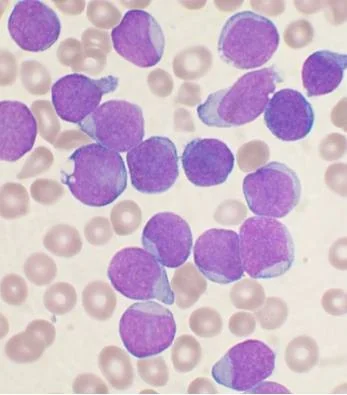
Blood film and bone marrow show lymphoblasts (no Auer rods). Immunophenotyping confirms the lymphoid lineage and distinguishes B-cell (majority) from T-cell ALL; the terminal deoxynucleotidyl transferase (TdT) marker is positive, helping distinguish ALL from AML and from mature lymphoid malignancies. Cytogenetics identifies prognostically important abnormalities - the Philadelphia chromosome (BCR-ABL1-positive ALL) carries a worse prognosis in adults but is now treatable with a tyrosine kinase inhibitor alongside chemotherapy. Lumbar puncture is performed for CNS staging and to deliver intrathecal chemotherapy.
Management
Multi-agent chemotherapy given in phases (induction, consolidation, maintenance, typically over about 2 years), with CNS-directed prophylaxis (intrathecal chemotherapy +/- cranial irradiation in high-risk cases) built into every regimen given the high risk of CNS relapse if omitted. Allogeneic stem cell transplant for high-risk or relapsed disease. Targeted therapy (TKI for Philadelphia-positive disease; immunotherapy such as CAR-T cell therapy or blinatumomab for relapsed/refractory disease) has substantially improved outcomes in recent years.3
Chronic lymphocytic leukaemia (CLL)
CLL is the commonest leukaemia overall in the Western world, almost exclusively a disease of older adults (median age at diagnosis around 70), and is caused by clonal proliferation of mature but dysfunctional B lymphocytes.
Clinical features
- Often entirely asymptomatic, discovered incidentally on a routine FBC showing lymphocytosis - a distinctive feature compared with the other three leukaemias
- Painless lymphadenopathy and splenomegaly when symptomatic
- Recurrent infections - from hypogammaglobulinaemia, as the abnormal clone crowds out normal, antibody-producing B cells
- "B" symptoms (fever, night sweats, weight loss) in more advanced disease
- Warm autoimmune haemolytic anaemia - a recognised and important association, sometimes the presenting feature
Investigations
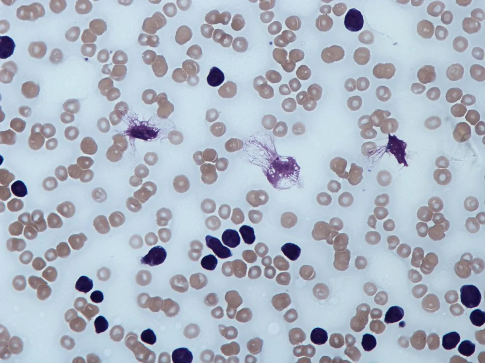
FBC - lymphocytosis, often marked. The blood film shows small, mature-appearing lymphocytes and characteristic smudge (smear) cells - fragile lymphocytes damaged during film preparation - a classic and highly suggestive finding.
Immunophenotyping (flow cytometry) confirms the diagnosis, classically CD5+, CD19+, CD23+ - the co-expression of CD5 (usually a T-cell marker) on B cells is a defining immunophenotypic feature. Cytogenetics/FISH, particularly 17p deletion (TP53 loss), identifies high-risk disease that responds poorly to standard chemoimmunotherapy and needs a targeted approach.
Staging and management
Staged clinically using the Binet (UK) or Rai (US) systems, based on the number of lymphoid areas involved and the presence of anaemia/thrombocytopenia.
- "Watch and wait" - many patients with early-stage, asymptomatic CLL are simply monitored, since treatment does not improve survival in this group and may add toxicity without benefit
- Indications to treat: progressive bone marrow failure, massive or progressive lymphadenopathy/splenomegaly, autoimmune cytopenias not responding to steroids, or significant B symptoms
- Targeted agents - BTK inhibitors (e.g. ibrutinib) and the BCL2 inhibitor venetoclax have transformed treatment, particularly for 17p-deleted/TP53-mutated disease that responds poorly to chemoimmunotherapy
- Chemoimmunotherapy (e.g. rituximab-based regimens) remains an option for fit patients with standard-risk disease
Chronic myeloid leukaemia (CML)
CML is defined by the Philadelphia chromosome t(9;22), producing the BCR-ABL1 fusion kinase, and is treated with tyrosine kinase inhibitors that have transformed its prognosis dramatically. Because its biology and treatment overlap so closely with the other myeloproliferative neoplasms, CML is covered in full detail in that article rather than repeated here.
Shared acute leukaemia emergencies
Prognosis
Prognosis varies enormously by leukaemia type, age and molecular/cytogenetic risk profile. AML in fit younger adults with favourable cytogenetics can achieve long-term cure rates over 60-70%, but overall survival in older or unfit patients remains poor.2 ALL in children has excellent outcomes, with cure rates around 90%; outcomes in adults are less favourable but have improved substantially with targeted and immunotherapies.3
CLL often follows an indolent course over many years, and many patients die of unrelated causes rather than their leukaemia - though 17p-deleted/TP53-mutated disease and Richter transformation carry a significantly worse prognosis. CML, once uniformly fatal within a few years, now carries a near-normal life expectancy for most chronic-phase patients treated with modern tyrosine kinase inhibitors.
References
- Dohner H, Weisdorf DJ, Bloomfield CD. Acute Myeloid Leukemia. N Engl J Med. 2015. Available here
- Sanz MA, Fenaux P, Tallman MS et al. Management of acute promyelocytic leukemia: updated recommendations. Blood. 2019. Available here
- Terwilliger T, Abdul-Hay M. Acute lymphoblastic leukemia: a comprehensive review. Blood Cancer J. 2017. Available here
- Hallek M. Chronic lymphocytic leukemia: 2020 update on diagnosis, risk stratification and treatment. Am J Hematol. 2019. Available here
- Paulo Henrique Orlandi Mourao, CC BY-SA 3.0, via Wikimedia Commons. Available here
- VashiDonsk, CC BY-SA 3.0, via Wikimedia Commons. Available here
- Dr Graham Beards, CC BY-SA 3.0, via Wikimedia Commons. Available here
- NICE Clinical Knowledge Summaries (CKS). Leukaemia - suspected. 2023. Available here
This article is written for revision and education. It is not clinical guidance and must not be used to make decisions about the care of a patient. Always check current NICE guidance and local protocols.