
Liver Cirrhosis: Diagnosis, Scoring and Management
Key points
- Cirrhosis: diffuse hepatic fibrosis with regenerative nodules replacing normal architecture - the irreversible endpoint of chronic liver injury.
- Commonest UK causes: alcohol-related liver disease, MASLD, and chronic viral hepatitis B and C.
- Compensated vs decompensated: compensated cirrhosis may be asymptomatic for years; decompensation is defined by ascites, variceal bleeding, encephalopathy or jaundice.
- Two consequences: portal hypertension (varices, ascites, splenomegaly) and loss of synthetic function (low albumin, raised INR, jaundice).
- Diagnosis: transient elastography (FibroScan) is first-line non-invasively; ultrasound shows a nodular liver, and biopsy is the gold standard but rarely needed.
- Scoring: Child-Pugh (bilirubin, albumin, INR, ascites, encephalopathy) grades severity; MELD guides transplant prioritisation.
- Surveillance: six-monthly ultrasound with or without AFP for hepatocellular carcinoma, and endoscopy to screen for varices.
- Management: treat the underlying cause, prevent and treat complications, and refer for transplantation once decompensated.
Introduction
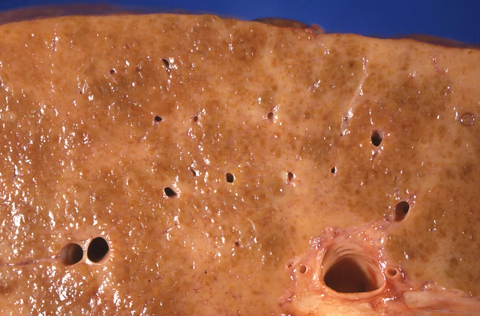
Cirrhosis is the final common pathway of chronic liver injury, in which persistent inflammation drives diffuse fibrosis and the formation of regenerative nodules, replacing the normal hepatic architecture.1 The consequences flow from two distinct problems: mechanical distortion of blood flow causing portal hypertension, and progressive loss of functioning hepatocyte mass causing hepatic synthetic failure.
Liver disease is one of the few major causes of death still rising in the UK, and is now the leading cause of death in people aged 35-49. Cirrhosis is conventionally divided into two clinically distinct stages, which differ enormously in prognosis:
| Stage | Features | Median survival |
|---|---|---|
| Compensated | Often asymptomatic; the liver maintains adequate function. May be found incidentally through abnormal LFTs or imaging | Over 12 years |
| Decompensated | Defined by the development of ascites, variceal bleeding, hepatic encephalopathy or jaundice | Around 2 years |
The transition from compensated to decompensated disease is the pivotal clinical event, occurring at a rate of roughly 5-7% per year, and often precipitated by an identifiable trigger such as infection, alcohol, a GI bleed or a hepatotoxic drug.
Causes
| Category | Causes |
|---|---|
| Commonest in the UK | Alcohol-related liver disease; MASLD (metabolic dysfunction-associated steatotic liver disease); chronic hepatitis B and C |
| Autoimmune | Autoimmune hepatitis, primary biliary cholangitis (AMA positive, middle-aged women, pruritus), primary sclerosing cholangitis (associated with ulcerative colitis) |
| Genetic and metabolic | Haemochromatosis, Wilson disease, alpha-1 antitrypsin deficiency, cystic fibrosis, glycogen storage disorders |
| Vascular | Budd-Chiari syndrome (hepatic vein thrombosis), right heart failure and constrictive pericarditis (cardiac cirrhosis) |
| Drugs and toxins | Methotrexate, amiodarone, isoniazid, methyldopa |
| Biliary | Secondary biliary cirrhosis from chronic biliary obstruction |
| Other | Cryptogenic (increasingly recognised as burnt-out MASLD), chronic hepatitis D, schistosomiasis (worldwide) |
Clinical features
Compensated cirrhosis is frequently asymptomatic, and non-specific fatigue may be the only complaint. Signs fall into two groups, reflecting the two underlying problems.
Stigmata of chronic liver disease
- Hands and nails: palmar erythema, Dupuytren contracture, leuconychia (from hypoalbuminaemia), clubbing, asterixis (a flapping tremor, indicating encephalopathy)
- Face: jaundice (sclerae first, visible when bilirubin exceeds about 50 µmol/L), parotid enlargement (in alcohol-related disease), foetor hepaticus (a sweet, musty breath)
- Skin: spider naevi (more than 5 is abnormal; they fill from a central arteriole outwards, distinguishing them from telangiectasia), bruising and petechiae, excoriations from pruritus
- Endocrine: gynaecomastia, testicular atrophy, loss of body hair, amenorrhoea - reflecting impaired oestrogen metabolism
- General: muscle wasting and sarcopenia, which is common, prognostically important and often overlooked
Signs of portal hypertension
- Ascites, with shifting dullness and a fluid thrill
- Splenomegaly, causing hypersplenism with thrombocytopenia
- Caput medusae - distended periumbilical veins draining away from the umbilicus
- Varices - oesophageal, gastric and rectal (rectal varices are distinct from haemorrhoids)
- Peripheral oedema
Investigations
Blood tests
- Liver enzymes are often only mildly abnormal, and may even be normal in established cirrhosis - never use normal LFTs to exclude it
- Albumin: low - a marker of synthetic function
- Prothrombin time / INR: prolonged - the most sensitive marker of synthetic function, as clotting factors have a short half-life
- Bilirubin: raised as function declines
- Platelets: low - thrombocytopenia from hypersplenism and reduced thrombopoietin production is often the earliest clue to cirrhosis
- Sodium: low - dilutional hyponatraemia indicates advanced disease and predicts poor outcome
- Urea and creatinine - watch for hepatorenal syndrome; urea may be misleadingly low from reduced hepatic synthesis
- Alpha-fetoprotein (AFP) as part of HCC surveillance
Establishing the cause
A non-invasive liver screen should be performed in every patient with newly identified cirrhosis: hepatitis B and C serology, autoantibodies (ANA, ASMA, AMA, anti-LKM1) with immunoglobulins, ferritin and transferrin saturation, caeruloplasmin (if under 40), alpha-1 antitrypsin level, coeliac serology, and a careful alcohol and drug history.2
Imaging and fibrosis assessment
- Transient elastography (FibroScan): NICE recommends this as the first-line test to diagnose cirrhosis in at-risk groups - people with hepatitis C, heavy alcohol use, or MASLD with advanced fibrosis scores. Readings are falsely elevated by acute hepatitis, cholestasis, congestion and recent food intake3
- Ultrasound: shows a small, nodular, coarse-textured liver, often with caudate lobe hypertrophy, splenomegaly, ascites and reversed or reduced portal vein flow on Doppler. Also the modality for HCC surveillance
- CT or MRI: for characterising focal lesions and assessing vascular anatomy
- Liver biopsy: the gold standard, but now rarely required given non-invasive alternatives. Used where the cause remains unclear or results would change management. A transjugular route is used if there is ascites or coagulopathy
- Upper GI endoscopy: to screen for and grade oesophageal varices at diagnosis, with repeat intervals guided by findings
Severity scoring
Two scores are used routinely and are commonly examined.1
Child-Pugh score
Grades cirrhosis severity using five variables, each scored 1-3, remembered as "A BEAN": Albumin, Bilirubin, Encephalopathy, Ascites, INR (prothrombin time).
| Variable | 1 point | 2 points | 3 points |
|---|---|---|---|
| Bilirubin (µmol/L) | Under 34 | 34-50 | Over 50 |
| Albumin (g/L) | Over 35 | 28-35 | Under 28 |
| INR | Under 1.7 | 1.7-2.3 | Over 2.3 |
| Ascites | None | Mild (diuretic-responsive) | Moderate to severe (refractory) |
| Encephalopathy | None | Grade I-II | Grade III-IV |
Total scores give Child-Pugh A (5-6 points), roughly 100% one-year survival; B (7-9), around 80%; and C (10-15), around 45%. The grade also guides drug safety, since many medicines require dose adjustment or avoidance in Child-Pugh B and C.
MELD score
The Model for End-stage Liver Disease uses bilirubin, INR and creatinine (with sodium in MELD-Na) to produce a continuous score predicting 3-month mortality. Because it is objective and continuous, it is used to prioritise patients on the liver transplant waiting list. In the UK, the UKELD score (bilirubin, INR, creatinine and sodium) is used, with a value of 49 or above indicating that transplantation offers a survival benefit and thus eligibility for listing.
Complications
The complications of cirrhosis follow logically from portal hypertension and synthetic failure, and each has its own management pathway:
| Complication | Key points |
|---|---|
| Variceal haemorrhage | Screen endoscopically. Primary prophylaxis with a non-selective beta-blocker (propranolol, carvedilol) or band ligation. Acute bleeding: terlipressin plus prophylactic antibiotics before endoscopy, then band ligation |
| Ascites | Salt restriction, spironolactone (first-line, as hyperaldosteronism drives it) with furosemide added. Large-volume paracentesis with albumin cover for tense ascites. TIPS for refractory ascites |
| Spontaneous bacterial peritonitis (SBP) | Diagnostic ascitic tap in every patient with ascites and any deterioration. Ascitic neutrophils above 250 cells/mm³ is diagnostic. Treat with IV antibiotics plus albumin; then lifelong prophylaxis |
| Hepatic encephalopathy | Identify and treat the precipitant (infection, GI bleed, constipation, electrolytes, sedatives). Lactulose first-line, rifaximin for prevention of recurrence. Do not restrict protein |
| Hepatorenal syndrome | Functional renal failure from splanchnic vasodilatation. Diagnosis of exclusion after stopping diuretics and nephrotoxics and giving albumin. Treat with terlipressin and albumin; transplantation is definitive |
| Hepatocellular carcinoma | Six-monthly ultrasound surveillance, with or without AFP, in all patients with cirrhosis |
| Coagulopathy | A rebalanced haemostasis: both procoagulant and anticoagulant factors fall, so a raised INR does not reliably indicate bleeding risk, and these patients can still develop thrombosis |
| Portal vein thrombosis | Consider with sudden decompensation; assess with Doppler ultrasound |
| Other | Hepatopulmonary syndrome, portopulmonary hypertension, cirrhotic cardiomyopathy, malnutrition and sarcopenia, osteoporosis |
Management
Treat the underlying cause
This is the single most effective intervention and can allow a decompensated liver to recompensate: complete alcohol abstinence, antiviral therapy for hepatitis B or C (with hepatitis C now curable in over 95%), weight loss and metabolic control in MASLD, immunosuppression in autoimmune hepatitis, venesection in haemochromatosis, and chelation in Wilson disease.
General measures and surveillance
- Six-monthly ultrasound (± AFP) for hepatocellular carcinoma surveillance3
- Endoscopic variceal screening, with prophylaxis as indicated
- Nutrition: these patients are often profoundly malnourished. Provide a high-calorie, high-protein diet with late-evening snacks to limit overnight catabolism. Never restrict protein, even in encephalopathy
- Vaccination: hepatitis A and B, influenza, pneumococcus and COVID-19
- Avoid hepatotoxic and precipitating drugs: NSAIDs (risk of renal failure, bleeding and fluid retention), opioids and benzodiazepines (precipitate encephalopathy), and aminoglycosides. Paracetamol is safe at reduced doses (2 g daily)
- Osteoporosis assessment, and treatment of pruritus with colestyramine
- Alcohol abstinence in all patients, whatever the cause of the cirrhosis
Liver transplantation
Transplantation is the only definitive treatment for end-stage disease. Referral is indicated for decompensated cirrhosis, a UKELD of 49 or above, hepatocellular carcinoma within transplant criteria, or intractable complications such as refractory ascites or recurrent encephalopathy. Contraindications include active alcohol or substance misuse without sustained abstinence, extrahepatic malignancy, active uncontrolled sepsis, and significant cardiorespiratory comorbidity. Five-year survival after transplantation is around 75-80%.
Red flags
Prognosis
Prognosis is determined by whether the cirrhosis is compensated or decompensated, and by whether the underlying cause can be removed. Compensated cirrhosis has a median survival exceeding 12 years, but once decompensation occurs this falls to around 2 years, and the presence of multiple complications shortens it further.1
Crucially, decompensation is not always a one-way street. Removing the driving insult - achieving alcohol abstinence, curing hepatitis C, or controlling autoimmune hepatitis - can allow a decompensated patient to recompensate, with substantial gains in survival. This makes identifying and treating the cause the highest-value intervention available, and gives a genuinely hopeful message to patients who are able to engage with it.
Adverse prognostic markers include a high Child-Pugh or MELD score, hyponatraemia, refractory ascites, hepatorenal syndrome, sarcopenia and recurrent encephalopathy. Once these appear, transplant assessment should be considered promptly rather than deferred.
References
- NICE NG50. Cirrhosis in over 16s: assessment and management. 2016. Available here
- British Society of Gastroenterology. Guidelines on the management of abnormal liver blood tests. Gut. 2018. Available here
- European Association for the Study of the Liver. EASL Clinical Practice Guidelines for decompensated cirrhosis. J Hepatol. 2018. Available here
- CDC/Dr Edwin P. Ewing, CC0, via Wikimedia Commons. Available here
- NICE Clinical Knowledge Summaries (CKS). Cirrhosis. 2023. Available here
- Tripathi D et al. UK guidelines on the management of variceal haemorrhage in cirrhotic patients. Gut. 2015. Available here
- NHS Blood and Transplant. Liver transplantation: patient selection criteria. Available here
This article is written for revision and education. It is not clinical guidance and must not be used to make decisions about the care of a patient. Always check current NICE guidance and local protocols.