
Stevens-Johnson Syndrome and Toxic Epidermal Necrolysis
Key points
- SJS and TEN: a single spectrum of rare, life-threatening mucocutaneous reactions, almost always drug-induced, causing widespread keratinocyte death, epidermal detachment and mucosal ulceration.
- Classification: by body surface area of epidermal detachment - SJS under 10%, SJS/TEN overlap 10-30%, TEN over 30% - the single factor that most determines severity and prognosis.
- Mechanism: a CD8+ T cell-mediated cytotoxic reaction against keratinocytes, with granulysin as a key mediator of the widespread apoptosis - distinct from staphylococcal scalded skin syndrome, which is toxin-mediated and splits higher in the epidermis.
- Common culprit drugs: allopurinol, anticonvulsants (carbamazepine, lamotrigine, phenytoin), sulfonamide antibiotics, NSAIDs (oxicams) and nevirapine account for the great majority of cases, with certain HLA subtypes conferring markedly increased individual risk.
- Clinical features: a flu-like prodrome, then painful, dusky-red or purpuric macules that coalesce and blister, a positive Nikolsky sign, and mucosal involvement (oral, ocular, genital) in over 90% of cases.
- SCORTEN: a validated severity score calculated within 24 hours of admission, combining age, malignancy, heart rate, body surface area detached, and serum urea, glucose and bicarbonate, to predict mortality.
- Management: immediate withdrawal of the causative drug is the single most important step, alongside early transfer to a burns unit or intensive care for supportive management as for a major burn.
- Prognosis: mortality rises steeply with SCORTEN and with the extent of skin detachment, and survivors can be left with long-term ocular, mucosal and skin sequelae.
Introduction
Stevens-Johnson syndrome (SJS) and toxic epidermal necrolysis (TEN) are rare but life-threatening conditions on a single clinical spectrum, characterised by widespread keratinocyte death causing epidermal detachment and mucosal ulceration. They are almost always triggered by a drug, and represent one of the most feared adverse drug reactions in medicine because of their high mortality and the severity of long-term sequelae in survivors.1
SJS and TEN are distinguished from each other only by the extent of body surface area affected, not by a different mechanism or drug list - they are the same disease at different points on a severity continuum. Recognising the earliest features, stopping the causative drug immediately, and arranging urgent specialist and often burns unit care are the interventions that most affect outcome.7
Aetiology
The overwhelming majority of cases are drug-induced, typically with onset 1-4 weeks after starting the causative drug (occasionally longer for some anticonvulsants).5
| Drug class | Examples |
|---|---|
| Anticonvulsants | Carbamazepine, lamotrigine, phenytoin, phenobarbital6 |
| Antibiotics | Sulfonamides (co-trimoxazole), and less commonly penicillins |
| Xanthine oxidase inhibitor | Allopurinol - one of the commonest identified causes in adults |
| NSAIDs | Oxicam derivatives (piroxicam) |
| Antiretrovirals | Nevirapine |
A small proportion of cases, particularly in children, follow infection rather than a drug, most notably Mycoplasma pneumoniae. Some authors now separate a distinct, generally milder mucositis-predominant syndrome following Mycoplasma infection (sometimes termed MIRM, mycoplasma-induced rash and mucositis) from classic drug-induced SJS/TEN, since it behaves somewhat differently and carries a better prognosis.8
Pathophysiology
SJS/TEN is a severe, delayed-type, T cell-mediated cytotoxic reaction against keratinocytes. Drug-specific CD8+ cytotoxic T lymphocytes are activated, and release granulysin, a cytotoxic protein now recognised as a key mediator of the massive, disseminated keratinocyte apoptosis seen in TEN - its concentration in blister fluid correlates with disease severity.3 Fas-FasL interactions and perforin/granzyme pathways contribute further to this widespread keratinocyte death, which occurs at the full thickness of the epidermis, causing it to separate from the underlying dermis.
Clinical features
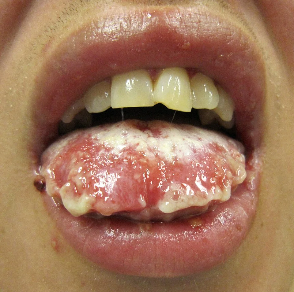
A 1-3 day flu-like prodrome (fever, malaise, sore throat, conjunctival irritation) often precedes the rash, which can make early presentations easy to mistake for a simple viral illness before the characteristic skin and mucosal changes appear.
The rash begins as painful, dusky-red or purpuric macules, sometimes with a targetoid appearance, starting on the trunk and face and spreading. Lesions coalesce and the epidermis becomes necrotic, forming flaccid blisters that rupture to leave denuded, raw dermis. A positive Nikolsky sign (gentle lateral pressure causes the epidermis to shear away) is characteristic.
Mucosal involvement occurs in over 90% of cases and often precedes or accompanies the skin changes - painful oral erosions, conjunctivitis or corneal ulceration, and genital ulceration are all common, and can be the presenting feature that brings the patient to medical attention before the skin signs are fully developed.
| Category | Body surface area detached |
|---|---|
| Stevens-Johnson syndrome (SJS) | Under 10% |
| SJS/TEN overlap | 10-30% |
| Toxic epidermal necrolysis (TEN) | Over 30% |
Clinical examination
- Estimate body surface area detached - using the same techniques as for burns assessment (rule of nines, or the patient's palm representing roughly 1%), since this defines the SJS/TEN category and feeds into SCORTEN
- Nikolsky sign - test gently on apparently uninvolved skin at the margin of affected areas
- Mucosal examination - oral cavity, conjunctivae and eyelids, and genital/perianal skin, all of which are frequently involved and easily under-assessed if attention focuses only on the skin
- Systemic assessment - temperature, hydration status, respiratory involvement (tracheobronchial mucosal sloughing can occur in severe TEN), and signs of secondary infection
- Careful, contemporaneous drug history - onset relative to any new medication in the preceding 1-4 weeks is central to identifying the causative agent
Differential diagnosis
- Staphylococcal scalded skin syndrome - toxin-mediated, intraepidermal splitting, mucosal sparing, and occurs mainly in young children; see Impetigo
- Erythema multiforme major - typical target lesions predominantly on the extremities, generally less extensive skin detachment, and more often triggered by infection (particularly herpes simplex) than by drugs
- Staphylococcal or streptococcal toxic shock syndrome - diffuse erythema without the same degree of epidermal necrosis and detachment, with prominent hypotension and multi-organ involvement
- Generalised bullous fixed drug eruption - can mimic SJS/TEN closely but tends to be less severe systemically and recurs at the same sites on re-exposure to the causative drug
- Acute graft-versus-host disease - considered in a recent transplant recipient with a similar widespread erythematous, blistering eruption
- Autoimmune blistering diseases (pemphigus vulgaris, bullous pemphigoid) - generally more gradual onset, without the same acute systemic prodrome, and distinguished by biopsy with direct immunofluorescence
Investigations
Investigation serves three purposes: confirming the diagnosis, calculating severity, and monitoring for complications.
- Skin biopsy with frozen section - shows full-thickness epidermal necrosis and helps confirm the diagnosis quickly, particularly useful in distinguishing SJS/TEN from staphylococcal scalded skin syndrome when the picture is unclear
- FBC, U&Es, LFTs, glucose and bicarbonate - feed directly into the SCORTEN score and guide fluid and metabolic management
- Blood, skin and line cultures - if secondary infection is suspected, given sepsis is the leading cause of death
- Mycoplasma serology/PCR - in children or where an infective trigger is suspected rather than a clear drug cause
SCORTEN
A validated severity score, calculated within the first 24 hours of admission, awarding one point for each of the following present, with mortality rising steeply as the score increases.2
- Age over 40 years
- Presence of malignancy
- Heart rate over 120 beats per minute
- Initial epidermal detachment over 10% of body surface area
- Serum urea over 10 mmol/L
- Serum glucose over 14 mmol/L
- Serum bicarbonate under 20 mmol/L
Management
- Early transfer to a burns unit or intensive care setting - for patients with significant body surface area detachment, since supportive management mirrors that of a major burn: fluid and electrolyte replacement, temperature regulation, meticulous wound care, and early nutritional support
- Specialist ophthalmology review - essential for any ocular involvement, to reduce the risk of long-term scarring, symblepharon (adhesion between the conjunctiva and eyelid) and visual loss
- Urology/gynaecology input - for genital involvement, to reduce the risk of scarring and stricture
- Careful, proactive infection surveillance rather than routine prophylactic antibiotics, since sepsis is the leading cause of death but antibiotic use should be guided by clinical and microbiological evidence rather than given blanket
- Analgesia - pain is often severe and under-treated relative to its severity
- Adjunctive systemic therapy - ciclosporin and, in some centres, intravenous immunoglobulin or systemic corticosteroids are used, though the evidence base is less robust than for the supportive measures above, and their use should be directed by a specialist centre experienced in managing this condition rather than adopted as routine first-line treatment1
Complications
- Sepsis - the leading cause of death, from loss of the normal skin barrier over a large body surface area
- Fluid and electrolyte imbalance, and acute kidney injury
- Respiratory failure - from tracheobronchial mucosal sloughing in severe TEN
- Ocular complications - corneal scarring, symblepharon, and in severe cases permanent visual impairment or blindness
- Genital and mucosal strictures - from scarring of ulcerated mucosal surfaces
- Long-term skin changes - dyspigmentation, scarring, and nail or hair loss
- Psychological impact - considerable, given the severity, disfigurement and life-threatening nature of the acute illness
Red flags
Prognosis
Mortality rises steeply with SCORTEN, from around 3% with a score of 0-1 to over 90% with a score of 5 or more, and overall mortality for TEN (over 30% body surface area detachment) is substantially higher than for SJS.2
Survivors, particularly of TEN, can be left with significant long-term sequelae - chronic dry eyes, corneal scarring and visual impairment, oral and genital mucosal strictures, and lasting skin pigment change - all of which mean follow-up after the acute illness should include specialist ophthalmology and dermatology review rather than being considered complete on hospital discharge. The prominent role of drug withdrawal and supportive care, rather than any single definitive treatment, in determining outcome is the central practical message of this topic.
References
- Creamer D, Walsh SA, Dziewulski P et al. UK guidelines for the management of Stevens-Johnson syndrome/toxic epidermal necrolysis in adults 2016. British Journal of Dermatology. 2016. Available here
- Bastuji-Garin S, Fouchard N, Bertocchi M et al. SCORTEN: a severity-of-illness score for toxic epidermal necrolysis. Journal of Investigative Dermatology. 2000. Available here
- Chung WH, Hung SI, Yang JY et al. Granulysin is a key mediator for disseminated keratinocyte death in Stevens-Johnson syndrome and toxic epidermal necrolysis. Nature Medicine. 2008. Available here
- Chung WH, Hung SI, Hong HS et al. Medical genetics: a marker for Stevens-Johnson syndrome. Nature. 2004. Available here
- Mockenhaupt M, Viboud C, Dunant A et al. Stevens-Johnson syndrome and toxic epidermal necrolysis: assessment of medication risks with emphasis on recently marketed drugs. Journal of Investigative Dermatology. 2008. Available here
- MHRA / GOV.UK. Antiepileptic drugs: risk of severe cutaneous adverse reactions. Available here
- DermNet NZ. Toxic epidermal necrolysis. Available here
- Canavan TN, Mathes EF, Frieden I, Shinkai K. Mycoplasma pneumoniae-induced rash and mucositis as a syndrome distinct from Stevens-Johnson syndrome and erythema multiforme: a systematic review. Journal of the American Academy of Dermatology. 2015. Available here
This article is written for revision and education. It is not clinical guidance and must not be used to make decisions about the care of a patient. Always check current NICE guidance and local protocols.