
Skin Manifestations of Systemic Disease
Key points
- Why this matters: the skin is often the most visible and accessible clue to an underlying systemic disease, and recognising these signs can bring forward a diagnosis by months or years.
- Acanthosis nigricans: velvety, hyperpigmented thickening of the flexures, most commonly a marker of insulin resistance and obesity, but occasionally a paraneoplastic sign (classically gastric adenocarcinoma) when rapid in onset, extensive, or affecting mucosa.
- Necrobiosis lipoidica: well-demarcated, yellow-brown, atrophic plaques with visible telangiectasia on the shins, strongly (though not exclusively) associated with diabetes.
- Pyoderma gangrenosum: a rapidly progressive, intensely painful ulcer with a violaceous, undermined border, associated with inflammatory bowel disease, rheumatoid arthritis and haematological malignancy - a diagnosis of exclusion, not a wound infection.
- Dermatomyositis: the heliotrope rash, Gottron's papules and shawl sign mark an underlying inflammatory myopathy, and in adults carry a genuine association with occult malignancy that must be actively screened for.
- Vasculitic (palpable) purpura: small vessel vasculitis affecting the skin, which can be isolated or reflect an underlying systemic vasculitis, drug reaction or infection, and needs assessment for other organ involvement.
- Xanthomata: visible lipid deposits in the skin (eruptive, tendon, palmar, and xanthelasma around the eyes) that mark an underlying lipid disorder, sometimes a severe genetic dyslipidaemia.
- Others worth knowing: livedo reticularis (antiphospholipid syndrome, vasculitis) and pretibial myxoedema (Graves' disease) complete the core list of cutaneous markers tested in this topic.
Introduction
Many systemic diseases produce recognisable changes in the skin, sometimes before the underlying condition is otherwise apparent. Because the skin is directly visible and easily examined, these signs offer a genuine opportunity for earlier diagnosis - a chance easily missed if a skin finding is dismissed as cosmetic or incidental rather than pursued as a clue to what is happening internally.
This note covers the cutaneous markers of systemic disease most often tested: acanthosis nigricans, necrobiosis lipoidica, pyoderma gangrenosum, the cutaneous signs of dermatomyositis, vasculitic purpura, and a shorter round-up of xanthomata, livedo reticularis and pretibial myxoedema. Erythema nodosum, another classic marker of systemic disease (particularly sarcoidosis and inflammatory bowel disease), has its own dedicated article.
Acanthosis nigricans
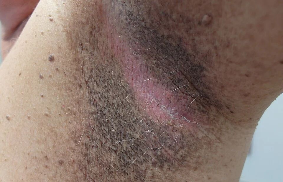
Acanthosis nigricans is a velvety, hyperpigmented, thickened area of skin, most often affecting the axillae, neck and groin, caused by keratinocyte and dermal fibroblast proliferation driven by high circulating insulin acting on insulin-like growth factor receptors.1
| Cause | Notes |
|---|---|
| Obesity and insulin resistance / type 2 diabetes | By far the commonest cause, reflecting compensatory hyperinsulinaemia |
| Polycystic ovary syndrome | Related to underlying insulin resistance |
| Endocrine disorders | Cushing syndrome, acromegaly, hypothyroidism |
| Drugs | Nicotinic acid, systemic corticosteroids, combined oral contraceptives |
| Malignant acanthosis nigricans | A rare paraneoplastic phenomenon, classically with gastric adenocarcinoma; suggested by rapid onset, extensive distribution, involvement of mucosa or palms, and accompanying weight loss |
Management addresses the underlying cause - weight loss and improved glycaemic control for the common insulin-resistance-related pattern - rather than the skin change itself, which has no direct treatment beyond emollients for comfort.
Necrobiosis lipoidica
Necrobiosis lipoidica presents as well-demarcated, yellow-brown, atrophic plaques with a shiny surface and visible telangiectasia, almost always on the shins, and is strongly (though not exclusively) associated with diabetes mellitus - a minority of cases occur in people without diabetes.2
Lesions are often asymptomatic but can ulcerate, particularly after minor trauma, given the atrophic, thinned skin overlying them. There is no specific curative treatment; potent topical or intralesional corticosteroids can help early inflammatory lesions, and good glycaemic control, while sensible for general reasons, does not reliably resolve established plaques.
Pyoderma gangrenosum
Pyoderma gangrenosum is a rare, neutrophilic dermatosis causing rapidly progressive, intensely painful ulceration, typically on the legs. It begins as a small pustule or nodule that breaks down within days into an ulcer with a violaceous, undermined border and a boggy, purulent base - pain is often disproportionate to the apparent size of the lesion.3
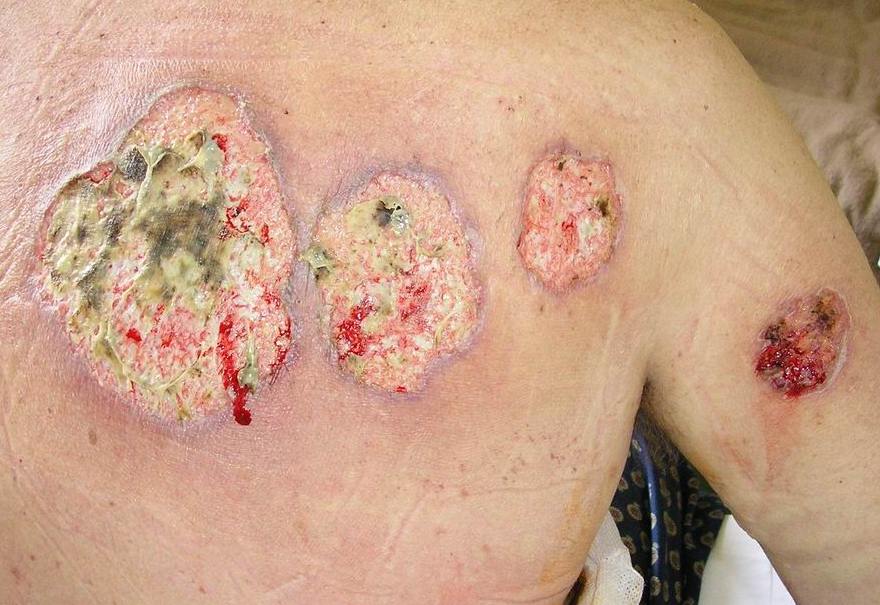
- Inflammatory bowel disease (Crohn's disease and ulcerative colitis) - the commonest identified association
- Rheumatoid arthritis and other inflammatory arthritides
- Haematological malignancy, particularly myeloid disorders
- Idiopathic - no underlying cause found in a substantial proportion of patients
Management is with systemic immunosuppression (corticosteroids, ciclosporin, or biologic therapy targeting the underlying associated disease) rather than surgical debridement or antibiotics alone, and any underlying systemic disease should be actively sought and treated.
Dermatomyositis
Dermatomyositis is an idiopathic inflammatory myopathy with characteristic skin signs that often precede or accompany proximal muscle weakness.4
- Heliotrope rash - a violaceous discolouration of the upper eyelids, often with periorbital oedema
- Gottron's papules - violaceous, scaly papules over the metacarpophalangeal and interphalangeal joints of the hands (the flat, non-papular version over the same sites is Gottron's sign)
- Shawl sign - erythema across the upper back, shoulders and posterior neck in a shawl-like distribution, often worsened by sun exposure
- 'Mechanic's hands' - rough, cracked, hyperkeratotic skin on the lateral aspects of the fingers
- Nailfold changes - dilated capillary loops and ragged cuticles, visible on careful examination or dermoscopy
Proximal muscle weakness (difficulty rising from a chair, climbing stairs, or raising the arms above the head) usually accompanies or follows the skin signs, though a subset of patients have skin disease alone ('amyopathic dermatomyositis').
Vasculitic (palpable) purpura
Cutaneous small vessel vasculitis produces palpable purpura - non-blanching, raised, purple lesions, typically on the lower legs - caused by inflammation and damage to small dermal vessels with resulting extravasation of red cells.6
- IgA vasculitis (Henoch-Schonlein purpura) - the commonest vasculitis in children, with palpable purpura, abdominal pain, arthralgia and renal involvement, often following an upper respiratory infection
- ANCA-associated vasculitis - granulomatosis with polyangiitis and microscopic polyangiitis can present with cutaneous purpura alongside renal, respiratory and other systemic involvement
- Drug-induced vasculitis - a recognised reaction to several drug classes
- Infection-associated vasculitis - particularly some chronic infections and immune complex-mediated processes
- Cutaneous small vessel vasculitis confined to the skin - a diagnosis of exclusion once systemic causes have been considered
Any patient with palpable purpura should be assessed for systemic involvement - urinalysis for haematuria/proteinuria, blood pressure, renal function, and symptoms suggesting respiratory, gastrointestinal or joint involvement - since the skin finding can be the presenting feature of a systemic vasculitis needing urgent specialist input.
Other cutaneous markers of systemic disease
| Sign | Association |
|---|---|
| Eruptive xanthomata | Sudden crops of small, yellow-red papules, usually on the buttocks and extensor limbs, from severe hypertriglyceridaemia |
| Tendon xanthomata | Firm nodules within the Achilles tendon and finger extensor tendons, characteristic of familial hypercholesterolaemia |
| Xanthelasma | Yellowish plaques around the eyelids; can occur with normal lipids but often reflects an underlying dyslipidaemia, particularly when appearing at a younger age7 |
| Livedo reticularis | A net-like, mottled, violaceous discolouration of the skin, seen in antiphospholipid syndrome, vasculitis, and some connective tissue diseases, as well as benign physiological cold-related forms |
| Pretibial myxoedema | Non-pitting, waxy, thickened plaques on the shins in Graves' disease, from glycosaminoglycan deposition; despite the name, it occurs with hyperthyroidism, not hypothyroidism8 |
Clinical examination
- Distribution and morphology of the specific sign, matched against the patterns above
- Full skin examination, since these signs are frequently found incidentally during examination for an unrelated complaint
- Systemic examination directed by the specific sign - joints and abdomen for pyoderma gangrenosum (IBD, arthritis), proximal muscle strength for dermatomyositis, urinalysis and blood pressure for vasculitic purpura, and a general endocrine and metabolic assessment for acanthosis nigricans and xanthomata
- Lymph nodes and a general cancer screen where a paraneoplastic process is suspected (malignant acanthosis nigricans, adult-onset dermatomyositis)
Investigations
Investigation is directed by the specific skin sign towards its most likely underlying cause, rather than a single generic work-up.
- Acanthosis nigricans - HbA1c, fasting glucose and insulin; consider endoscopy/imaging if malignant acanthosis nigricans is suspected
- Necrobiosis lipoidica - HbA1c/glucose, though biopsy is occasionally needed if the diagnosis is uncertain
- Pyoderma gangrenosum - a diagnosis of exclusion: wound culture and biopsy to rule out infection and malignancy, plus investigation for associated disease (colonoscopy if IBD is suspected, FBC and blood film for haematological malignancy)
- Dermatomyositis - creatine kinase and other muscle enzymes, myositis-specific and myositis-associated autoantibodies, electromyography, muscle MRI or biopsy, and age-appropriate malignancy screening
- Vasculitic purpura - FBC, renal function, urinalysis, ANCA, complement levels, and skin biopsy with direct immunofluorescence to characterise the vasculitis and identify IgA deposition if Henoch-Schonlein purpura is suspected
- Xanthomata - full fasting lipid profile, and consideration of familial hypercholesterolaemia genetic testing if tendon xanthomata or a strong family history are present
Management
In every condition covered here, the priority is identifying and treating the underlying systemic disease, since the skin sign itself is a marker rather than the primary problem. Direct skin-specific treatment (emollients for acanthosis nigricans, topical or intralesional corticosteroids for early necrobiosis lipoidica, immunosuppression for pyoderma gangrenosum, sun protection and topical treatment for dermatomyositis rash) provides symptomatic relief but does not substitute for addressing the cause. Specialist referral - dermatology, endocrinology, gastroenterology, rheumatology or haematology as relevant - is appropriate once the likely underlying association is identified.
Red flags
Prognosis
Prognosis for each of these conditions is governed almost entirely by the underlying systemic disease rather than the skin sign itself. Acanthosis nigricans related to insulin resistance improves with weight loss and glycaemic control; pyoderma gangrenosum and vasculitis respond to treatment of the associated inflammatory bowel disease, arthritis, or vasculitic process; and dermatomyositis outcome depends heavily on whether an associated malignancy is found and treated.
The consistent lesson across this topic is that these skin findings function as diagnostic signposts rather than isolated dermatological problems, and the greatest value in recognising them lies in the systemic work-up they should trigger rather than in any treatment directed at the skin alone.
References
- Brady MF, Rawla P. Acanthosis Nigricans. StatPearls. 2023. Available here
- Reid SD, Ladizinski B, Lee K et al. Update on necrobiosis lipoidica: a review of etiology, diagnosis, and treatment options. Journal of the American Academy of Dermatology. 2013. Available here
- George C, Deroide F, Rustin M. Pyoderma gangrenosum - a guide to diagnosis and management. Clinical Medicine. 2019. Available here
- Dalakas MC. Inflammatory muscle diseases. New England Journal of Medicine. 2015. Available here
- Hill CL, Zhang Y, Sigurgeirsson B et al. Frequency of specific cancer types in dermatomyositis and polymyositis: a population-based study. The Lancet. 2001. Available here
- Fraticelli P, Benfaremo D, Gabrielli A. Diagnosis and management of leukocytoclastic vasculitis. Internal and Emergency Medicine. 2021. Available here
- NICE NG238. Cardiovascular disease: risk assessment and reduction, including lipid modification. 2023. Available here
- DermNet NZ. Pretibial myxoedema. Available here
This article is written for revision and education. It is not clinical guidance and must not be used to make decisions about the care of a patient. Always check current NICE guidance and local protocols.