
Skin Ulcers
Key points
- The big three: venous, arterial and neuropathic disease account for the great majority of chronic leg and foot ulcers, distinguished mainly by site, pain, and the state of the surrounding skin and pulses.
- Venous ulcers: the commonest cause (around 70%), from chronic venous hypertension; shallow, irregular, exudative ulcers in the gaiter area (around the medial malleolus), with surrounding haemosiderin staining, lipodermatosclerosis and varicose eczema.
- Arterial ulcers: from peripheral arterial disease; punched-out, deep, painful ulcers at distal sites and pressure points, with cold peripheries, weak or absent pulses, and pain worse on elevation and relieved by dependency.
- Neuropathic ulcers: typically diabetic, occurring painlessly over pressure points (metatarsal heads, heel) because of lost protective sensation, often with a surrounding callus and preserved perfusion.
- ABPI: the ankle-brachial pressure index is the key test distinguishing these causes and determining whether compression bandaging is safe (broadly normal 0.9-1.3; below 0.8 needs caution; below 0.5 is an urgent vascular referral, and compression is contraindicated).
- Mixed ulcers: venous and arterial disease coexist in a meaningful proportion of older patients, requiring modified, reduced compression under specialist guidance rather than either standard approach alone.
- Other causes: vasculitis, pyoderma gangrenosum and malignancy (including Marjolin ulcer arising within a chronic wound) can all present as a non-healing ulcer and should be considered when a lesion does not fit the typical venous, arterial or neuropathic pattern.
- Red flag: a raised, rolled or indurated edge, an ulcer that fails to respond to appropriate treatment, or one arising within a long-standing scar needs biopsy to exclude malignant transformation.
Introduction
Chronic skin ulceration, particularly of the lower leg and foot, is common and has a substantial impact on quality of life, mobility and, in the case of arterial and neuropathic disease, limb survival. The three commonest causes - venous, arterial and neuropathic disease - have distinct mechanisms and, crucially, opposite management priorities in some respects, which is why correctly distinguishing them before starting treatment matters so much.
The central practical danger in this topic is applying compression bandaging - the correct treatment for a venous ulcer - to a limb with significant arterial insufficiency, where it can precipitate critical ischaemia. This is why the ankle-brachial pressure index (ABPI) is checked before compression is started in essentially every case of chronic leg ulceration.8
Venous ulcers
Venous ulcers are the commonest cause of chronic leg ulceration, arising from chronic venous hypertension due to valvular incompetence in the deep or superficial venous system, or previous deep vein thrombosis. Sustained high pressure in the venous system causes capillary damage, fibrin deposition, and impaired oxygen and nutrient delivery to the skin, eventually leading to breakdown and ulceration.1
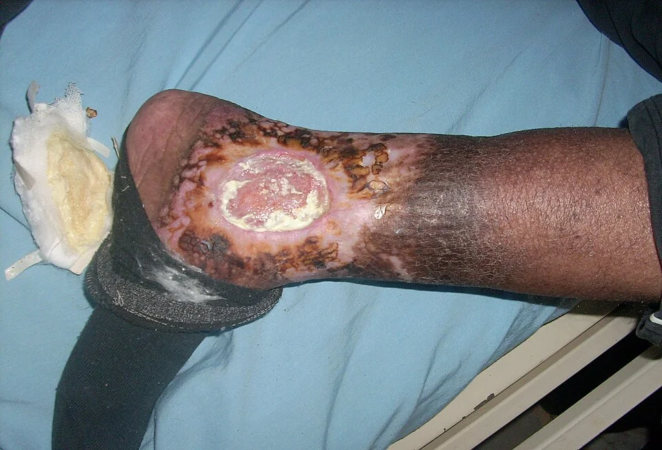
- Site - the 'gaiter area', particularly around the medial malleolus
- Appearance - shallow, irregular border, moist with a granulating or fibrinous base, often heavily exudative
- Surrounding skin - haemosiderin staining (brown discolouration from extravasated red cells), varicose eczema, lipodermatosclerosis (woody induration and narrowing of the lower leg, sometimes described as an 'inverted champagne bottle' shape), and atrophie blanche (white, atrophic scarred patches)
- Pain - typically mild to moderate, and characteristically improved by leg elevation
Management
- Multi-layer compression bandaging - the cornerstone of treatment, but only after confirming an adequate ABPI, since compression on a limb with significant arterial insufficiency can cause tissue necrosis6
- Leg elevation when resting, to reduce venous pressure
- Treatment of underlying superficial venous incompetence (endovenous ablation or surgery) once the ulcer has healed, reducing recurrence
- Emollients and appropriate topical treatment for associated varicose eczema
- Long-term compression hosiery after healing, to reduce recurrence, which is otherwise common
Arterial ulcers
Arterial ulcers result from peripheral arterial disease causing insufficient blood flow to maintain tissue viability, usually in the context of established atherosclerosis and its risk factors (smoking, diabetes, hypertension, hyperlipidaemia).2,5
- Site - distal areas and pressure points furthest from the blood supply - toes, heel, lateral malleolus, and over the metatarsal heads
- Appearance - well-defined, 'punched-out' edges, often deep enough to expose tendon or bone, with a pale, poorly granulating base
- Surrounding skin - cool, pale or dusky, hairless, shiny, with weak or absent peripheral pulses
- Pain - often severe, characteristically worse on elevation and at night, and relieved by hanging the leg over the side of the bed or standing - the opposite pattern to venous ulcers
Management
- Urgent vascular assessment for significant disease, with imaging (duplex ultrasound, CT or MR angiography) to define the anatomy
- Revascularisation - angioplasty, stenting or bypass surgery where appropriate, to restore perfusion and allow healing
- Best medical therapy - smoking cessation, antiplatelet therapy, statin, and optimisation of blood pressure and diabetic control
- Compression bandaging is contraindicated in the presence of significant arterial disease, since it further reduces perfusion and can precipitate critical limb ischaemia
Neuropathic ulcers
Neuropathic ulcers, most often seen in diabetes, result from loss of protective sensation due to peripheral neuropathy, combined with repetitive unnoticed pressure or minor trauma over bony prominences of the foot.4
- Site - pressure-bearing areas of the foot - the plantar surface over the metatarsal heads, the heel, and over the toes
- Appearance - punched-out, often surrounded by a thick callus, typically painless despite sometimes being deep
- Perfusion - often relatively preserved (pulses may be normal), since the underlying problem is neuropathy rather than ischaemia, though coexisting peripheral arterial disease is common in longstanding diabetes and should always be checked for
- Sensation - reduced or absent protective sensation on monofilament testing is the defining feature
Management
- Pressure offloading - specialist footwear, total contact casting, or removable devices to redistribute pressure away from the ulcer
- Regular podiatry/chiropody and callus debridement
- Optimising glycaemic control
- Screening for and treating any coexisting peripheral arterial disease, since a purely neuropathic ulcer in isolation is less common than one with a mixed neuroischaemic component in practice
- Structured diabetic foot multidisciplinary team involvement for any ulcer that fails to improve, given the high risk of progression to infection and amputation if undertreated
Mixed and other ulcers
Mixed venous-arterial ulcers, with features of both, are common in older patients with widespread vascular disease, and management requires a modified, reduced level of compression guided by the ABPI and ideally by specialist tissue viability or vascular input, rather than either standard venous or arterial management alone.
A smaller proportion of chronic ulcers have other causes, each covered in more detail elsewhere: pyoderma gangrenosum (a rapidly progressive, painful ulcer with a violaceous, undermined edge, associated with inflammatory bowel disease and other systemic conditions - see Skin Manifestations of Systemic Disease), vasculitic ulceration, and malignancy, including squamous cell carcinoma arising within a chronic wound or scar (a Marjolin ulcer - see Squamous Cell Carcinoma and Bowen Disease). Pressure ulcers, from sustained pressure over bony prominences in an immobile patient, are covered in their own dedicated article.
Clinical examination
| Feature | Venous | Arterial | Neuropathic |
|---|---|---|---|
| Typical site | Gaiter area (medial malleolus) | Distal - toes, heel, lateral malleolus | Pressure points - metatarsal heads, heel |
| Edge/base | Irregular, shallow, granulating | Punched-out, deep, pale base | Punched-out, often callused edge |
| Pain | Mild-moderate, better on elevation | Severe, worse on elevation/at night | Often painless (reduced sensation) |
| Surrounding skin | Haemosiderin staining, lipodermatosclerosis, eczema | Pale, cool, hairless, shiny | Callus, often otherwise unremarkable |
| Pulses | Normal | Weak or absent | Often normal (unless coexisting arterial disease) |
- Ankle-brachial pressure index (ABPI) - measured in every patient with a chronic leg ulcer before considering compression
- Peripheral pulses - dorsalis pedis, posterior tibial, popliteal and femoral
- Monofilament and vibration testing - to assess for peripheral neuropathy
- Wound characteristics - size, depth, base, edge and surrounding skin, documented and measured to track progress
- Signs of infection - increasing pain, spreading erythema, malodour, or systemic upset
Investigations
- ABPI - systolic pressure at the ankle divided by the higher of the two brachial systolic pressures; roughly 0.9-1.3 is normal, under 0.8 warrants caution with compression, and under 0.5 indicates severe arterial disease needing urgent vascular referral and no compression
- Duplex ultrasound - assesses venous reflux/incompetence in suspected venous disease, and arterial flow and stenosis in suspected arterial disease
- Wound swab - only if clinical infection is suspected; routine swabbing of a colonised but clinically uninfected ulcer is not helpful and can lead to unnecessary antibiotic use
- Biopsy - for any ulcer that is atypical, fails to heal despite appropriate treatment, or has a raised or indurated edge, to exclude malignancy or an alternative diagnosis such as vasculitis or pyoderma gangrenosum
- HbA1c, FBC, renal function and other bloods as directed by the suspected underlying cause
Management principles
- Treat the underlying cause - compression for venous disease (once arterial disease is excluded), revascularisation for arterial disease, offloading for neuropathic disease
- Wound bed preparation - debridement of non-viable tissue where appropriate, and dressing selection matched to exudate level rather than a single dressing for every ulcer
- Manage infection appropriately - systemic antibiotics for clinical infection (spreading erythema, systemic upset), not for asymptomatic bacterial colonisation
- Address risk factors - smoking cessation, glycaemic and blood pressure control, and nutrition, all of which influence healing
- Analgesia appropriate to the cause, particularly for the often severe pain of arterial ulcers
- Multidisciplinary input - tissue viability nursing, vascular surgery, diabetology/podiatry and dermatology as relevant to the underlying cause
Complications
- Cellulitis and, less commonly, osteomyelitis, particularly with neuropathic or arterial ulcers where infection can spread more readily
- Chronic non-healing ulceration, with significant impact on mobility and quality of life
- Amputation - a real risk with arterial and neuropathic ulcers if not managed proactively
- Malignant transformation (Marjolin ulcer) within a chronic, longstanding wound
- Recurrence after healing, particularly for venous ulcers if long-term compression hosiery is not maintained
Red flags
Prognosis
Venous ulcers generally heal well with correctly applied compression therapy, though recurrence is common (up to half within a year) without maintenance compression hosiery and attention to underlying venous incompetence.3
Arterial and neuropathic ulcers carry a less certain outlook and a genuine risk of progression to limb-threatening infection or ischaemia if not managed proactively; diabetic foot ulceration in particular is associated with a substantial risk of amputation and, notably, with mortality rates over subsequent years comparable to some cancers, underscoring why prompt multidisciplinary diabetic foot care is taken so seriously.7
References
- NICE Clinical Knowledge Summaries. Leg ulcer - venous. Available here
- NICE Clinical Knowledge Summaries. Peripheral arterial disease. Available here
- SIGN 120. Management of chronic venous leg ulcers. 2010. Available here
- NICE NG19. Diabetic foot problems: prevention and management. 2015, updated 2019. Available here
- NICE CG147. Peripheral arterial disease: diagnosis and management. 2012. Available here
- O'Meara S, Cullum N, Nelson EA, Dumville JC. Compression for venous leg ulcers. Cochrane Database of Systematic Reviews. 2012. Available here
- Boulton AJ, Vileikyte L, Ragnarson-Tennvall G, Apelqvist J. The global burden of diabetic foot disease. The Lancet. 2005. Available here
- DermNet NZ. Leg ulcers. Available here
This article is written for revision and education. It is not clinical guidance and must not be used to make decisions about the care of a patient. Always check current NICE guidance and local protocols.