
Ventricular Tachycardia and Ventricular Fibrillation
Key points
- Ventricular tachycardia: three or more consecutive ventricular beats at over 100 per minute. Sustained VT lasts over 30 seconds or causes haemodynamic compromise.
- The cardinal rule: a broad complex tachycardia is ventricular tachycardia until proven otherwise. Around 80% are VT, rising above 95% with previous myocardial infarction.
- Ventricular fibrillation: chaotic, disorganised ventricular activity with no cardiac output. It is a cardiac arrest rhythm requiring immediate unsynchronised defibrillation.
- Monomorphic VT: uniform QRS complexes, usually from re-entry around myocardial scar after infarction.
- Polymorphic VT: varying QRS morphology. With a long QT this is torsades de pointes, treated with magnesium; with a normal QT, suspect acute ischaemia.
- Unstable VT: shock, syncope, myocardial ischaemia or heart failure require immediate synchronised DC cardioversion.
- Stable VT: amiodarone 300 mg IV over 20-60 minutes, then 900 mg over 24 hours. Never give verapamil to an undiagnosed broad complex tachycardia.
- Prevention: an implantable cardioverter defibrillator for secondary prevention after a survived arrest, and for primary prevention in selected patients with an ejection fraction of 35% or below.
Introduction
Ventricular arrhythmias arise below the bundle of His. Because the impulse spreads slowly from myocyte to myocyte rather than through the fast His-Purkinje system, the QRS complex is broad - 120 ms or more.
They occupy one end of a spectrum of urgency. Ventricular ectopic beats are common and usually benign. Non-sustained VT may be an incidental finding or a marker of significant underlying disease. Sustained monomorphic VT may be tolerated for hours or may cause immediate collapse. Ventricular fibrillation is cardiac arrest, and untreated it is fatal within minutes.
| Term | Definition |
|---|---|
| Ventricular ectopic beat | A single premature broad complex beat, usually followed by a compensatory pause |
| Bigeminy / trigeminy | An ectopic beat following every one or every two sinus beats |
| Non-sustained VT | Three or more consecutive ventricular beats over 100/min, lasting under 30 seconds |
| Sustained VT | VT lasting over 30 seconds, or causing haemodynamic compromise requiring earlier intervention |
| Monomorphic VT | All QRS complexes have the same morphology - a single re-entrant circuit or focus |
| Polymorphic VT | QRS morphology varies beat to beat |
| Torsades de pointes | Polymorphic VT in the context of a prolonged QT interval, with a twisting axis |
| Ventricular flutter | A regular sinusoidal ventricular rhythm at around 300/min with no distinguishable QRS and T |
| Ventricular fibrillation | Chaotic, disorganised ventricular activity with no cardiac output |
Aetiology
Structural heart disease
Most sustained monomorphic VT arises from re-entry around a region of myocardial scar, most commonly the border zone of a previous infarct, where slow conduction through surviving muscle fibres within fibrous tissue creates the substrate for a circuit.
- Ischaemic heart disease - by far the commonest cause. VT within 48 hours of an acute infarct reflects acute ischaemia; VT occurring later reflects established scar and carries a worse prognosis.
- Dilated cardiomyopathy
- Hypertrophic cardiomyopathy - a leading cause of sudden death in young athletes
- Arrhythmogenic right ventricular cardiomyopathy - fibrofatty replacement of the right ventricle, with VT of left bundle branch block morphology
- Valvular heart disease, particularly severe aortic stenosis
- Myocarditis and cardiac sarcoidosis
- Congenital heart disease after surgical repair, with circuits around surgical scar
- Chagas disease - an important cause worldwide
Structurally normal hearts
- Idiopathic VT - right ventricular outflow tract VT (LBBB morphology with an inferior axis, responsive to adenosine and beta-blockers) and fascicular VT (RBBB with left axis deviation, characteristically verapamil-sensitive). Both carry a good prognosis and are curable by ablation.
- Long QT syndrome - congenital or acquired, causing torsades de pointes
- Brugada syndrome - a sodium channelopathy with coved ST elevation in V1-V2 and a risk of polymorphic VT, classically at night or with fever
- Catecholaminergic polymorphic VT - bidirectional or polymorphic VT triggered by exercise or emotion in young people
- Short QT syndrome - rare
Reversible precipitants
- Acute myocardial ischaemia or infarction - the commonest trigger of VF
- Electrolyte disturbance - hypokalaemia, hypomagnesaemia, hypocalcaemia and hyperkalaemia
- Drugs - QT-prolonging agents, class Ic antiarrhythmics in structural heart disease, digoxin toxicity, and cocaine
- Hypoxia and acidosis
- Hypothermia
- Electrocution and commotio cordis - a blow to the chest during the vulnerable phase of repolarisation
- Reperfusion - accelerated idioventricular rhythm is common after successful reperfusion and is usually benign
Clinical features
Presentation ranges from asymptomatic non-sustained runs found on a monitor to cardiac arrest. What determines the difference is the rate, the duration, and above all the state of the underlying ventricle.
Symptoms
- Palpitations - rapid and usually regular in monomorphic VT
- Presyncope and syncope - syncope without prodrome, or syncope on exertion, is a red flag for a ventricular arrhythmia and must not be dismissed as a simple faint
- Chest pain and breathlessness
- Cardiac arrest - VF and pulseless VT present as sudden collapse with no output
- Asymptomatic - non-sustained VT is often found incidentally on ambulatory monitoring
Examination
The purpose is to establish stability and to look for signs of AV dissociation, which effectively confirm the diagnosis.
Look also for evidence of structural heart disease, scars from previous cardiac surgery or a device, and signs of heart failure or hypoperfusion.
Distinguishing VT from SVT with aberrancy
This distinction is academically interesting and examinable, but in practice the presence of any doubt should lead to treatment as VT. The features below increase confidence rather than replacing that rule.
| Feature | Why it matters |
|---|---|
| Previous myocardial infarction or structural heart disease | The single most powerful predictor - raises the probability of VT above 95% |
| AV dissociation | P waves marching through independently; cannon a waves clinically. Effectively diagnostic. |
| Capture beats | An occasional narrow QRS where a sinus impulse conducts normally through the ventricles |
| Fusion beats | A hybrid complex from simultaneous sinus and ventricular activation |
| Very broad QRS (over 160 ms) | Wider than typical bundle branch block |
| Extreme (northwest) axis | Negative QRS in both lead I and aVF |
| Concordance across the chest leads | All precordial complexes entirely positive or entirely negative |
| QRS morphology not typical of RBBB or LBBB | Aberrancy should look like a recognisable bundle branch block pattern |
| Age over 35 | Prior probability of structural disease is higher |
Investigations
During the arrhythmia
Obtain a 12-lead ECG if the patient is stable enough - it is far more informative than a rhythm strip and is invaluable to the electrophysiologist later. In an unstable patient, treatment comes first.
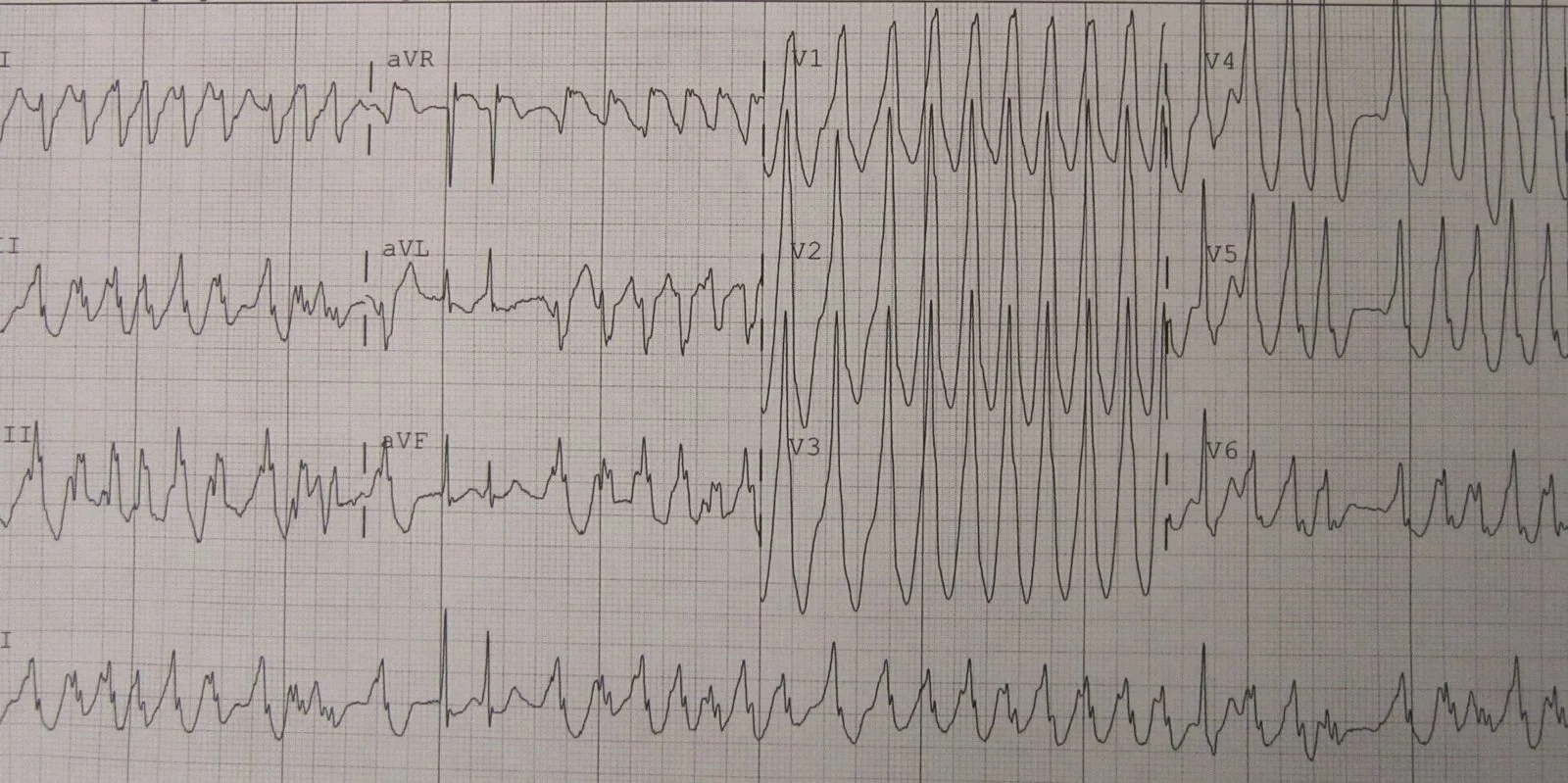
- Monomorphic VT - regular broad complex tachycardia at 100-250/min with uniform QRS morphology
- Polymorphic VT - continuously varying QRS morphology and axis
- Torsades de pointes - polymorphic VT with a characteristic twisting of the axis about the baseline, in the context of a long QT
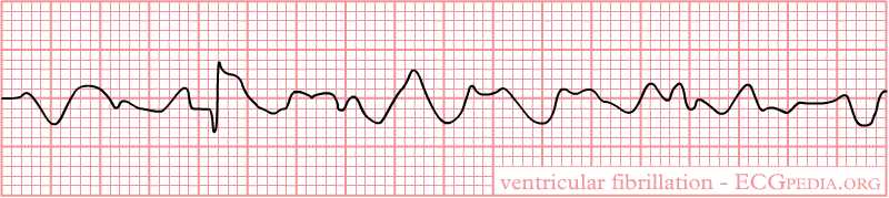
- Ventricular fibrillation - chaotic, irregular waveforms of varying amplitude with no identifiable QRS complexes. Coarse VF has a better prognosis than fine VF, which may be mistaken for asystole.
Immediate bloods and bedside tests
- U&Es, magnesium and calcium - correcting these is part of the treatment, not just the workup. Aim for potassium above 4.0 mmol/L and magnesium above 1.0 mmol/L.
- Troponin - to identify acute ischaemia as the precipitant, remembering that VT and defibrillation both raise it
- Arterial blood gas - hypoxia and acidosis
- Drug levels - digoxin in particular
- Toxicology - cocaine and other stimulants in young patients
- Thyroid function and FBC
Establishing the substrate
- Resting 12-lead ECG in sinus rhythm - Q waves from previous infarction, QTc, Brugada pattern, epsilon waves and T inversion in V1-V3 (ARVC), left ventricular hypertrophy
- Echocardiogram - left ventricular ejection fraction is the key number, since it drives ICD decisions
- Coronary angiography - to identify and treat ischaemia in anyone with a plausible ischaemic substrate
- Cardiac MRI - detects scar, infiltration, myocarditis and ARVC, and characterises the substrate for ablation
- Ambulatory monitoring and exercise testing - to quantify ectopic burden and provoke catecholamine-dependent arrhythmias
- Electrophysiological study - for risk stratification in selected patients and to map circuits before ablation
- Genetic testing and family screening - where an inherited channelopathy or cardiomyopathy is suspected
Acute management
Pulseless VT and ventricular fibrillation
VT with a pulse: unstable
Any adverse feature - shock, syncope, myocardial ischaemia or heart failure - requires synchronised DC cardioversion under sedation or general anaesthesia, up to three attempts. Synchronisation matters: an unsynchronised shock landing on the T wave can induce VF.
If three shocks fail, give amiodarone 300 mg IV over 10-20 minutes, shock again, and follow with an infusion of 900 mg over 24 hours.1
VT with a pulse: stable
- Amiodarone 300 mg IV over 20-60 minutes through a large vein or, preferably, a central line, followed by 900 mg over 24 hours
- Correct electrolytes - potassium to above 4.0 mmol/L and magnesium to above 1.0 mmol/L
- Treat ischaemia - if acute coronary syndrome is the driver, reperfusion is the definitive antiarrhythmic
- Seek expert help early, and if drug treatment fails, proceed to synchronised cardioversion
Polymorphic VT
Management depends entirely on the QT interval in sinus rhythm.
| QT in sinus rhythm | Diagnosis | Treatment |
|---|---|---|
| Prolonged | Torsades de pointes | Magnesium sulfate 2 g IV over 10 minutes; stop all QT-prolonging drugs; correct potassium and magnesium; consider overdrive pacing or isoprenaline. Defibrillate if pulseless. |
| Normal | Usually acute myocardial ischaemia | Treat the ischaemia urgently - angiography and revascularisation. Beta-blockers and amiodarone; magnesium is not the answer here. |
| Normal, exercise-triggered, young patient | Catecholaminergic polymorphic VT | Beta-blockers, avoid exertion, refer for inherited cardiac conditions assessment and consider an ICD |
Long-term management
Treat the substrate
- Revascularisation where ischaemia is demonstrated
- Optimal heart failure therapy - a beta-blocker, an ACE inhibitor or ARB, a mineralocorticoid receptor antagonist and an SGLT2 inhibitor. These reduce sudden death as well as improving symptoms.
- Correct and maintain electrolytes, and review every QT-prolonging drug
- Treat the underlying cardiomyopathy or channelopathy
Implantable cardioverter defibrillators
An ICD does not prevent arrhythmia; it terminates it. It is the most effective intervention for preventing sudden arrhythmic death in selected patients.2
| Category | Typical indication |
|---|---|
| Secondary prevention | Survived cardiac arrest due to VT or VF; sustained VT with syncope or significant haemodynamic compromise; sustained VT with an ejection fraction of 35% or below |
| Primary prevention | Ischaemic or non-ischaemic cardiomyopathy with an ejection fraction of 35% or below and NYHA class I-III symptoms, despite at least 3 months of optimal medical therapy |
| Inherited conditions | High-risk hypertrophic cardiomyopathy, long QT syndrome, Brugada syndrome, ARVC or CPVT, guided by validated risk scores |
Where the QRS is broad and there is heart failure, a cardiac resynchronisation therapy defibrillator (CRT-D) may be more appropriate, combining biventricular pacing with defibrillation.
Drugs and ablation
- Beta-blockers - the mainstay, reducing sudden death across almost every substrate
- Amiodarone - reduces arrhythmia burden and ICD shocks, but does not improve survival and carries substantial long-term toxicity
- Sotalol - an alternative, with QT monitoring
- Mexiletine - occasionally used, particularly in long QT type 3
- Catheter ablation - for scar-related VT causing recurrent ICD shocks, for idiopathic outflow tract and fascicular VT (where it is often curative), and increasingly earlier in the disease course
- Avoid class Ic agents (flecainide, propafenone) in structural or ischaemic heart disease - the CAST trial demonstrated increased mortality
Practical consequences
- Driving - the DVLA imposes substantial restrictions after sustained VT, cardiac arrest and ICD implantation, and Group 2 licences are usually permanently revoked. Check current guidance for every patient.3
- Family screening - essential where an inherited cause is suspected, and after any sudden cardiac death in someone under 40
- Psychological support - ICD shocks are painful and frightening, and anxiety and depression are common after them
- Resuscitation and device deactivation planning - important in end-of-life care, where repeated shocks in a dying patient are distressing and futile
Complications
- Sudden cardiac death - the principal outcome VT and VF cause
- Hypoxic brain injury - the main determinant of quality of life in survivors of cardiac arrest
- Heart failure - from repeated arrhythmia, from the underlying substrate, and from a very frequent ventricular ectopic burden (ectopy-induced cardiomyopathy)
- Electrical storm - three or more episodes of sustained VT, VF or appropriate ICD shocks within 24 hours. A medical emergency requiring sedation, beta-blockade, amiodarone and consideration of urgent ablation.
- Injury from syncope
- Inappropriate ICD shocks - from AF with a rapid ventricular response, sinus tachycardia or lead fracture
- Antiarrhythmic drug toxicity - particularly amiodarone
- Psychological morbidity and loss of livelihood
Red flags
Prognosis
Prognosis is determined far more by the state of the ventricle than by the arrhythmia itself.
Idiopathic VT in a structurally normal heart - outflow tract and fascicular VT - carries an excellent prognosis, rarely causes sudden death, and is curable by ablation in the great majority of cases.
VT associated with structural heart disease carries a substantially worse outlook, with the risk of sudden death rising as ejection fraction falls. Sustained VT late after myocardial infarction identifies a high-risk group in whom an ICD improves survival.
Ventricular fibrillation outside hospital remains highly lethal, with overall survival to discharge in the UK of the order of one in ten. Outcome is dominated by three factors that are all modifiable at a population level: whether the arrest was witnessed, whether bystander CPR was given, and the time to first defibrillation - survival falls by roughly 10% for every minute of delay. This is the argument for public access defibrillators and for teaching basic life support widely.
VF within the first 48 hours of an acute myocardial infarction is a marker of acute ischaemia rather than a fixed arrhythmic substrate. It does not by itself indicate an ICD, provided revascularisation is achieved, and its prognosis is considerably better than that of VF occurring later.
References
- Resuscitation Council UK. Adult advanced life support guidelines. Available here
- NICE TA314. Implantable cardioverter defibrillators and cardiac resynchronisation therapy for arrhythmias and heart failure. 2014. Available here
- DVLA. Assessing fitness to drive: a guide for medical professionals. Available here
- Zeppenfeld K, Tfelt-Hansen J, de Riva M et al. 2022 ESC Guidelines for the management of patients with ventricular arrhythmias and the prevention of sudden cardiac death. European Heart Journal. 2022. Available here
- Echt DS, Liebson PR, Mitchell LB et al. Mortality and morbidity in patients receiving encainide, flecainide, or placebo: the Cardiac Arrhythmia Suppression Trial. New England Journal of Medicine. 1991. Available here
- BNF. Amiodarone hydrochloride - indications, cautions and monitoring. Available here
- NICE NG106. Chronic heart failure in adults: diagnosis and management. 2018. Available here
- British Heart Foundation. Out-of-hospital cardiac arrest outcomes. Available here
This article is written for revision and education. It is not clinical guidance and must not be used to make decisions about the care of a patient. Always check current NICE guidance and local protocols.