
Dilated and Restrictive Cardiomyopathy
Key points
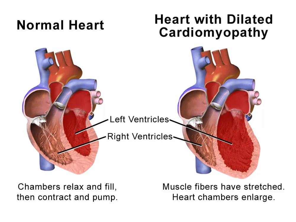
- Dilated cardiomyopathy: a dilated left ventricle with impaired systolic function, not explained by coronary disease or abnormal loading.
- Causes worth hunting: alcohol, tachycardia, thyroid disease, haemochromatosis, thiamine deficiency and peripartum disease are all potentially reversible.
- Genetics: around a third are inherited. Titin (TTN) is commonest; lamin A/C (LMNA) carries a high arrhythmic risk and a low threshold for a defibrillator.
- DCM management: the four pillars of heart failure therapy, devices where indicated, and treating the underlying cause.
- Restrictive cardiomyopathy: stiff, non-compliant ventricles that fill poorly, with normal or near-normal systolic function and normal chamber size.
- Commonest restrictive cause in the UK: cardiac amyloidosis - AL from a plasma cell dyscrasia, or ATTR in older people.
- The amyloid clue: thick walls on echocardiography with LOW voltages on the ECG. In hypertrophy from any other cause the voltages are high.
- Never miss: constrictive pericarditis, which mimics restrictive cardiomyopathy and is curable by pericardiectomy.
Introduction
The cardiomyopathies are diseases of the heart muscle itself, rather than the consequence of coronary disease, hypertension or valve disease. They are classified by the structural and functional phenotype they produce.1
| Type | Ventricle | Function |
|---|---|---|
| Dilated (DCM) | Dilated, thin-walled | Impaired systolic function |
| Hypertrophic (HCM) | Thick-walled, small cavity | Preserved systolic function, impaired filling |
| Restrictive (RCM) | Normal size, stiff | Preserved systolic function, severely impaired filling |
| Arrhythmogenic (ACM/ARVC) | Fibrofatty replacement, usually right ventricle | Arrhythmia predominates over pump failure |
| Non-dilated left ventricular | Normal size with scar or hypokinesia | A newer category in the 2023 ESC classification |
This article covers dilated and restrictive cardiomyopathy. Hypertrophic cardiomyopathy is dealt with separately, as is myocarditis, which is one of the commonest routes into dilated cardiomyopathy.
The reason these matter for a student is not their frequency - both are relatively uncommon - but that each contains a subset of reversible causes. A dilated ventricle from alcohol, thyrotoxicosis, thiamine deficiency or an unrecognised tachycardia can recover substantially if the cause is found and removed.
Aetiology
Dilated cardiomyopathy
| Category | Causes |
|---|---|
| Genetic (around a third) | Titin (TTN) truncating variants - the commonest. Lamin A/C (LMNA) - important because it causes conduction disease and a high risk of ventricular arrhythmia. Also desmin, and dystrophin in Duchenne and Becker muscular dystrophy. |
| Idiopathic | No cause identified after full assessment - still a large group |
| Toxic | Alcohol (a major and potentially reversible cause), cocaine and amphetamines, anthracyclines (doxorubicin), trastuzumab, some tyrosine kinase inhibitors, clozapine |
| Infective | Viral myocarditis - Coxsackie B, parvovirus B19, adenovirus, HIV, SARS-CoV-2. Chagas disease (Trypanosoma cruzi) - the commonest cause worldwide in endemic South American regions. |
| Metabolic and endocrine | Thyrotoxicosis and hypothyroidism, haemochromatosis, thiamine deficiency (wet beriberi), selenium deficiency, phaeochromocytoma, acromegaly |
| Peripartum | Onset in the last month of pregnancy or within 5 months of delivery |
| Tachycardia-induced | Persistent atrial fibrillation or other sustained tachyarrhythmia; often fully reversible with rate or rhythm control |
| Autoimmune | Systemic lupus erythematosus, sarcoidosis, systemic sclerosis, giant cell myocarditis |
Restrictive cardiomyopathy
Far less common, and dominated in UK practice by cardiac amyloidosis.
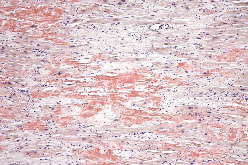
- Amyloidosis - AL amyloid from a plasma cell dyscrasia, and ATTR amyloid from transthyretin, either hereditary or the wild-type form that increasingly presents in men over 70
- Sarcoidosis - granulomatous infiltration, often with conduction disease and ventricular arrhythmia
- Haemochromatosis - iron deposition, which characteristically produces a mixed restrictive and dilated picture
- Endomyocardial fibrosis - a major cause in tropical regions
- Loeffler endocarditis - eosinophilic infiltration in hypereosinophilic syndrome
- Radiotherapy to the chest, and previous mediastinal irradiation
- Systemic sclerosis, Fabry disease, and the glycogen and lysosomal storage disorders
Risk factors
- Family history of cardiomyopathy, heart failure under 60, unexplained sudden death, or a pacemaker in a young relative
- Alcohol excess - both amount and duration
- Previous anthracycline or trastuzumab chemotherapy, and previous chest radiotherapy
- Pregnancy and the puerperium - particularly with multiple pregnancy, pre-eclampsia, older maternal age and African ethnicity
- Persistent tachyarrhythmia, including inadequately rate-controlled atrial fibrillation
- Haemochromatosis, or a family history of it
- Plasma cell dyscrasia - myeloma or MGUS, raising the risk of AL amyloidosis
- Age over 70 and male sex - for wild-type ATTR amyloidosis
- Recent viral illness, HIV, and travel or residence in Chagas-endemic regions
Clinical features
Dilated cardiomyopathy
Presents as heart failure with reduced ejection fraction, and the symptoms are those of the syndrome: exertional breathlessness, orthopnoea, paroxysmal nocturnal dyspnoea, fatigue and ankle swelling.
It may also present with arrhythmia - palpitations, syncope or cardiac arrest - or with a thromboembolic event from mural thrombus in the poorly contracting ventricle. Some are found incidentally through an abnormal ECG or a chest X-ray showing cardiomegaly.
Restrictive cardiomyopathy
The hallmark is predominantly right-sided congestion out of proportion to left-sided symptoms. Patients complain of marked fatigue and exercise intolerance, ankle swelling, abdominal distension from ascites, and early satiety - while their lungs stay relatively clear.
Breathlessness reflects an inability to increase cardiac output rather than pulmonary congestion, because the stiff ventricle cannot fill more when demand rises. Atrial fibrillation is common as the atria dilate against high filling pressures.
Clinical examination
Dilated cardiomyopathy
- Displaced, diffuse apex beat - the volume-loaded, dilated ventricle
- Third heart sound (S3) producing a gallop rhythm
- Functional mitral and tricuspid regurgitation - pansystolic murmurs from annular stretching, not from primary valve disease
- Raised JVP, basal crackles, hepatomegaly and peripheral oedema
- Low-volume pulse, sometimes pulsus alternans in severe impairment
- Atrial fibrillation is common
Restrictive cardiomyopathy
- Markedly raised JVP with a sharp y descent - the ventricle fills rapidly at first, then stops abruptly
- Kussmaul sign - the JVP rises rather than falls on inspiration
- Hepatomegaly, ascites and peripheral oedema, often marked
- Apex beat is normal or impalpable - the ventricle is not enlarged, which is a useful contrast with dilated cardiomyopathy
- A third or fourth heart sound may be present
- In amyloidosis, look for the extracardiac features above and for postural hypotension
Differential diagnosis
For dilated cardiomyopathy, the essential exclusion is ischaemic heart disease - a dilated poorly contracting ventricle from previous infarction is far commoner than a true cardiomyopathy, and needs coronary assessment. Also consider valvular disease, hypertensive heart disease, and takotsubo cardiomyopathy after emotional or physical stress.
Other differentials for a restrictive picture include hypertrophic cardiomyopathy, hypertensive heart disease, chronic liver disease and nephrotic syndrome for the oedema and ascites, and right heart failure from any cause.
Investigations
- ECG - in DCM: poor R wave progression, left bundle branch block, atrial fibrillation, ventricular ectopy. In amyloidosis: low voltages with a pseudo-infarct pattern, conduction disease, and atrial fibrillation.
- Chest X-ray - cardiomegaly and pulmonary congestion in DCM; the heart size may be normal in restriction
- Echocardiography - the primary test. DCM: a dilated left ventricle with a reduced ejection fraction and functional regurgitation. RCM: normal-sized ventricles with biatrial enlargement, marked diastolic dysfunction, and in amyloid a thickened 'speckled' myocardium with reduced longitudinal strain sparing the apex (apical sparing).
- Cardiac MRI - characterises the myocardium and often gives the diagnosis. Amyloid shows a distinctive diffuse subendocardial late gadolinium enhancement; sarcoidosis patchy mid-wall enhancement; myocarditis subepicardial enhancement.
- Bloods - FBC, U&Es, LFTs, TFTs, ferritin and transferrin saturation (haemochromatosis), NT-proBNP, troponin, HIV serology, autoantibodies, and serum free light chains with serum and urine immunofixation where AL amyloid is suspected
- Bone scintigraphy (DPD or PYP) - takes up avidly in ATTR amyloidosis and allows diagnosis without biopsy, provided a plasma cell dyscrasia has been excluded
- Coronary angiography or CT coronary angiography - to exclude ischaemic heart disease as the cause of a dilated ventricle
- Ambulatory ECG monitoring - for arrhythmia and risk stratification
- Genetic testing and family screening - for dilated cardiomyopathy with a family history, and for hereditary ATTR
- Endomyocardial biopsy - reserved for cases where the diagnosis remains unclear, or where giant cell myocarditis or AL amyloid needs tissue confirmation
Management
Dilated cardiomyopathy
Treatment is standard heart failure therapy for reduced ejection fraction, applied fully and titrated to target doses - the four pillars of an ACE inhibitor or ARNI, a beta-blocker, a mineralocorticoid receptor antagonist and an SGLT2 inhibitor, with loop diuretics for congestion.2
- Treat the cause - complete alcohol abstinence, rate or rhythm control for tachycardia-induced disease, venesection for haemochromatosis, thiamine for beriberi, correction of thyroid dysfunction
- Device therapy - cardiac resynchronisation for a broad QRS with persisting symptoms; an ICD for primary prevention in selected patients, and at a lower threshold in LMNA disease
- Anticoagulation - for atrial fibrillation or documented mural thrombus
- Cardiac rehabilitation, vaccination and specialist heart failure follow-up
- Advanced therapies - transplantation or a left ventricular assist device for end-stage disease
Restrictive cardiomyopathy
Management is largely treatment of the underlying disease plus careful symptom control, because no therapy reverses the myocardial stiffness itself.
- Diuretics for congestion, used cautiously - these patients are preload dependent, and over-diuresis causes a sharp fall in cardiac output, hypotension and renal impairment
- Rate control and anticoagulation for atrial fibrillation, which is common and poorly tolerated
- AL amyloidosis - urgent haematology referral for chemotherapy or autologous stem cell transplantation directed at the underlying plasma cell clone. The cardiac prognosis depends on suppressing light chain production.
- ATTR amyloidosis - tafamidis, a transthyretin stabiliser, reduces mortality and hospitalisation; newer silencing therapies are emerging
- Haemochromatosis - venesection and iron chelation
- Sarcoidosis - corticosteroids and immunosuppression, with a low threshold for a device given the arrhythmia risk
- Cardiac transplantation in selected patients
Complications
- Progressive heart failure - the common endpoint of both, and the usual cause of death
- Ventricular arrhythmia and sudden cardiac death - particularly in LMNA dilated cardiomyopathy and in cardiac sarcoidosis
- Atrial fibrillation - very common in both, and badly tolerated in restriction where atrial contraction contributes so much to filling
- Mural thrombus and systemic embolism - from a dilated, poorly contracting ventricle, or from a dilated fibrillating atrium
- Conduction disease and complete heart block - characteristic of LMNA disease, cardiac sarcoidosis and amyloidosis
- Functional mitral and tricuspid regurgitation - from annular dilatation, worsening the failure
- Cardiorenal syndrome and cardiac cirrhosis - from chronic venous congestion
- Cardiac cachexia and progressive functional decline
Red flags
Prognosis
Prognosis in dilated cardiomyopathy has improved markedly with modern heart failure therapy and devices. Outcome depends chiefly on the cause and on whether it can be removed: alcohol-related disease with complete abstinence, and tachycardia-induced disease with rhythm control, can show substantial or complete recovery of ejection fraction. Idiopathic and genetic forms treated with full four-pillar therapy now have far better survival than the older textbook figures suggest.
Restrictive cardiomyopathy carries a worse outlook, and it is dominated by the underlying disease. Untreated AL amyloidosis with cardiac involvement historically had a median survival measured in months, though modern chemotherapy directed at the plasma cell clone has changed this substantially where it is caught early. ATTR amyloidosis progresses more slowly, and tafamidis reduces mortality and hospitalisation.
The recurring theme across both is that the label matters less than the mechanism. Establishing why the muscle has failed identifies the patients who can recover, the families who need screening, and the minority whose disease is curable rather than merely manageable - which is why the diagnostic workup is worth doing properly rather than settling for 'heart failure'.
References
- Arbelo E, Protonotarios A, Gimeno JR et al. ESC Guidelines for the management of cardiomyopathies. European Heart Journal. 2023. Available here
- NICE NG106. Chronic heart failure in adults: diagnosis and management. 2018. Available here
- Maurer MS, Schwartz JH, Gundapaneni B et al. Tafamidis treatment for patients with transthyretin amyloid cardiomyopathy (ATTR-ACT). New England Journal of Medicine. 2018. Available here
- Garcia MJ. Constrictive pericarditis versus restrictive cardiomyopathy. Journal of the American College of Cardiology. 2016. Available here
- Sliwa K, Petrie MC, van der Meer P et al. Clinical presentation, management, and 6-month outcomes in women with peripartum cardiomyopathy. European Heart Journal. 2020. Available here
- NICE TA696. Tafamidis for treating transthyretin amyloidosis with cardiomyopathy. Available here
- Cardiomyopathy UK. Dilated and restrictive cardiomyopathy information. Available here
- BNF. Furosemide - indications and dosing. Available here
This article is written for revision and education. It is not clinical guidance and must not be used to make decisions about the care of a patient. Always check current NICE guidance and local protocols.