
Nephrotic Syndrome: The Triad, the Causes and the Complications
Key points
- Nephrotic syndrome: a triad of proteinuria over 3.5 g/24 hours, hypoalbuminaemia (under 30 g/L) and oedema, usually with hyperlipidaemia. It reflects damage to the glomerular filtration barrier - specifically the podocyte.
- Nephrotic versus nephritic: nephrotic is a protein leak with bland urine; nephritic is inflammation with haematuria, red cell casts, hypertension and an acute fall in GFR. The distinction directs the whole work-up.
- Commonest cause in children: minimal change disease (around 90% of childhood cases). It is steroid-responsive in the great majority, so children are usually treated empirically without a biopsy.
- Commonest causes in adults: focal segmental glomerulosclerosis and membranous nephropathy among primary causes, and diabetic nephropathy overall. Adults need a biopsy.
- Membranous nephropathy: associated with anti-PLA2R antibodies, and secondary to malignancy (especially solid tumours), hepatitis B, lupus, and drugs such as penicillamine, gold and NSAIDs.
- Thrombotic risk: nephrotic syndrome is a hypercoagulable state from urinary loss of antithrombin III and altered clotting factors - classically causing renal vein thrombosis and venous thromboembolism.
- Infection risk: urinary loss of immunoglobulins and complement predisposes to encapsulated organisms, classically pneumococcal peritonitis in children.
- Management: salt and fluid restriction with loop diuretics for oedema, an ACE inhibitor or ARB to reduce proteinuria, a statin, thromboprophylaxis where risk is high, and disease-specific immunosuppression.
Introduction
Nephrotic syndrome is defined by a classic triad, and all three components should be sought before the label is applied:1
- Proteinuria - more than 3.5 g per 24 hours in adults (equivalent to a urine protein:creatinine ratio above 300-350 mg/mmol, or ACR above about 250 mg/mmol)
- Hypoalbuminaemia - serum albumin below about 30 g/L
- Oedema - typically periorbital on waking, then dependent and, in severe cases, generalised with ascites and pleural effusions
Hyperlipidaemia is so frequently present that many texts include it as a fourth component.3 The underlying lesion is damage to the glomerular filtration barrier - and specifically to the podocyte and its foot processes, which form the final and most selective layer of that barrier. When podocytes are injured, the barrier's charge and size selectivity fails and albumin escapes into the urine.
Pathophysiology
Understanding the consequences of losing large amounts of protein in the urine explains every complication of the syndrome, and is far more useful than memorising them.
| Protein lost or mechanism | Consequence |
|---|---|
| Albumin | Hypoalbuminaemia, reducing plasma oncotic pressure and allowing fluid to move into the interstitium - oedema. Secondary activation of the renin-angiotensin-aldosterone system from reduced effective circulating volume causes further sodium and water retention, which is now thought to contribute at least as much as the oncotic effect. |
| Antithrombin III, protein S, and increased hepatic synthesis of fibrinogen and factors V and VIII | A hypercoagulable state - venous thromboembolism and, characteristically, renal vein thrombosis |
| Immunoglobulins (especially IgG) and complement components (factor B) | Increased susceptibility to infection, particularly with encapsulated organisms - Streptococcus pneumoniae, Haemophilus influenzae - classically causing spontaneous bacterial peritonitis and cellulitis in children |
| Compensatory hepatic lipoprotein synthesis (and reduced lipoprotein lipase activity) | Hyperlipidaemia with raised LDL cholesterol and triglycerides, contributing to long-term cardiovascular risk |
| Thyroxine-binding globulin, vitamin D-binding protein and transferrin | Abnormal thyroid function tests, vitamin D deficiency with secondary hyperparathyroidism, and an iron-resistant microcytic anaemia |
| Loss of protein-bound drug carriage | Altered pharmacokinetics of highly protein-bound drugs, so standard doses may produce exaggerated effects |
Causes
Causes are conventionally divided into primary (idiopathic) glomerular disease and secondary causes, and the likely histology depends heavily on the patient's age.
| Disease | Typical patient | Histology | Notes |
|---|---|---|---|
| Minimal change disease | Children (around 90% of childhood nephrotic syndrome); also a minority of adults | Light microscopy is normal; electron microscopy shows podocyte foot process effacement. Immunofluorescence is negative. | Highly steroid-responsive - over 90% of children remit. May follow a viral illness or immunisation; occasionally associated with Hodgkin lymphoma or NSAIDs. Relapses are common. |
| Focal segmental glomerulosclerosis (FSGS) | Adults, particularly of Black African or African-Caribbean ancestry; also children | Sclerosis affecting some glomeruli (focal) and part of each affected glomerulus (segmental) | The commonest primary cause in adults. Secondary FSGS occurs with obesity, HIV, heroin use, sickle cell disease, reflux nephropathy and reduced renal mass. Often steroid-resistant, and recurs in transplants. |
| Membranous nephropathy | Older adults (peak in the fifth to sixth decade) | Diffuse thickening of the glomerular basement membrane with subepithelial immune complex deposits, giving a 'spike and dome' appearance on silver staining; granular IgG and C3 on immunofluorescence | Anti-PLA2R antibodies are present in around 70-80% of primary cases and are now central to diagnosis and monitoring. Follows a 'rule of thirds': a third remit spontaneously, a third persist, a third progress. Must exclude secondary causes. |
| Membranoproliferative (mesangiocapillary) GN | Children and young adults | Mesangial proliferation with 'tram-track' double contouring of the basement membrane | Often a mixed nephrotic-nephritic picture. Associated with hepatitis C, cryoglobulinaemia, endocarditis and complement dysregulation (C3 glomerulopathy, with a low C3). |
| Category | Causes |
|---|---|
| Metabolic | Diabetic nephropathy - the commonest cause of nephrotic-range proteinuria overall in the UK |
| Systemic autoimmune | Lupus nephritis (particularly class V, membranous lupus nephritis), rheumatoid arthritis, systemic sclerosis, Sjogren syndrome |
| Infection | Hepatitis B (classically membranous), hepatitis C (membranoproliferative), HIV (collapsing FSGS), syphilis, malaria (classically quartan malarial nephropathy), infective endocarditis |
| Malignancy | Solid tumours - especially lung, breast, colon and stomach - with membranous nephropathy; Hodgkin lymphoma with minimal change disease. In an older adult with new membranous nephropathy, an occult malignancy must be considered. |
| Drugs | NSAIDs, penicillamine, gold, captopril, lithium, and anti-TNF agents |
| Amyloidosis and paraproteinaemia | AL amyloidosis and myeloma-related renal disease - suspect where there is a paraprotein, macroglossia or cardiac involvement |
| Genetic | Congenital nephrotic syndrome (nephrin and podocin mutations), Alport syndrome, Fabry disease |
| Pre-eclampsia | An important obstetric cause of proteinuria and oedema |
Clinical features
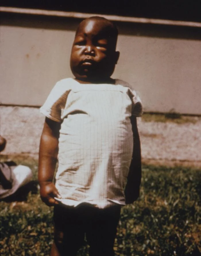
Patients typically present with oedema, which is the feature that brings them to medical attention, and often describe swelling that is worst around the eyes on waking and around the ankles by the end of the day.
- Periorbital oedema, characteristically prominent on waking, because the tissue there is loose and gravity has acted overnight - this is a classic presenting sign in children and often prompts an initial diagnosis of allergy
- Peripheral pitting oedema of the ankles and legs, and sacral oedema in bed-bound patients
- Generalised oedema (anasarca) in severe cases, with ascites, pleural effusions and scrotal or vulval oedema
- Frothy urine - a genuinely useful symptom that patients often mention if asked directly
- Weight gain over days to weeks, from fluid retention
- Breathlessness from pleural effusions, ascites splinting the diaphragm, or (rarely) pulmonary embolism
- Fatigue and lethargy
- Xanthelasma or other signs of hyperlipidaemia in longstanding disease
- Muehrcke's lines - paired transverse white bands across the nails, a sign of chronic hypoalbuminaemia
- Features of the underlying cause - rash and arthralgia in lupus, diabetic retinopathy, lymphadenopathy in lymphoma, macroglossia in amyloidosis
Blood pressure may be normal, low (from intravascular depletion) or high, and its interpretation is important: marked hypertension with haematuria should shift suspicion towards a nephritic process or a mixed picture.
Investigations
Confirming the syndrome
- Urine dipstick - heavy proteinuria (3+ or 4+); check for blood, whose presence raises the possibility of a nephritic or mixed process
- Urine protein quantification - urine protein:creatinine ratio (PCR) or ACR on an early morning sample has largely replaced the 24-hour collection; PCR above 300-350 mg/mmol corresponds to nephrotic-range proteinuria2
- Serum albumin - below 30 g/L
- U&Es and eGFR - to assess renal function and detect associated AKI
- Urine microscopy - looking for red cell casts (which point to a nephritic component) and oval fat bodies or lipid casts (seen in heavy proteinuria)
- Lipid profile - typically raised total and LDL cholesterol and triglycerides
- Full blood count and coagulation screen
Identifying the cause
- HbA1c and fundoscopy - for diabetic nephropathy; the presence of retinopathy makes a diabetic cause much more likely
- Immunology - ANA and anti-dsDNA (lupus), complement C3 and C4 (low in lupus and in membranoproliferative GN/C3 glomerulopathy), ANCA, and anti-PLA2R antibodies for membranous nephropathy
- Serum and urine electrophoresis with serum free light chains - for myeloma and amyloidosis
- Virology - hepatitis B and C, and HIV serology, all of which cause specific patterns and change management
- Age-appropriate malignancy screening in adults with membranous nephropathy - chest imaging, and investigation guided by symptoms; this is a genuine and frequently examined association
- Renal tract ultrasound - to assess kidney size and exclude obstruction, and to confirm two kidneys before biopsy
Renal biopsy
Management
Management has two strands that proceed in parallel: general (supportive) measures that apply to all patients regardless of histology, and disease-specific immunosuppression determined by the biopsy.4,5
General measures
| Problem | Management |
|---|---|
| Oedema | Dietary sodium restriction (roughly under 2 g sodium daily) and fluid restriction are the foundation. Loop diuretics (furosemide) are first line, often needing higher doses because gut oedema impairs absorption and hypoalbuminaemia reduces delivery to the tubule - so intravenous administration may be required. Add a thiazide-like diuretic or amiloride for synergy if needed. Aim for a gradual weight loss of about 0.5 to 1 kg daily - faster diuresis risks intravascular depletion and AKI. Daily weights are the key monitoring tool. |
| Proteinuria | ACE inhibitor or ARB reduces intraglomerular pressure and proteinuria independently of blood pressure, and is used in nearly all patients (with the exception of children being treated for steroid-responsive minimal change disease, in whom remission is the goal). Monitor U&Es and potassium. |
| Hyperlipidaemia | Statin therapy, given the substantial cardiovascular risk of sustained nephrotic syndrome |
| Thrombotic risk | Prophylactic anticoagulation is considered where risk is high - particularly with albumin below about 20 to 25 g/L, membranous nephropathy (the highest-risk histology), immobility or previous thromboembolism. Treat established thrombosis with full anticoagulation. Avoid immobility, and use compression stockings. |
| Infection risk | Pneumococcal vaccination (and annual influenza vaccination); a high index of suspicion for spontaneous bacterial peritonitis and cellulitis; prompt antibiotics for suspected infection. Live vaccines are contraindicated during immunosuppression. |
| Nutrition | Adequate but not excessive protein intake, with dietetic input; vitamin D replacement where deficient |
| Monitoring | Daily weight, fluid balance, blood pressure, U&Es and albumin during the acute phase, with urine PCR to track response |
Disease-specific treatment
| Diagnosis | Treatment |
|---|---|
| Minimal change disease | High-dose oral corticosteroids (e.g. prednisolone 60 mg/m2 daily in children, then tapered over weeks to months). Over 90% of children remit, usually within 2 to 4 weeks. Frequently relapsing or steroid-dependent disease is treated with steroid-sparing agents - levamisole, ciclosporin, tacrolimus, mycophenolate or rituximab. |
| FSGS | Corticosteroids for primary disease, though response is less reliable and often requires prolonged courses; calcineurin inhibitors or rituximab in resistant disease. Secondary FSGS is treated by addressing the cause (weight loss, antiretroviral therapy for HIV) with renin-angiotensin blockade rather than immunosuppression. |
| Membranous nephropathy | Supportive treatment alone for at least 6 months in low-risk patients, because roughly a third remit spontaneously. Immunosuppression is reserved for those with persistent heavy proteinuria, declining renal function or high anti-PLA2R titres: rituximab is now commonly first line, with the alternative Ponticelli regimen (alternating corticosteroid and cyclophosphamide) or a calcineurin inhibitor. |
| Membranoproliferative GN | Treat the underlying cause (antiviral therapy for hepatitis C, antibiotics for endocarditis); immunosuppression or complement-directed therapy for primary complement-mediated disease |
| Diabetic nephropathy | Not immunosuppression - renin-angiotensin blockade, SGLT2 inhibition, glycaemic and blood pressure control; see the diabetic nephropathy article |
| Lupus nephritis | Immunosuppression guided by the ISN/RPS class - typically mycophenolate or cyclophosphamide with corticosteroids, increasingly with belimumab or voclosporin |
| Secondary to malignancy, drugs or infection | Treat or remove the cause - the nephrotic syndrome frequently resolves when the tumour is treated or the drug withdrawn |
Complications
Red flags
Prognosis
Prognosis depends almost entirely on the underlying histology and the response to treatment, so the biopsy result (or, in children, the steroid response) is what determines the outlook rather than the severity of the initial oedema.
Minimal change disease in children has an excellent prognosis. Over 90% remit with corticosteroids, and progression to chronic kidney disease is rare. The main problem is relapse, which occurs in the majority at least once, and a subset become frequently relapsing or steroid-dependent, requiring steroid-sparing agents and carrying the cumulative burden of treatment toxicity rather than of renal failure.
FSGS has the least favourable outlook among the primary causes. Steroid resistance is common, and a substantial proportion progress to end-stage renal disease over 5 to 20 years; primary FSGS also has an important tendency to recur in a transplanted kidney, sometimes within days. Achieving remission of proteinuria is the strongest predictor of preserved renal function.
Membranous nephropathy follows the classic 'rule of thirds' - approximately a third undergo spontaneous remission, a third have persistent proteinuria with stable function, and a third progress to renal failure. This is precisely why low-risk patients are managed supportively for at least 6 months before committing them to immunosuppression, and why anti-PLA2R titres are useful for identifying those who will not remit spontaneously.
Across all causes, the strongest predictors of a poor renal outcome are persistent nephrotic-range proteinuria, impaired renal function at presentation, hypertension and interstitial fibrosis on biopsy. Conversely, achieving and sustaining remission of proteinuria - by whatever means - is consistently associated with preserved kidney function and reduced cardiovascular and thrombotic risk.
References
- KDIGO 2021 Clinical Practice Guideline for the Management of Glomerular Diseases. Available here
- NICE Clinical Knowledge Summaries. Proteinuria and nephrotic syndrome. Available here
- Hull RP, Goldsmith DJA. Nephrotic syndrome in adults. BMJ. 2008. Available here
- UK Kidney Association. Clinical practice guidelines - glomerular disease. Available here
- BNF. Prednisolone, furosemide and rituximab. Available here
- CDC Public Health Image Library (PHIL ID 3894), public domain, via Wikimedia Commons. Available here
This article is written for revision and education. It is not clinical guidance and must not be used to make decisions about the care of a patient. Always check current NICE guidance and local protocols.