
Empyema: Pus in the Pleural Space
Key points
- Empyema: pus in the pleural space, or pleural fluid with a positive Gram stain or culture. It will not resolve with antibiotics alone.
- The commonest cause: pneumonia. Around 40% of hospitalised patients with pneumonia develop an effusion, and roughly 1 in 10 of those effusions becomes complicated or frankly purulent.
- The decisive test: pleural fluid pH. A pH below 7.2 in the context of infection means a complicated parapneumonic effusion and mandates chest drain insertion.
- Three stages: exudative (free-flowing, drains easily), fibrinopurulent (loculated by fibrin septations), and organising (a thick pleural peel trapping the lung).
- Organisms: community-acquired disease is dominated by the Streptococcus anginosus (milleri) group, Streptococcus pneumoniae, Staphylococcus aureus and anaerobes; hospital-acquired by MRSA and Gram-negatives.
- Antibiotics: prolonged, typically 2 to 6 weeks, with anaerobic cover. Aminoglycosides are avoided because they penetrate the pleural space poorly and are inactivated by low pH.
- Intrapleural therapy: combined tPA (alteplase) and DNase (dornase alfa) improves drainage and reduces the need for surgery. Neither agent works alone.
- Surgical referral: if there is no improvement after 5 to 7 days of drainage and antibiotics, refer for VATS decortication.
Introduction
An empyema is pus in the pleural space. In practice the term is used slightly more broadly, and pleural infection sits on a spectrum with three points along it, all of which are managed as a single continuum of severity.
| Category | Pleural fluid findings | Management |
|---|---|---|
| Simple parapneumonic effusion | Clear fluid, pH above 7.2, normal glucose, negative Gram stain and culture | Antibiotics for the pneumonia; the effusion resolves |
| Complicated parapneumonic effusion | pH below 7.2, glucose below 3.3 mmol/L, raised LDH, or a positive Gram stain or culture | Chest drain plus antibiotics |
| Empyema | Frank pus | Chest drain plus antibiotics, and often intrapleural fibrinolytics or surgery |
The distinction matters because the two ends behave completely differently: a simple parapneumonic effusion resolves with the antibiotics given for the pneumonia, whereas a complicated one or an empyema will not resolve without drainage, however good the antibiotic. Failing to sample an effusion in a patient with pneumonia who is not improving is a recurrent and entirely avoidable cause of prolonged illness.
The three pathological stages
- Exudative stage (first few days) - sterile inflammatory fluid crosses the inflamed visceral pleura. The fluid is free-flowing and drains easily.
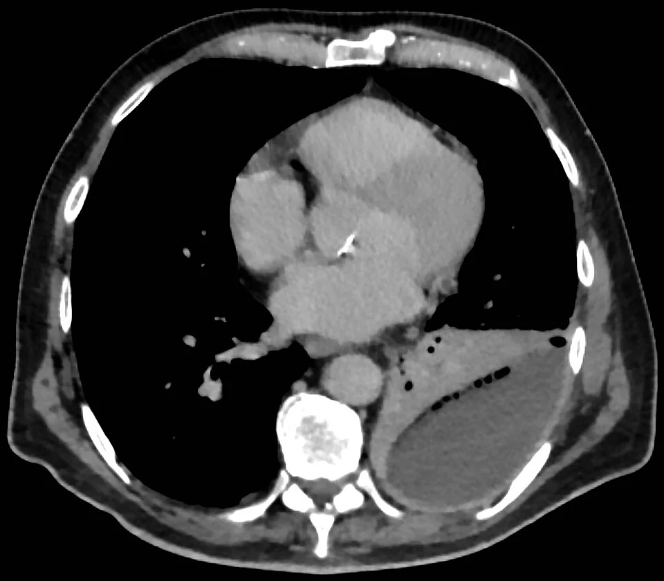
- Fibrinopurulent stage (days to 1 to 2 weeks) - bacterial invasion drives neutrophil influx and fibrin deposition, forming septations and loculations. Fluid becomes viscous, glucose and pH fall as bacteria and neutrophils consume glucose and generate lactate, and drainage becomes difficult. This is the stage at which fibrinolytics help.
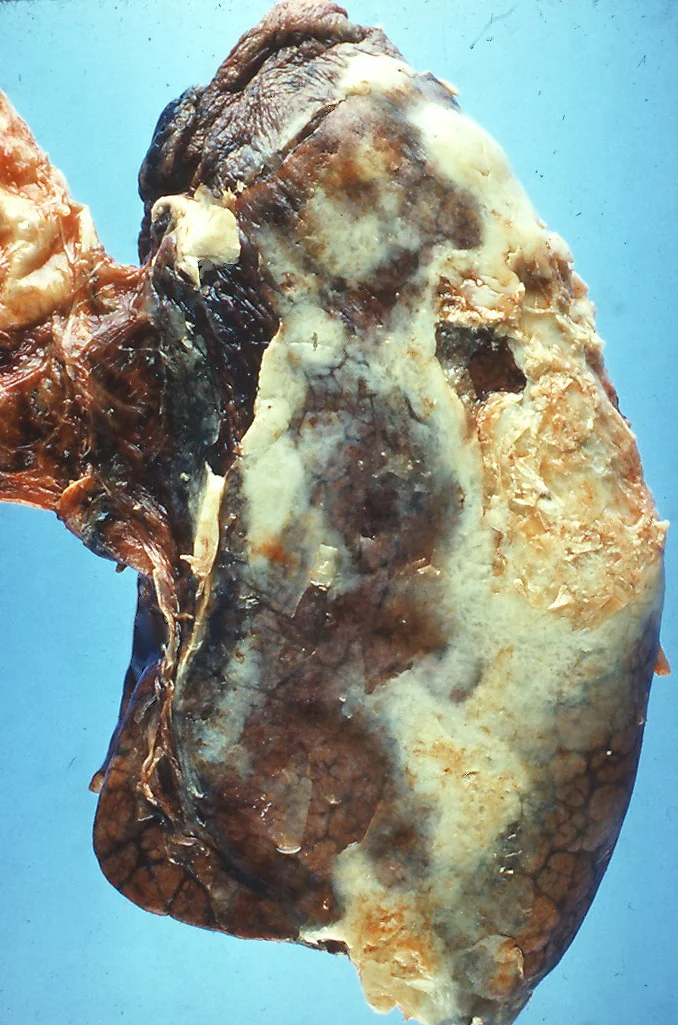
- Organising stage (after 2 to 4 weeks) - fibroblasts lay down a thick, inelastic pleural peel encasing the lung. The lung can no longer expand, and surgical decortication is the only remedy.
Causes and risk factors
Causes
- Pneumonia - around 50 to 60% of cases, and the dominant cause
- Thoracic surgery or trauma - including penetrating injury and retained haemothorax
- Oesophageal rupture - a surgical emergency in which pleural fluid amylase is very high and the pH very low
- Iatrogenic - after chest drain insertion, pleural aspiration or thoracoscopy
- Spread from a subphrenic or hepatic abscess
- Tuberculosis - producing a more indolent, lymphocytic empyema, often with marked pleural thickening
- Bronchial obstruction by tumour with a post-obstructive pneumonia
- No identifiable preceding pneumonia - in a substantial minority, particularly with aspiration and anaerobic infection
Risk factors
- Alcohol excess and intravenous drug use
- Diabetes mellitus
- Aspiration - reduced conscious level, stroke, seizures, oesophageal disease
- Poor dental hygiene - a source of anaerobic organisms and a genuinely useful thing to examine for
- Immunosuppression, including corticosteroids and chemotherapy
- Gastro-oesophageal reflux
- Malnutrition and low serum albumin
- Delayed presentation or delayed antibiotic treatment of pneumonia
Organisms
| Setting | Typical organisms |
|---|---|
| Community-acquired | Streptococcus anginosus (milleri) group - a particular association with empyema and abscess formation. Also Streptococcus pneumoniae, Staphylococcus aureus, and anaerobes (Fusobacterium, Prevotella, Bacteroides), especially after aspiration. |
| Hospital-acquired | MRSA, Gram-negative organisms including Escherichia coli, Klebsiella, Enterobacter and Pseudomonas, and enterococci. Substantially higher mortality. |
| Other | Mycobacterium tuberculosis, and fungi in the immunocompromised |
Fluid culture is negative in up to 40% of cases, often because antibiotics have already been given. Inoculating pleural fluid into blood culture bottles at the bedside substantially improves the yield and should be routine.
Clinical features
The classic presentation is a patient with pneumonia who is not getting better. Suspicion should rise steeply when fever persists or returns after 48 to 72 hours of appropriate antibiotics, or when the CRP fails to fall.
Symptoms
- Persistent or recurrent fever, classically swinging
- Pleuritic chest pain
- Breathlessness, worsening as the collection enlarges
- Productive cough and, if a bronchopleural fistula develops, expectoration of large volumes of purulent fluid
- Weight loss, anorexia, night sweats and profound fatigue - anaerobic empyema in particular may present insidiously over weeks and be mistaken for malignancy or tuberculosis
- Rigors and features of sepsis in fulminant disease
Signs
- Stony dull percussion note with reduced or absent breath sounds and reduced vocal resonance over the collection
- Reduced chest expansion on the affected side
- Fever and tachycardia, with hypotension in sepsis
- Finger clubbing in chronic empyema
- Poor dentition - look for it, as it points to an anaerobic source
- Empyema necessitans - the collection tracking through the chest wall to form a subcutaneous swelling or discharging sinus. Rare, but classically associated with tuberculosis and actinomycosis.
Investigations
Imaging
- Chest radiograph - shows the effusion. An empyema may appear D-shaped with a convex medial border because it is loculated against the chest wall, unlike the concave meniscus of a free effusion.
- Thoracic ultrasound - essential. It confirms fluid, demonstrates septations and echogenic debris that indicate a complicated collection, and selects the drainage site. Septations on ultrasound predict a poorer response to drainage alone.
- Contrast-enhanced CT thorax - shows the split pleura sign: enhancement and separation of the thickened visceral and parietal pleura by the collection. CT also distinguishes empyema (lenticular, compresses lung, obtuse angles with the chest wall) from a lung abscess (spherical, within the lung, acute angles, thick irregular wall), and identifies an underlying bronchial obstruction.
Pleural fluid
Every effusion complicating pneumonia that is large enough to sample should be sampled, under ultrasound guidance.
- Appearance - frank pus is diagnostic; send nothing further to decide about drainage, and insert the drain
- pH - measured on a blood gas analyser. Below 7.2 mandates chest drain insertion. Do not send pus through a gas analyser, as it damages the machine and the answer is already obvious.
- Glucose - below 3.3 mmol/L supports a complicated effusion
- LDH and protein - raised, confirming an exudate
- Gram stain, culture and sensitivities - including anaerobic culture, plus inoculation into blood culture bottles
- AFB smear and culture, and adenosine deaminase if tuberculosis is suspected
- Cytology - to exclude an underlying malignancy
- Amylase - very high in oesophageal rupture
Blood tests
- FBC - neutrophilia, and anaemia of chronic disease in prolonged illness
- CRP - high and, critically, used serially to track response
- U&Es - urea contributes to the RAPID prognostic score
- Albumin - low albumin is both a risk factor and a poor prognostic marker
- Blood cultures - positive in around 15%
- HIV test, and consider immunodeficiency in recurrent or unusual infection
Management
Treatment rests on three pillars: drainage, prolonged antibiotics, and nutritional support, with escalation to fibrinolytics or surgery when drainage fails.1
Drainage
- Insert a chest drain for frank pus, a positive Gram stain or culture, or a pH below 7.2 in the context of infection
- Small-bore drains (10 to 14 French) are as effective as large-bore drains and considerably more comfortable, so a large drain is not required simply because the fluid is thick
- Ultrasound guidance is mandatory
- Flush the drain regularly with saline to prevent blockage, which is the commonest technical cause of failure
- Monitor the response with daily observations, CRP, drain output and repeat imaging - a drain that has stopped draining while the patient remains unwell is either blocked or badly sited
Antibiotics
- Start empirically after cultures are taken, and always include anaerobic cover
- Community-acquired: co-amoxiclav, or cefuroxime with metronidazole. In penicillin allergy, clindamycin with a fluoroquinolone or a cephalosporin according to local guidance.
- Hospital-acquired: broader cover including MRSA and Pseudomonas - for example piperacillin-tazobactam or meropenem with vancomycin, guided by local policy
- Do not use aminoglycosides - they penetrate the pleural space poorly and are inactivated by the low pH of infected pleural fluid
- Duration is prolonged, typically 2 to 6 weeks, with an intravenous phase followed by oral treatment, guided by clinical and CRP response
- Rationalise once cultures are available, and involve microbiology early
Intrapleural fibrinolytics
Surgery
- Refer to thoracic surgery if there is no clinical improvement after 5 to 7 days of appropriate drainage and antibiotics
- Video-assisted thoracoscopic surgery (VATS) - breaks down loculations, evacuates the collection and allows the lung to re-expand. Preferred where the patient is fit enough.
- Open thoracotomy and decortication - for organised empyema with a thick pleural peel
- Open thoracic window or rib resection drainage - for patients too unfit for major surgery, allowing long-term open drainage
- Early surgical discussion rather than late referral improves outcomes, and it is worth flagging a poorly responding patient to the surgeons before day 7 rather than after
Supportive care
- Nutritional support - patients are frequently catabolic and hypoalbuminaemic, and this independently affects outcome. Involve dietetics early.
- VTE prophylaxis - risk is high with sepsis and immobility
- Analgesia sufficient to allow deep breathing and physiotherapy
- Chest physiotherapy and early mobilisation
- Treat the source - dental review where poor dentition is implicated, swallow assessment where aspiration is suspected
- Bronchoscopy if there is any suggestion of underlying bronchial obstruction
- Smoking cessation and alcohol support where relevant
Complications
- Sepsis and septic shock
- Fibrothorax and trapped lung - an organised pleural peel restricting lung expansion, causing lasting breathlessness and a restrictive defect, and requiring decortication
- Bronchopleural fistula - a communication between the airway and pleural space, presenting with a persistent air leak and expectoration of purulent fluid
- Empyema necessitans - the collection discharging through the chest wall
- Lung abscess
- Osteomyelitis of the ribs, mediastinitis and pericarditis by direct spread
- Metastatic infection - brain abscess and infective endocarditis, particularly with the Streptococcus anginosus group
- Prolonged hospitalisation and deconditioning - median stay is measured in weeks
- Death - overall mortality around 20%, and higher in hospital-acquired infection and in older patients with comorbidity
Red flags
Prognosis
Pleural infection remains a serious illness. Mortality is around 20%, rising in older patients, those with significant comorbidity, and those with hospital-acquired infection. Around one in five patients ultimately requires surgery, and median hospital stay is measured in weeks rather than days.
Prognosis depends heavily on how early drainage is instituted. An effusion drained in the exudative stage resolves quickly and completely; one left until the organising stage requires decortication and may leave permanent restriction. This is why the threshold for sampling an effusion complicating pneumonia should be very low, and why the pH result should be chased actively rather than filed.
Most survivors recover well, although breathlessness and reduced exercise tolerance may persist for months and some residual pleural thickening is common on follow-up imaging. Patients should be followed up with a chest radiograph and clinical review to confirm resolution, to identify a trapped lung needing decortication, and to exclude an underlying malignancy that presented as infection.
References
- British Thoracic Society. Pleural disease guideline. 2023. Available here
- Rahman NM, Maskell NA, West A et al. Intrapleural use of tissue plasminogen activator and DNase in pleural infection (MIST2). NEJM. 2011. Available here
- Rahman NM, Kahan BC, Miller RF et al. A clinical score (RAPID) to identify those at risk for poor outcome at presentation in patients with pleural infection. Chest. 2014. Available here
- NICE CG191. Pneumonia in adults: diagnosis and management. 2014, updated 2023. Available here
- Maskell NA, Davies CWH, Nunn AJ et al. UK controlled trial of intrapleural streptokinase for pleural infection (MIST1). NEJM. 2005. Available here
- BNF. Co-amoxiclav, metronidazole and alteplase - indications and dosing. Available here
- Hellerhoff, CC BY-SA 4.0, via Wikimedia Commons. Available here
- Yale Rosen, CC BY-SA 2.0, via Wikimedia Commons. Available here
This article is written for revision and education. It is not clinical guidance and must not be used to make decisions about the care of a patient. Always check current NICE guidance and local protocols.