
Brain Abscess: Diagnosis and Management
Key points
- Brain abscess: a focal collection of pus within the brain parenchyma, usually bacterial, arising from direct spread from a contiguous focus, haematogenous seeding, trauma, or neurosurgery.
- Classic triad: headache, fever and focal neurological deficit - present together in only a minority, so its absence does not exclude the diagnosis.
- Commonest sources: contiguous spread from otitis media/mastoiditis or sinusitis, and haematogenous spread from a distant source such as endocarditis or a dental infection.
- Imaging hallmark: a ring-enhancing lesion with a hypodense/hyperintense centre and surrounding oedema on contrast CT or MRI.
- Management: combines neurosurgical drainage (aspiration or excision) with prolonged, high-dose broad-spectrum IV antibiotics.
- Steroids: used cautiously for significant surrounding oedema causing mass effect, but can reduce antibiotic penetration and should not replace source control.
- Seizures: common both at presentation and as a long-term complication, given the cortical location of many abscesses.
- Do not perform lumbar puncture: in suspected brain abscess - the risk of herniation from a focal mass lesion is significant, and CSF is rarely diagnostic.
Introduction
A brain abscess is a focal, encapsulated collection of pus within the brain parenchyma, most often bacterial in origin. It behaves as a space-occupying lesion with an infective cause, which means its clinical picture combines features of raised intracranial pressure and focal neurological deficit with signs of infection - though any of these components can be subtle or absent, particularly early in the illness or in immunocompromised patients.1
Brain abscess is a useful topic because it forces you to think about how infection reaches the brain - a skill that also underlies meningitis and encephalitis - and because its imaging appearance (the ring-enhancing lesion) has an important differential of its own that is frequently tested.
Incidence has changed with medical practice. Abscesses arising from contiguous ear and sinus infection have become less common in countries with ready access to antibiotics, while those following neurosurgery, penetrating trauma or occurring in immunocompromised hosts now account for a greater share. The practical consequence is that the likely organism depends heavily on the clinical context, and empirical cover should be chosen with the presumed route of entry in mind rather than applied uniformly.
Aetiology
Organisms reach the brain by one of several routes, and identifying the likely route helps predict both the causative organism and where to look for a primary source.3
- Direct (contiguous) spread - from otitis media or mastoiditis (typically producing a temporal lobe or cerebellar abscess), sinusitis (typically frontal lobe), or dental infection; the commonest route overall
- Haematogenous spread - from a distant infective source such as infective endocarditis, bronchiectasis or lung abscess, or intra-abdominal/pelvic sepsis; abscesses from this route are more likely to be multiple and located at the grey-white matter junction (following the pattern of embolic spread)
- Penetrating trauma or neurosurgery - direct inoculation of organisms into the brain
- Congenital cardiac shunts (right-to-left) - allow organisms to bypass pulmonary filtering and reach the brain haematogenously; an important association in younger patients with cyanotic congenital heart disease
- Immunocompromise - HIV and other causes of immunosuppression predispose to abscess from organisms that would not typically cause disease in an immunocompetent host (e.g. Toxoplasma gondii, Nocardia, fungal pathogens)
Common organisms include Streptococcus species (including the Streptococcus anginosus/'milleri' group), Staphylococcus aureus (particularly after trauma or neurosurgery, or with endocarditis), and anaerobes, especially with a dental or sinus source; abscesses are often polymicrobial. In the immunocompromised, Toxoplasma gondii and fungal pathogens become important considerations.
Clinical features
Presentation reflects the combined effects of a mass lesion and infection, and can evolve over days to a few weeks.
The tempo is explained by how an abscess matures. Over roughly the first two weeks, organisms seed the brain and provoke a poorly demarcated cerebritis - inflamed, oedematous tissue without a discrete cavity. Only afterwards does a collagen capsule form, walling the pus off into the discrete, ring-enhancing lesion recognised on imaging. This is why the illness can smoulder with headache and malaise before localising signs appear, and why early cerebritis may show only ill-defined change on CT rather than a classic ring.
It also has a therapeutic consequence. An abscess in the cerebritis phase is not yet a drainable collection, so early cases may be managed with antibiotics and close imaging surveillance until a capsule forms - whereas an established, encapsulated abscess penetrates poorly to antibiotics and generally needs drainage.
Location shapes the presentation in ways that are worth anticipating. A frontal abscess may cause personality change and subtle cognitive slowing long before any motor sign appears; a temporal abscess, classically arising from otitis media, may produce a dysphasia or a visual field defect; and a cerebellar abscess causes ataxia and, because of the confined posterior fossa, can obstruct the fourth ventricle and cause acute hydrocephalus with relatively little warning.
- Headache - often the earliest and most consistent symptom, may be localised to the side of the abscess
- Fever - present in only around half of patients, so its absence does not exclude an abscess
- Focal neurological deficit - reflecting the specific location: hemiparesis, dysphasia, ataxia (cerebellar abscess), depending on site
- Seizures - common, given the frequent cortical/subcortical location of abscesses
- Signs of raised intracranial pressure - nausea, vomiting, papilloedema, declining conscious level, particularly as the abscess enlarges or surrounding oedema increases
- Signs of the primary source - ear discharge/pain (otitis media), sinus tenderness, a new murmur or peripheral stigmata of endocarditis, dental pain or recent dental work
Clinical examination
- Conscious level and vital signs, including temperature
- Full neurological examination for a focal deficit localising to the abscess site
- Fundoscopy for papilloedema
- Ear, nose and dental examination for a contiguous source
- Cardiovascular examination for a murmur or peripheral stigmata suggesting infective endocarditis as a haematogenous source
- Signs of immunosuppression or risk factors for it
Differential diagnosis
The imaging appearance of a brain abscess - a ring-enhancing lesion with surrounding oedema - has an important differential of its own.
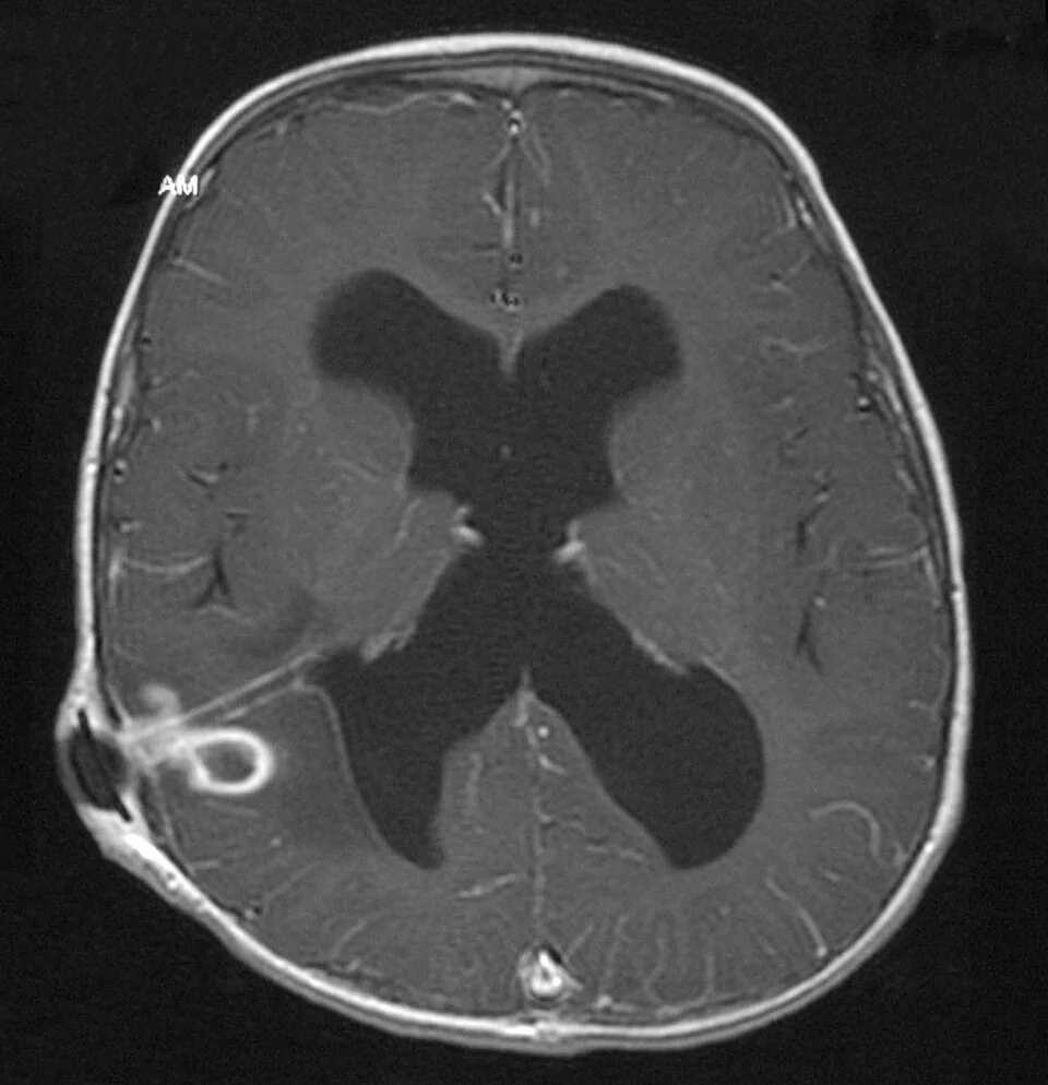
- Primary or metastatic brain tumour - particularly glioblastoma or a solitary metastasis, which can closely mimic an abscess radiologically; clinical context (fever, source of infection) and specific MRI sequences (diffusion-weighted imaging - abscess pus typically shows restricted diffusion, unlike most tumour necrosis) help distinguish them
- Toxoplasmosis - typically multiple ring-enhancing lesions in the immunocompromised (particularly advanced HIV), a key differential in that population
- Resolving haematoma
- Demyelinating lesion (tumefactive multiple sclerosis) - can occasionally show ring enhancement
- Radiation necrosis - in a patient with previous cranial radiotherapy
Investigations
- Contrast-enhanced CT or, preferably, MRI brain - the key investigation, showing a ring-enhancing lesion with a hypodense (CT) or T2-hyperintense (MRI) centre and surrounding vasogenic oedema; diffusion-weighted MRI typically shows restricted diffusion within the abscess cavity, helping distinguish it from a necrotic tumour
- Blood cultures - before antibiotics where possible, particularly if a haematogenous source (endocarditis) is suspected
- Inflammatory markers (FBC, CRP, ESR) - often raised, though normal values do not exclude the diagnosis
- Echocardiography - if infective endocarditis is a plausible source
- Imaging/assessment of the presumed source - CT sinuses/mastoids, dental review, as guided by clinical suspicion
- Stereotactic aspiration - provides definitive microbiological diagnosis (Gram stain and culture of the pus) and is usually both diagnostic and therapeutic
- HIV test - if risk factors are present or the presentation/imaging pattern suggests an opportunistic cause
Management
Source control
Neurosurgical drainage - usually stereotactic (image-guided) aspiration, or occasionally surgical excision - is central to management, both to reduce mass effect and to obtain pus for culture that guides antibiotic choice. Small abscesses (typically under around 2-2.5 cm) in a patient who is clinically stable may occasionally be managed with antibiotics alone under close imaging surveillance, but drainage is preferred whenever feasible.
Antibiotics
Broad-spectrum, high-dose IV antibiotics are started empirically, tailored to the likely source and later refined by culture results, and continued for a prolonged course - typically 4-6 weeks or longer, reflecting poor antibiotic penetration into the abscess cavity and the risk of relapse with a shorter course.
Response to treatment is judged on a combination of clinical improvement, falling inflammatory markers and, critically, serial imaging. Abscess cavities shrink slowly, and the ring of enhancement can persist for weeks or months after the infection is controlled, so residual enhancement alone is not evidence of treatment failure. Conversely, an enlarging cavity, new lesions or clinical deterioration should prompt reconsideration of the organism, the antibiotic choice, and whether further drainage is needed.
Duration is guided by response rather than a fixed rule, and the switch from intravenous to oral therapy is made in discussion with microbiology. Because courses run for many weeks, most patients are managed through an outpatient parenteral antibiotic therapy service with regular monitoring of renal function, liver function and drug levels.
- Empirical regimen commonly combines a third-generation cephalosporin (e.g. ceftriaxone) with metronidazole for anaerobic cover, particularly with a dental or sinus source
- Add vancomycin or another anti-staphylococcal agent if post-traumatic, post-neurosurgical, or endocarditis-related abscess is suspected
- Adjust according to culture and sensitivity results once available from aspirated pus or blood cultures
- Treat the primary source - definitive management of otitis media/mastoiditis, sinusitis, dental infection, or endocarditis, since failure to address the source risks relapse
Managing raised intracranial pressure and seizures
- Dexamethasone - can be used for significant surrounding oedema causing mass effect or threatening herniation, but used cautiously and for the shortest effective duration, since it can reduce antibiotic penetration into the abscess and blunt the radiological/clinical response used to judge treatment success
- Antiepileptic treatment - for seizures at presentation; prophylactic use in patients who have not seized is not routine but is considered in some high-risk presentations
- Serial imaging - to monitor response to treatment and abscess size over the treatment course
Complications
- Raised intracranial pressure and herniation
- Rupture into the ventricular system (ventriculitis) - a serious and often life-threatening complication
- Seizures and post-abscess epilepsy
- Permanent focal neurological deficit
- Recurrence, particularly if the primary source is not adequately treated
- Death - mortality remains significant, particularly with delayed diagnosis or ventricular rupture
Red flags
Because the classic triad is unreliable and inflammatory markers may be normal, the diagnosis is often made by taking the possibility seriously in a patient with the right risk profile rather than by any single finding. The features below should prompt contrast imaging rather than a period of observation.
Prognosis
With prompt neurosurgical drainage and appropriately targeted, prolonged antibiotic therapy, most patients survive, though mortality remains significant, particularly with delayed diagnosis, multiple abscesses, or rupture into the ventricular system. A substantial proportion of survivors are left with residual seizures or a focal neurological deficit, related to the size and location of the abscess and how promptly it was treated. Close collaboration between neurosurgery, microbiology and the team managing the primary source (ENT, cardiology, dentistry) improves both survival and long-term outcome.2
Follow-up imaging is continued beyond the end of antibiotic treatment, since relapse typically occurs within the first few months and is more likely where the primary source was not definitively treated. Patients should be advised to seek review promptly if headache, fever or focal symptoms return.
Because seizures occur in a substantial proportion of patients both at presentation and later, all survivors need a clear plan regarding antiepileptic treatment and its duration, and explicit driving advice with DVLA notification where a seizure has occurred. Cognitive and neuropsychological sequelae are common with frontal or temporal abscesses and are easily overlooked when attention is focused on the infection, so follow-up should include an assessment of cognition and function rather than imaging alone.
References
- Brouwer MC, Tunkel AR, McKhann GM, van de Beek D. Brain abscess. New England Journal of Medicine. 2014. Available here
- NICE CKS. Meningitis and brain abscess (referenced within neurology/infection guidance). Available here
- Sonneville R, Ruimy R, Benzonana N et al. An update on bacterial brain abscess in immunocompetent patients. Clinical Microbiology and Infection. 2017. Available here
This article is written for revision and education. It is not clinical guidance and must not be used to make decisions about the care of a patient. Always check current NICE guidance and local protocols.