
Bronchiectasis: The Vicious Cycle, Finding the Cause and Airway Clearance
Key points
- Bronchiectasis: permanent, abnormal dilatation of the bronchi with bronchial wall thickening, caused by a self-sustaining cycle of impaired mucus clearance, infection and inflammation.
- Presentation: chronic cough productive of large volumes of purulent sputum every day, recurrent chest infections, haemoptysis and breathlessness.
- Signs: coarse inspiratory crackles that do not clear on coughing, wheeze, and finger clubbing in a minority.
- Diagnosis: high-resolution CT of the chest is the gold standard. The key sign is a bronchus wider than its accompanying artery - the signet ring sign.
- Causes: around 40% are idiopathic. Always exclude the treatable ones: immunodeficiency, ABPA, cystic fibrosis, primary ciliary dyskinesia and proximal obstruction.
- Organisms: Haemophilus influenzae is commonest. Chronic Pseudomonas aeruginosa colonisation marks more severe disease and a worse prognosis.
- Management: daily airway clearance physiotherapy is the cornerstone, with prolonged 14-day antibiotic courses for exacerbations, vaccination and pulmonary rehabilitation.
- A drug to avoid: nebulised DNase (dornase alfa) helps in cystic fibrosis but causes harm in non-cystic fibrosis bronchiectasis and must not be used.
Introduction
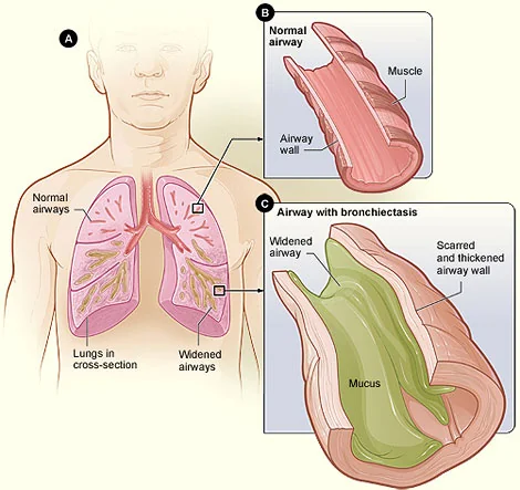
Bronchiectasis is permanent, abnormal dilatation of the bronchi accompanied by bronchial wall thickening and impaired clearance of secretions. Unlike asthma and COPD, the structural damage is irreversible - the aim of treatment is to interrupt the cycle that produced it and prevent further destruction, not to reverse what has already happened.
Once considered a disease of the pre-antibiotic era, bronchiectasis is being diagnosed with increasing frequency as CT scanning has become routine. UK prevalence is now estimated at around 500 per 100,000 and rises steeply with age.3
The vicious cycle
The pathogenesis is best understood as a self-perpetuating cycle, and every element of management is aimed at breaking it at one point or another.
- An initial insult - infection, obstruction or an inherited defect - impairs mucociliary clearance
- Retained secretions permit bacterial colonisation of the airway
- Colonisation drives chronic neutrophilic inflammation, with release of elastase and other proteases
- Proteolytic damage destroys the bronchial wall, causing further dilatation and worse clearance, which returns the patient to step one
Causes
Around 40% of cases are idiopathic, but a cause should be sought in every patient because a meaningful minority have something specifically treatable - and identifying it changes management entirely.1
| Category | Examples and clues |
|---|---|
| Post-infective | The commonest identified cause. Severe childhood pneumonia, whooping cough, measles, and tuberculosis (which classically causes upper lobe disease and traction bronchiectasis) |
| Cystic fibrosis | Suspect in any young patient, especially with upper lobe disease, malabsorption, infertility, sinusitis or a family history |
| Primary ciliary dyskinesia | Kartagener syndrome is the triad of bronchiectasis, chronic sinusitis and situs inversus. Also causes infertility (immotile sperm) and neonatal respiratory distress. |
| Immunodeficiency | Common variable immunodeficiency and other hypogammaglobulinaemias, HIV, and secondary immunosuppression from haematological malignancy or chemotherapy. Highly treatable with immunoglobulin replacement. |
| Allergic bronchopulmonary aspergillosis | Characteristically central or proximal bronchiectasis in an asthmatic, with a very high IgE and eosinophilia |
| Obstruction | Bronchial carcinoma, an inhaled foreign body (think of this in a child or in localised disease), lymph node compression. Always consider in focal, single-lobe bronchiectasis. |
| Connective tissue disease | Rheumatoid arthritis most commonly, and Sjogren syndrome. May precede the joint disease. |
| Inflammatory bowel disease | Particularly ulcerative colitis |
| Aspiration and reflux | Recurrent aspiration from swallowing disorders, achalasia or gastro-oesophageal reflux |
| Alpha-1 antitrypsin deficiency | Usually with emphysema |
| Yellow nail syndrome | Yellow dystrophic nails, lymphoedema and pleural effusions |
| Idiopathic | Around 40%, and a diagnosis made only after the above have been excluded |
Clinical features
Symptoms
- Chronic productive cough - the defining symptom. Daily production of large volumes of purulent sputum, often worse in the morning and on changing position, and frequently measured in cupfuls rather than teaspoons.
- Recurrent chest infections - typically several courses of antibiotics a year
- Haemoptysis - occurring in up to half of patients at some point, from hypertrophied and fragile bronchial arteries. Occasionally massive.
- Breathlessness and wheeze
- Pleuritic chest pain
- Fatigue, weight loss and reduced exercise tolerance
- Chronic rhinosinusitis - very common, and a pointer towards ciliary dyskinesia or immunodeficiency
Examination
- Coarse inspiratory crackles - typically bibasal, and characteristically do not clear on coughing, unlike the crackles of simple bronchitis
- Wheeze, from associated airflow obstruction
- Finger clubbing - present in a minority, and less common than the textbooks suggest
- Large volumes of purulent sputum in the sputum pot, which is worth asking to see
- Cachexia in advanced disease
- Signs of cor pulmonale - raised JVP, peripheral oedema, a loud P2 - in end-stage disease
- Signs of an underlying cause - situs inversus with dextrocardia, rheumatoid joint deformity, nasal polyps, or yellow nails
Investigations
Confirming the diagnosis
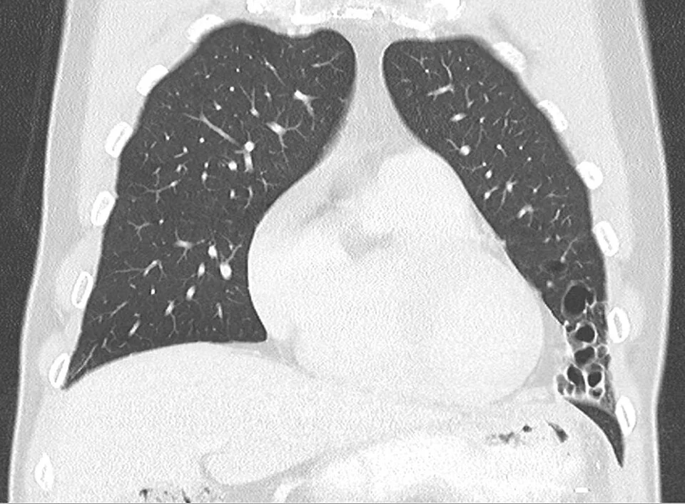
High-resolution CT of the chest is the gold standard and is required for the diagnosis. A chest radiograph may be normal even in significant disease and cannot exclude it.
- The signet ring sign - the dilated bronchus is wider than its accompanying pulmonary artery, so the pair in cross-section resembles a ring and stone. This is the cardinal sign.
- Lack of normal tapering of the bronchi towards the periphery
- Visible bronchi within 1 cm of the pleural surface, where they are normally too small to see
- Bronchial wall thickening, mucus plugging (the tree-in-bud pattern) and, in advanced disease, cystic change with air-fluid levels
- On a chest radiograph, look for tramline shadows, ring shadows and crowded, poorly defined vessel markings
Looking for the cause
A minimum work-up is recommended in every newly diagnosed patient, with further tests directed by the clinical picture:1
- Serum immunoglobulins (IgG, IgA, IgM) and serum electrophoresis - to detect hypogammaglobulinaemia
- Specific antibody responses to pneumococcal polysaccharide vaccine - to detect a functional antibody deficiency with normal total levels
- Total IgE, Aspergillus-specific IgE and Aspergillus precipitins - for ABPA
- Full blood count - eosinophilia in ABPA, neutrophilia in infection, lymphopenia in immunodeficiency
- Sputum culture including for mycobacteria and fungi - to identify chronic colonising organisms, and specifically to look for Pseudomonas and non-tuberculous mycobacteria
- Cystic fibrosis testing - sweat test and genotyping in younger patients, upper lobe disease, malabsorption, male infertility or Staphylococcus aureus colonisation
- Primary ciliary dyskinesia testing - nasal nitric oxide screening then ciliary brush biopsy, in those with a history of neonatal respiratory distress, chronic sinusitis, otitis media or infertility
- Rheumatoid factor and anti-CCP, and other connective tissue serology where indicated
- Alpha-1 antitrypsin and HIV testing where suggested by the picture
- Bronchoscopy - if the disease is focal, to exclude an obstructing lesion or foreign body
Assessing severity
- Spirometry - usually an obstructive pattern, and used to track decline
- Oxygen saturations, and an arterial blood gas if there is chronic hypoxaemia
- Body mass index - low BMI is a marker of severity
- The Bronchiectasis Severity Index combines age, BMI, FEV1, hospital admissions, exacerbation frequency, breathlessness score, Pseudomonas colonisation and radiological extent to predict mortality and admission
Management
Airway clearance and mucoactive treatment
- Active cycle of breathing technique, autogenic drainage, postural drainage and percussion, or oscillating positive expiratory pressure devices such as a flutter valve or Acapella
- Nebulised hypertonic saline or mannitol - to hydrate secretions and improve clearance, usually preceded by a bronchodilator
- Carbocisteine - an oral mucolytic that may help some patients
- Pulmonary rehabilitation - for anyone with breathlessness limiting activity
- Adequate hydration and, where obstruction is demonstrated, a bronchodilator before physiotherapy
Managing exacerbations
- An exacerbation is a deterioration in three or more of: cough, sputum volume or consistency, sputum purulence, breathlessness, exercise tolerance, fatigue, or haemoptysis
- Send a sputum sample before starting antibiotics, then treat empirically without waiting for the result
- Treat for 14 days, not the 5 to 7 used in other chest infections - shorter courses relapse
- Base the choice on previous sputum results. Amoxicillin or doxycycline for Haemophilus influenzae; ciprofloxacin is the oral agent for Pseudomonas aeruginosa
- Intravenous antibiotics for severe exacerbations, resistant organisms or failure of oral treatment - often given at home through an outreach service
- Intensify physiotherapy during an exacerbation, since retained secretions are the reason the patient has deteriorated
Long-term treatment
- Long-term macrolide (azithromycin three times a week) - for patients with 3 or more exacerbations a year. It reduces exacerbations through both antimicrobial and anti-inflammatory effects. Before starting, exclude non-tuberculous mycobacterial infection (monotherapy would induce resistance), and check an ECG for QT prolongation and liver function.
- Inhaled antibiotics - nebulised colistin, gentamicin or tobramycin for chronic Pseudomonas colonisation with frequent exacerbations
- Eradication therapy on first isolation of Pseudomonas - typically oral ciprofloxacin with or without a nebulised antibiotic, since preventing chronic colonisation alters the whole trajectory of the disease
- Vaccination - annual influenza, plus pneumococcal and COVID-19
- Treat the underlying cause - immunoglobulin replacement for antibody deficiency, corticosteroids for ABPA, CFTR modulators for cystic fibrosis, reflux and aspiration management
- Inhaled corticosteroids are not routinely indicated unless there is coexisting asthma or COPD; they increase the risk of infection including non-tuberculous mycobacteria
- Nutritional support, and smoking cessation
- Surgical resection - for localised disease with severe symptoms despite optimal medical treatment
- Lung transplantation - for selected patients with end-stage disease
Complications
- Recurrent exacerbations and progressive lung function decline
- Chronic Pseudomonas aeruginosa colonisation - associated with more exacerbations, worse quality of life, faster decline and higher mortality
- Massive haemoptysis - a life-threatening emergency from erosion of hypertrophied bronchial arteries, treated with airway protection, positioning the bleeding side down, tranexamic acid and bronchial artery embolisation
- Respiratory failure and cor pulmonale
- Pneumonia, empyema and lung abscess
- Pneumothorax
- Non-tuberculous mycobacterial infection - increasingly common and difficult to treat, requiring prolonged multi-drug therapy
- Secondary AA amyloidosis - rare now, from decades of chronic inflammation, presenting with proteinuria and renal failure
- Depression, anxiety and social isolation - the burden of daily sputum production, physiotherapy and repeated infections is considerable and consistently under-recognised
Red flags
Prognosis
Bronchiectasis varies enormously in behaviour. Many patients with limited, well-managed disease have a normal life expectancy and few limitations. At the other end, extensive disease with chronic Pseudomonas colonisation and frequent exacerbations carries a substantially reduced life expectancy, with mortality driven by progressive respiratory failure.
Adverse prognostic factors are captured by the Bronchiectasis Severity Index: older age, low BMI, reduced FEV1, previous hospital admissions, frequent exacerbations, breathlessness, Pseudomonas colonisation and radiologically extensive disease.
What is genuinely encouraging is how much of this is modifiable. Daily airway clearance, prompt and adequately long treatment of exacerbations, eradication of Pseudomonas at first isolation, vaccination, pulmonary rehabilitation and treatment of a reversible underlying cause together change the trajectory of the disease. The most common failing in practice is not a lack of available treatment but a failure to teach and reinforce physiotherapy - the one intervention the patient performs themselves, every day, for the rest of their life.
References
- British Thoracic Society. Guideline for bronchiectasis in adults. 2019. Available here
- Polverino E, Goeminne PC, McDonnell MJ et al. European Respiratory Society guidelines for the management of adult bronchiectasis. European Respiratory Journal. 2017. Available here
- NICE Clinical Knowledge Summaries. Bronchiectasis. Available here
- Chalmers JD, Goeminne P, Aliberti S et al. The Bronchiectasis Severity Index. American Journal of Respiratory and Critical Care Medicine. 2014. Available here
- O'Donnell AE, Barker AF, Ilowite JS, Fick RB. Treatment of idiopathic bronchiectasis with aerosolized recombinant human DNase I. Chest. 1998. Available here
- NICE NG117. Bronchiectasis (non-cystic fibrosis), acute exacerbation: antimicrobial prescribing. 2018. Available here
- BNF. Azithromycin and ciprofloxacin - indications and dosing. Available here
- National Heart, Lung and Blood Institute (NIH), public domain, via Wikimedia Commons. Available here
- Hellerhoff, CC BY-SA 4.0, via Wikimedia Commons. Available here
This article is written for revision and education. It is not clinical guidance and must not be used to make decisions about the care of a patient. Always check current NICE guidance and local protocols.