
Urinary Tract Calculi
Key points
- Urinary tract calculi: crystalline stones forming within the urinary tract when solute concentration exceeds solubility. Lifetime prevalence is around 10%, with a male predominance and a peak between 30 and 60 years.
- Commonest type: calcium oxalate (around 80%), followed by calcium phosphate, struvite, uric acid and cystine.
- Classic presentation: sudden severe colicky 'loin-to-groin' pain with the patient writhing and unable to lie still (in contrast to peritonitis), often with nausea, vomiting and non-visible haematuria.
- First-line imaging: non-contrast CT of the kidneys, ureters and bladder (CT KUB) within 24 hours. Ultrasound is used first in pregnancy and in children to avoid radiation.1
- Radiolucency: uric acid stones are radiolucent on plain X-ray (but visible on CT). Calcium stones are radio-opaque; struvite forms staghorn calculi.
- Analgesia: intramuscular or rectal NSAIDs (e.g. diclofenac) are first line, not opioids.
- Who passes spontaneously: most stones under 5 mm pass without intervention. Alpha-blockers (tamsulosin) may aid passage of distal stones of 5-10 mm.
- The emergency: an obstructed, infected kidney - fever plus obstruction requires urgent decompression by nephrostomy or stent, alongside antibiotics.
Introduction
Urinary tract calculi (urolithiasis) are crystalline aggregates that form when the urine becomes supersaturated with stone-forming solutes, or when the natural inhibitors of crystallisation are deficient. Stones may form anywhere in the tract but arise predominantly in the kidney, and cause symptoms chiefly when they pass into and obstruct the ureter.1
They are common - lifetime prevalence is around 10%, with a male-to-female ratio of roughly 2:1 and a peak incidence between 30 and 60 years. Recurrence is the rule rather than the exception, with around 50% recurring within 10 years, which is why metabolic assessment and prevention matter as much as treating the acute episode.
Types of stone
| Type | Proportion | Associations | X-ray appearance |
|---|---|---|---|
| Calcium oxalate | ~80% (most common overall) | Hypercalciuria, hyperoxaluria, low fluid intake, high oxalate diet (spinach, rhubarb, nuts, tea), Crohn's disease and ileal resection (enteric hyperoxaluria) | Radio-opaque |
| Calcium phosphate | ~10% | Hyperparathyroidism, renal tubular acidosis type 1, alkaline urine | Radio-opaque |
| Struvite (magnesium ammonium phosphate, 'triple phosphate') | ~5-10% | Urease-producing organisms - Proteus, Klebsiella, Pseudomonas - which alkalinise urine. Form staghorn calculi; commoner in women and with recurrent UTI | Radio-opaque |
| Uric acid | ~5-10% | Persistently acidic urine, gout, high purine diet, tumour lysis syndrome and myeloproliferative disease, chronic diarrhoea | RADIOLUCENT on plain X-ray (visible on CT) |
| Cystine | ~1% | Cystinuria - an autosomal recessive defect of tubular amino acid reabsorption. Presents young, often with recurrent stones and a family history | Faintly radio-opaque ('ground glass') |
| Drug-induced | Rare | Indinavir, and other protease inhibitors | Often radiolucent |
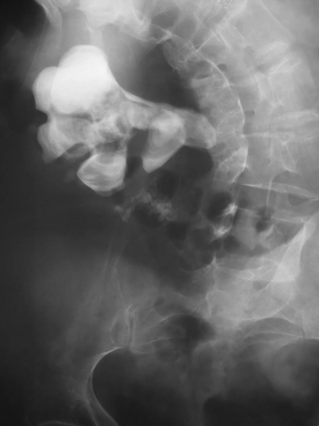
Risk factors
General
- Low fluid intake and dehydration - the single most important modifiable factor, producing concentrated urine
- Hot climates and occupations with high insensible losses
- Male sex and age 30-60
- Previous stones and a family history
- Obesity, diabetes and metabolic syndrome
- Diet - high salt, high animal protein, high oxalate; and, counterintuitively, a low calcium diet (which increases oxalate absorption and therefore risk)
- Sedentary lifestyle and immobilisation
Medical and anatomical
- Hypercalcaemia of any cause - especially primary hyperparathyroidism ('stones, bones, groans and psychiatric moans')
- Recurrent urinary tract infection with urease-producing organisms - struvite stones
- Gout and hyperuricaemia; tumour lysis syndrome
- Inflammatory bowel disease, ileal resection and malabsorption - enteric hyperoxaluria
- Renal tubular acidosis type 1 and medullary sponge kidney
- Cystinuria and primary hyperoxaluria - inherited causes presenting young
- Anatomical abnormalities causing stasis - PUJ obstruction, horseshoe kidney, vesicoureteric reflux, urinary diversion
- Drugs - loop diuretics, thiazides (protective for calcium stones but may unmask hypercalcaemia), carbonic anhydrase inhibitors (acetazolamide, topiramate), indinavir, excess vitamin D or calcium supplements
Clinical features
Renal colic is the classic presentation, produced by acute obstruction and distension of the collecting system rather than by the stone scraping the ureter.
- Sudden onset, severe, colicky pain radiating from loin to groin - waxing and waning in waves, often described as the worst pain the patient has experienced (and frequently compared to labour)
- The patient is restless and writhing, unable to find a comfortable position - a genuinely useful discriminator, since patients with peritonitis lie very still
- Nausea and vomiting - common and prominent
- Haematuria - usually non-visible (microscopic), present in around 90%; visible haematuria occurs in a minority. Note that absence of haematuria does not exclude a stone
- Radiation and lower urinary tract symptoms depend on stone position - a stone at the VUJ classically causes frequency, urgency and pain radiating to the tip of the penis, scrotum or labia
- Sweating, pallor and tachycardia from pain
- Fever and rigors - NOT a feature of uncomplicated colic; their presence means infection, and with obstruction that is an emergency
- Staghorn calculi are often asymptomatic or present insidiously with recurrent infection, haematuria or vague loin discomfort rather than acute colic
On examination there is usually loin tenderness but a soft, non-peritonitic abdomen. Marked abdominal tenderness with guarding should prompt reconsideration of the diagnosis.
Differential diagnosis
| Condition | Distinguishing features |
|---|---|
| Leaking abdominal aortic aneurysm | Age over 50, hypotension, pulsatile expansile mass, cardiovascular risk factors - as above |
| Pyelonephritis | Fever and rigors, constant rather than colicky pain, systemically unwell |
| Ectopic pregnancy | Any woman of childbearing age - pregnancy test is mandatory |
| Ovarian torsion or cyst accident | Pelvic pain, adnexal tenderness; ultrasound diagnostic |
| Testicular torsion | Referred pain may present as abdominal pain - always examine the testes in a boy or young man |
| Appendicitis / diverticulitis | Localised peritonism, patient lies still, altered bowel habit |
| Biliary colic | Right upper quadrant, related to fatty meals, deranged LFTs |
| Bowel obstruction | Distension, vomiting, absolute constipation, abnormal bowel sounds |
| Musculoskeletal pain / radiculopathy | Reproduced by movement or palpation, dermatomal distribution |
| Renal infarction | Markedly raised LDH, atrial fibrillation or thrombophilia |
Investigations
Bedside and laboratory
- Urine dipstick - haematuria in around 90%; also assess for nitrites and leucocytes suggesting concurrent infection
- Pregnancy test in all women of childbearing age - changes both the differential and the imaging
- FBC and CRP - looking for evidence of infection
- U&Es and creatinine - to detect AKI, especially important with a solitary kidney or bilateral stones
- Serum calcium, phosphate and urate - screening for hyperparathyroidism and gout; PTH if calcium is raised
- Blood cultures if febrile
- Urine culture if infection suspected
- Stone analysis - patients should be asked to sieve their urine and retain any stone passed, since its composition guides prevention
Imaging
- Non-contrast CT KUB is first line, and NICE recommends it within 24 hours of presentation. It has near-100% sensitivity and specificity, detects all stone types including radiolucent uric acid stones, shows the degree of obstruction, and identifies alternative diagnoses such as AAA
- Ultrasound is first line in pregnancy and in children, and is a reasonable initial test in young patients, to avoid ionising radiation. It detects hydronephrosis well but is far less sensitive for ureteric stones themselves
- Plain KUB X-ray - limited diagnostic role, but useful for monitoring known radio-opaque stones over time and for planning lithotripsy
- CT with contrast or MR urography - occasionally used where anatomy needs clarifying
Management
Acute management
- Analgesia first - NSAIDs are first line, given intramuscularly or rectally (e.g. diclofenac), and are more effective than opioids for renal colic because they reduce ureteric smooth muscle tone and renal pelvic pressure as well as providing analgesia. IV paracetamol is the alternative if NSAIDs are contraindicated; opioids are reserved for when both are unsuitable or insufficient
- Antiemetics and intravenous fluids if vomiting
- Antibiotics if there is any evidence of infection
- Assess for the emergency features - fever, AKI, solitary kidney, bilateral obstruction, uncontrolled pain
Conservative management and medical expulsive therapy
- Most stones under 5 mm pass spontaneously (around 80%), usually within 4 weeks; stones of 5-10 mm pass in roughly half of cases
- Watchful waiting with oral analgesia is appropriate for small distal stones in a well patient with controlled pain and no infection or renal impairment
- Alpha-blockers (tamsulosin) - medical expulsive therapy; NICE supports their use for distal ureteric stones of 5-10 mm to aid spontaneous passage. Benefit is modest and they are not recommended for smaller stones
- Advise a high fluid intake and to sieve urine to retrieve the stone
- Arrange follow-up imaging to confirm passage - a patient whose pain settles has not necessarily passed the stone, and a silently obstructed kidney can be lost
Surgical and interventional management
| Procedure | Typical indication |
|---|---|
| Extracorporeal shock wave lithotripsy (ESWL) | External shock waves fragment the stone. Suitable for renal stones under about 2 cm and some proximal ureteric stones. Contraindicated in pregnancy, in uncorrected coagulopathy and in distal obstruction |
| Ureteroscopy with laser lithotripsy | Endoscopic access via the urethra and bladder; preferred in pregnancy, in obese patients, in those on anticoagulants, and for distal ureteric stones. Usually leaves a temporary stent |
| Percutaneous nephrolithotomy (PCNL) | Percutaneous access directly into the kidney; the treatment of choice for large renal stones (over 2 cm) and staghorn calculi |
| Ureteric stent or percutaneous nephrostomy | Emergency decompression of an obstructed or infected system - a temporising measure, with definitive stone treatment once sepsis has resolved |
| Open or laparoscopic surgery | Now rare; reserved for complex anatomy or failure of the above |
Prevention of recurrence
- Fluid intake of 2.5 to 3 litres per day, aiming for pale urine and a urine output of over 2.5 L - by far the most important measure for all stone types
- Reduce dietary salt (which increases urinary calcium excretion) and moderate animal protein intake
- Maintain a normal dietary calcium intake - restricting calcium is counterproductive, as dietary calcium binds oxalate in the gut and reduces its absorption; only calcium supplements are discouraged
- Reduce oxalate-rich foods (spinach, rhubarb, nuts, chocolate, strong tea) in oxalate stone formers
- Thiazide diuretics - reduce urinary calcium excretion in recurrent calcium stone formers
- Potassium citrate - alkalinises urine; used in uric acid stones and in hypocitraturic calcium stone formers
- Allopurinol - for uric acid stones and hyperuricosuria
- Treat the underlying cause - parathyroidectomy in primary hyperparathyroidism, eradication of infection and complete stone clearance in struvite stones
- Weight loss and treatment of metabolic syndrome
Complications
- Obstruction and hydronephrosis, with permanent loss of renal function if unrelieved - irreversible damage begins within days to weeks
- Infection: pyelonephritis, pyonephrosis and urosepsis - the most dangerous acute complication
- Acute kidney injury, particularly with bilateral stones or a solitary kidney
- Chronic kidney disease from recurrent obstruction and infection
- Recurrent stone formation - around 50% within 10 years
- Ureteric stricture - from chronic impaction or from instrumentation
- Complications of treatment - stent discomfort, haematuria and 'steinstrasse' (a column of fragments obstructing the ureter after lithotripsy); bleeding and injury after PCNL
- Xanthogranulomatous pyelonephritis and a non-functioning kidney with chronic obstruction and infection
Red flags
Prognosis
The prognosis of an individual episode is good. Around 80% of stones under 5 mm pass spontaneously, generally within four weeks, and the great majority of patients require no more than analgesia, fluids and follow-up imaging to confirm passage. Larger stones increasingly require intervention, but modern endoscopic and lithotripsy techniques achieve high stone-free rates with low morbidity.
Recurrence is the dominant long-term issue. Roughly 50% of stone formers will have another stone within 10 years and up to 75% within 20 years without preventive measures. This is why stone analysis, metabolic assessment in selected patients, and above all sustained high fluid intake are central to management - fluid intake alone substantially reduces recurrence.
Renal outcome depends on obstruction and infection rather than on the stone itself. A briefly obstructed kidney recovers fully, but prolonged unrelieved obstruction causes irreversible loss of function, and an obstructed infected system can destroy a kidney within days and carries significant mortality. Patients with staghorn calculi, recurrent infection or anatomical abnormalities are those most likely to progress to chronic kidney disease, and warrant long-term urological follow-up.
References
- NICE NG118. Renal and ureteric stones: assessment and management. 2019. Available here
- NICE Clinical Knowledge Summaries. Renal or ureteric colic - acute. Available here
- European Association of Urology. Guidelines on Urolithiasis. Available here
- Turk C, Petrik A, Sarica K et al. EAU Guidelines on Diagnosis and Conservative Management of Urolithiasis. European Urology. 2016. Available here
- BNF. Tamsulosin and diclofenac - indications and cautions. Available here
- Nevit Dilmen, CC BY-SA 3.0, via Wikimedia Commons. Available here
This article is written for revision and education. It is not clinical guidance and must not be used to make decisions about the care of a patient. Always check current NICE guidance and local protocols.