
Phimosis and Paraphimosis
Key points
- Phimosis: inability to retract the foreskin over the glans. It is physiological and normal in young boys, and only pathological when caused by scarring.
- Physiological phimosis: the foreskin is naturally adherent at birth and separates gradually - only ~50% retract by age 10 and around 99% by age 17. Do not force retraction, which causes scarring and creates true phimosis.
- Pathological phimosis: caused by a scarred, white, fibrotic ring, most importantly from balanitis xerotica obliterans (BXO / lichen sclerosus) - an absolute indication for circumcision.
- Paraphimosis: the retracted foreskin cannot be returned over the glans. The tight band causes venous congestion, progressive oedema and, if unrelieved, ischaemia and necrosis of the glans.
- Paraphimosis is an emergency: a urological emergency requiring immediate reduction. Delay risks irreversible glans necrosis.
- The commonest cause: iatrogenic - the foreskin retracted for catheterisation, cleaning or examination and never replaced. This makes it largely preventable.
- Reducing a paraphimosis: analgesia first, then sustained manual compression to squeeze out oedema, then reduce the glans back through the ring. Adjuncts: ice, granulated sugar (osmotic), hyaluronidase.
- If reduction fails: emergency dorsal slit of the constricting band under local or general anaesthetic, with elective circumcision later.
Introduction
These two conditions are frequently confused but are entirely different clinical problems. Phimosis is an inability to retract the foreskin, is usually chronic, and is often entirely normal in childhood. Paraphimosis is an inability to replace an already-retracted foreskin, is acute, and is a urological emergency.1
A brief note on anatomy: the foreskin (prepuce) is a double layer of skin covering the glans, tethered underneath by the frenulum. At birth the inner surface is naturally adherent to the glans, and these adhesions separate spontaneously over childhood through desquamation and intermittent erections. Understanding this normal process is what prevents unnecessary intervention in boys.
Phimosis
Physiological phimosis
Non-retractility of the foreskin in a boy is normal, not a disease. The foreskin is non-retractile in almost all newborns, and separation is a gradual process continuing well into adolescence.
- At birth: retractile in only about 4%
- By age 3: roughly 90% are at least partially retractile
- By age 10: only about 50% fully retract
- By age 17: around 99% fully retract
- The foreskin looks healthy and supple, with no white scarring - the key distinguishing feature from pathological phimosis
- Ballooning of the foreskin during voiding is common, generally harmless, and resolves spontaneously; it is not in itself an indication for circumcision
Pathological phimosis
True pathological phimosis is caused by scarring of the preputial opening, producing a rigid, often visibly white fibrotic ring that will not stretch.
- Balanitis xerotica obliterans (BXO) - genital lichen sclerosus; the most important cause. Produces a characteristic white, thickened, scarred prepuce, and may involve the glans and urethral meatus causing meatal stenosis. It is an absolute indication for circumcision, and carries a small long-term association with penile squamous cell carcinoma, so it warrants follow-up
- Recurrent balanitis or balanoposthitis - repeated inflammation causing fibrosis
- Forced retraction in childhood, with subsequent scarring
- Diabetes mellitus - recurrent candidal balanitis is common and may be the presenting feature of undiagnosed diabetes; always check blood glucose in an adult with new phimosis or recurrent balanitis
- Poor hygiene, chronic irritation, and previous trauma or catheterisation
- Lichen planus and other dermatoses
Clinical features and complications
- Inability to retract the foreskin, with a visible tight or scarred ring
- Ballooning of the foreskin on voiding, spraying or a poor urinary stream
- Recurrent balanitis - pain, erythema, discharge and swelling
- Pain on erection or intercourse, and difficulty with hygiene
- Recurrent urinary tract infections
- Acute urinary retention in severe cases - a genuine emergency
- Difficulty with catheterisation
Management
- Reassurance and education for physiological phimosis in boys - explaining the natural history, avoiding forced retraction, and gentle hygiene. Most need nothing else
- Topical corticosteroid - a moderately potent steroid such as betamethasone 0.05% applied twice daily for 4-8 weeks, combined with gentle stretching, is effective in a substantial majority and often avoids surgery. This is first-line active treatment
- Treat underlying causes - antifungals or antibiotics for balanitis, and glycaemic control in diabetes
- Preputioplasty - a foreskin-preserving procedure widening the opening; an option where the foreskin is otherwise healthy
- Circumcision - indicated for BXO (absolute indication), recurrent balanitis, recurrent UTI, failed medical treatment, symptomatic pathological phimosis, and recurrent paraphimosis
- Check blood glucose in any adult presenting with new phimosis or recurrent balanitis
Paraphimosis
Paraphimosis occurs when a retracted foreskin cannot be returned to its normal position. The tight preputial ring sits proximal to the glans and acts as a constricting band.
Causes
- Iatrogenic - by far the commonest cause. The foreskin is retracted for urethral catheterisation, catheter care, cleaning or examination and is not replaced afterwards. This makes most cases entirely preventable
- Sexual activity or masturbation with failure to replace the foreskin
- Vigorous cleaning or forced retraction, particularly in children and in patients receiving nursing care
- Pre-existing phimosis or a tight preputial ring
- Penile piercing and, rarely, foreign bodies or constricting objects
- Chronic balanitis and oedematous states (heart failure, nephrotic syndrome) predisposing to swelling
- Cognitive impairment, dementia or reduced sensation (e.g. spinal injury, diabetic neuropathy), where the patient may not report discomfort
Clinical features
- Severe penile pain and distress
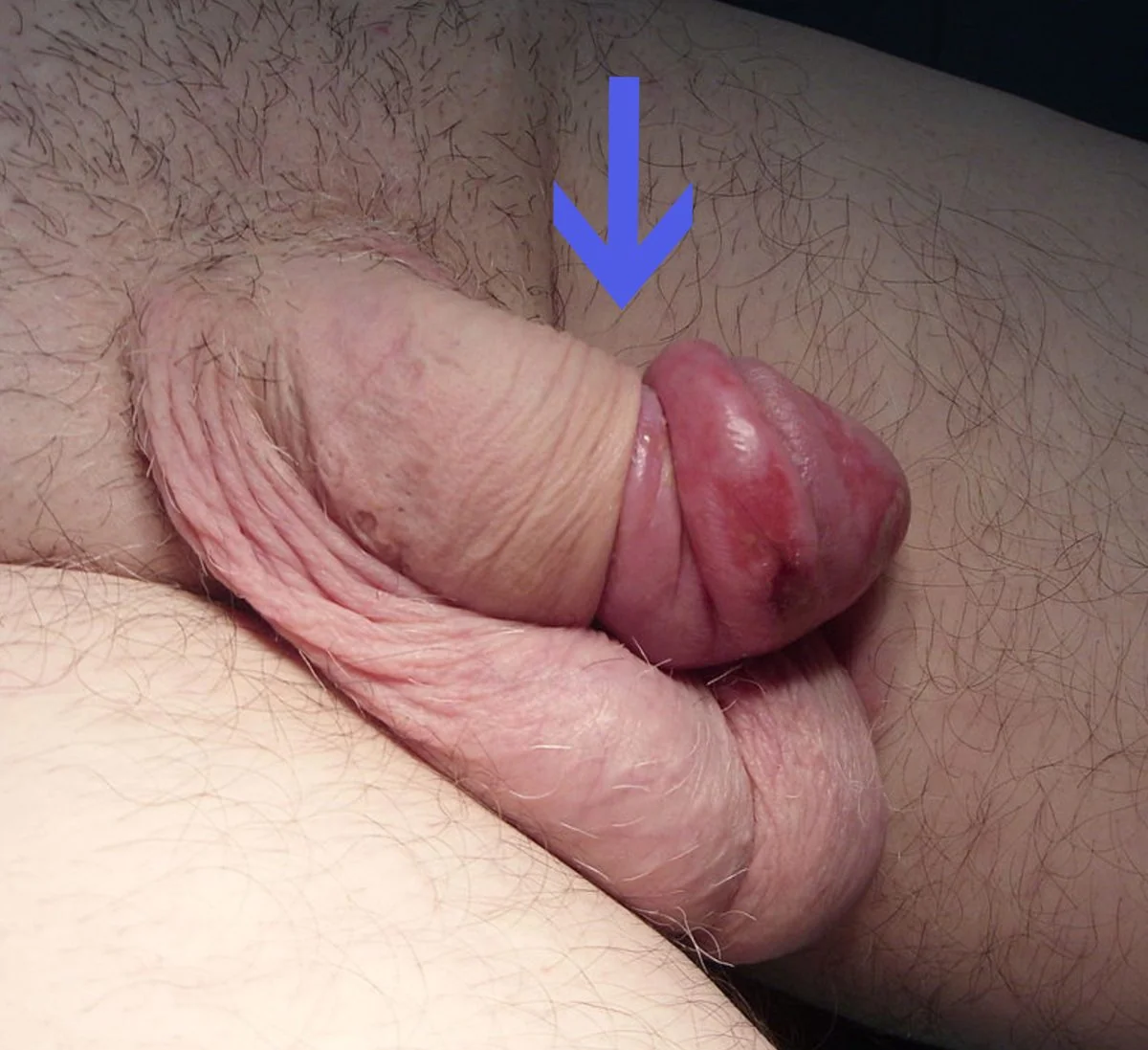
- Swollen, oedematous glans with a collar of oedematous foreskin proximal to it
- A palpable constricting band of tissue behind the corona
- The shaft proximal to the ring is typically normal - the swelling is distal to the constriction
- Progressive colour change - initially erythematous, then dusky, purple or blue, and finally black with necrosis if unrelieved
- Difficulty passing urine in severe cases
- In late presentations there may be ulceration or frank necrosis of the glans
Management - a stepwise approach
- Give adequate analgesia first - this is essential and frequently skimped. Options include oral or intravenous opioid, topical local anaesthetic gel, or a penile ring block with plain lidocaine (never use adrenaline-containing local anaesthetic on the penis, because of the risk of ischaemic necrosis from vasoconstriction of end arteries)
- Reduce the oedema - apply firm, sustained manual compression to the glans and swollen prepuce for 5 to 10 minutes. Patience matters more than force; the aim is to squeeze fluid back proximally through the ring
- Adjuncts to reduce swelling: an ice pack or cold compress (wrapped, not applied directly), granulated sugar applied to the glans for 1-2 hours to draw out fluid by osmosis, compressive bandaging, or injection of hyaluronidase into the oedematous tissue to disperse it
- Perform the reduction: place both thumbs on the glans and the index and middle fingers of both hands behind the constricting ring, then push the glans back through the ring with the thumbs while drawing the foreskin forward with the fingers
- If manual reduction fails - urgent urological referral for a dorsal slit. A longitudinal incision is made in the constricting band under local or general anaesthetic, immediately relieving the constriction. Elective circumcision is then usually performed later
- Occasionally the 'puncture technique' (multiple small punctures in the oedematous foreskin to allow fluid to escape) is used to aid reduction
- After successful reduction, ensure the foreskin is left in its normal forward position, treat any underlying balanitis, and consider referral for elective circumcision if paraphimosis is recurrent or there is significant underlying phimosis
Differential diagnosis
| Condition | Distinguishing features |
|---|---|
| Paraphimosis | Foreskin retracted behind the glans with a constricting band; swelling distal to the ring; severe pain |
| Balanitis / balanoposthitis | Foreskin in normal position; erythema, discharge, itch and tenderness of glans and prepuce; often candidal, especially in diabetes |
| Penile constriction from a foreign body | A ring, band or hair tourniquet visible; always inspect for a constricting object, particularly in children (hair-thread tourniquet) and in adults |
| Priapism | Persistent erection of the corpora with a typically soft glans; a different emergency requiring aspiration |
| Angio-oedema or allergic reaction | Diffuse swelling without a constricting band; possible urticaria elsewhere; may follow contact allergen |
| Cellulitis of the penis | Spreading erythema, warmth, fever, no constricting band |
| Fournier's gangrene | Necrosis, crepitus, systemic toxicity and pain out of proportion; a surgical emergency |
| Penile cancer | Chronic ulcerated or fungating lesion rather than acute swelling; may coexist with phimosis and be hidden beneath it |
| Insect bite or trauma | History; localised swelling without a ring |
Complications
Of phimosis
- Recurrent balanitis and balanoposthitis
- Recurrent urinary tract infection
- Urinary obstruction and acute retention
- Painful erection and sexual dysfunction
- Paraphimosis - if a tight foreskin is retracted
- Meatal stenosis - particularly with BXO involving the meatus
- Penile squamous cell carcinoma - a recognised long-term association with chronic phimosis and BXO, and one reason a phimosis that conceals the glans should not simply be ignored in an older man
- Difficulty with catheterisation and with hygiene
Of paraphimosis
- Glans ischaemia, necrosis and auto-amputation - the feared outcome of delayed treatment
- Ulceration and infection of the glans and prepuce
- Urinary retention from urethral compression
- Penile gangrene and Fournier's gangrene
- Recurrence - common if the underlying tight foreskin is not addressed
- Scarring and subsequent worsening phimosis
- Sexual dysfunction and psychological distress
- Complications of surgery - bleeding, infection, cosmetic dissatisfaction, and meatal stenosis after circumcision
Red flags
Prognosis
Physiological phimosis has an excellent prognosis and requires nothing more than reassurance in the great majority of boys. The natural history is one of steady spontaneous resolution, with around 99% of foreskins fully retractile by late adolescence. The main harm in this group comes from unnecessary intervention - forced retraction causing scarring, or circumcision performed for a normal developmental finding.
Pathological phimosis responds well to treatment. Topical corticosteroids with gentle stretching are successful in a substantial majority, avoiding surgery altogether, and circumcision is curative where it is needed. The exception is balanitis xerotica obliterans, which is a chronic, progressive scarring dermatosis: circumcision is effective for the preputial disease, but the condition can involve the glans and urethral meatus, sometimes requiring meatal surgery, and it carries a small but real long-term risk of penile squamous cell carcinoma. These patients therefore warrant ongoing follow-up rather than discharge.
Paraphimosis has an excellent outcome if treated promptly, and a poor one if not. Reduction achieved within hours virtually always preserves the glans completely, and manual reduction succeeds in most cases when adequate analgesia and patient, sustained compression are used. Delay is what causes harm: prolonged constriction leads to ischaemia, necrosis and, in the worst cases, loss of the glans, with the case illustrated above showing the degree of oedema that develops over just a few days. Recurrence is common unless the underlying tight foreskin is dealt with, which is why elective circumcision is usually offered afterwards. Given that most hospital cases are iatrogenic, the most valuable intervention of all remains the simplest - replacing the foreskin every time it is retracted.
References
- NICE Clinical Knowledge Summaries. Phimosis and paraphimosis. Available here
- British Association of Urological Surgeons (BAUS). Circumcision and foreskin conditions - patient information. Available here
- European Association of Urology. Guidelines on Paediatric Urology - phimosis. Available here
- Bragg BN, Kong EL, Leslie SW. Paraphimosis. StatPearls. 2023. Available here
- British Association of Dermatologists. Guidelines for the management of lichen sclerosus. Available here
- Drvgaikwad, CC BY 3.0, via Wikimedia Commons. Available here
This article is written for revision and education. It is not clinical guidance and must not be used to make decisions about the care of a patient. Always check current NICE guidance and local protocols.