
Lichen Planus
Key points
- Lichen planus: a chronic, T cell-mediated inflammatory disease targeting the skin and mucous membranes, classically causing itchy, violaceous, flat-topped, polygonal papules - the '6 Ps'.
- Wickham striae: fine, white, lace-like lines on the surface of papules, best seen with a drop of oil or under magnification, and near-pathognomonic for lichen planus.
- Distribution: flexor surfaces of the wrists and forearms, ankles and lower back; oral involvement (lacy white streaks on the buccal mucosa) is common and can occur alone.
- Koebner phenomenon: new lesions appearing along lines of skin trauma, shared with psoriasis and vitiligo.
- Associations: hepatitis C infection, lichenoid drug reactions (gold, antimalarials, thiazides, ACE inhibitors, NSAIDs, beta-blockers), and graft-versus-host disease can all produce an identical or near-identical picture.
- Oral lichen planus: carries a small but real risk of malignant transformation to oral squamous cell carcinoma, particularly the erosive subtype, and needs periodic review rather than a one-off diagnosis.
- Management: potent topical corticosteroids are first-line for skin disease; oral corticosteroids, retinoids or other immunosuppressants are reserved for severe, widespread or resistant disease.
- Prognosis: cutaneous lichen planus is usually self-limiting, resolving within 1-2 years, though it often leaves post-inflammatory hyperpigmentation; oral, nail and scalp disease can be considerably more chronic.
Introduction
Lichen planus is a chronic, inflammatory disease affecting the skin, nails, hair and mucous membranes, most commonly presenting in adults between 30 and 60. It is uncommon compared with eczema or psoriasis but is a favourite in exams because of its striking, memorable morphology and its list of associations, which is longer and more clinically important than its rarity might suggest.1
The name reflects its appearance: lichen for the flat, lichen-like plaques, and planus for their flat-topped surface. Distinct patterns affect the skin, the mouth, the genitals, the nails and the scalp, and while cutaneous disease is generally self-limiting, mucosal and scalp involvement can be considerably more persistent and carries its own specific risks.2
Pathophysiology
Lichen planus is a T cell-mediated autoimmune reaction directed against basal keratinocytes. Cytotoxic CD8+ T cells accumulate at the dermo-epidermal junction and trigger apoptosis of basal keratinocytes, which is seen histologically as a dense, band-like (lichenoid) lymphocytic infiltrate hugging the basal layer, with degenerating keratinocytes (colloid or Civatte bodies) within the epidermis.4
What triggers this T cell response is not fully understood, but in a proportion of patients an identifiable driver can be found - a virus, a drug, or an immune-mediated process such as graft-versus-host disease - all of which appear to alter self-antigens on basal keratinocytes enough to provoke the same cytotoxic T cell attack. In many patients, however, no trigger is ever identified and the disease is labelled idiopathic.
- Hepatitis C virus infection - a well-established association, particularly with oral lichen planus, and testing is worthwhile in at-risk patients6
- Lichenoid drug reactions - gold, antimalarials (hydroxychloroquine), thiazide diuretics, ACE inhibitors, beta-blockers and NSAIDs can all produce an eruption clinically and histologically similar to idiopathic lichen planus, sometimes only after months of treatment
- Graft-versus-host disease - chronic GVHD after allogeneic stem cell transplantation frequently produces lichen planus-like skin and oral lesions
- Dental amalgam (contact sensitivity to mercury) - implicated in some cases of oral lichen planus confined to mucosa directly adjacent to an amalgam filling
Clinical features
Classic cutaneous lichen planus is remembered by the '6 Ps': Pruritic, Planar (flat-topped), Polygonal, Purple (violaceous), Papules, and Plaques.
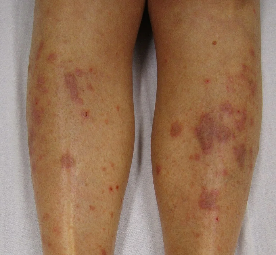
Lesions are typically a few millimetres across, intensely itchy, and found symmetrically on the flexor surfaces of the wrists and forearms, the shins and ankles, and the lower back. Close examination, ideally with a drop of oil or under magnification, reveals Wickham striae - fine, white, lace-like lines on the surface of well-developed papules - which are a highly specific finding.
Mucosal, nail and scalp variants
| Site | Features |
|---|---|
| Oral | Lacy, white, reticulate streaks on the buccal mucosa (Wickham striae equivalent); can also be erosive or ulcerative, which is more symptomatic and carries the greater malignant potential |
| Genital | Similar white lacy pattern or erosions on the vulva, glans or perianal skin; can cause scarring and stenosis if erosive and untreated |
| Nails | Longitudinal ridging, thinning, and in severe cases pterygium formation - permanent scarring where the proximal nail fold fuses with the nail bed |
| Scalp (lichen planopilaris) | Perifollicular erythema and scaling progressing to scarring alopecia; early treatment matters because established hair loss here is irreversible |
| Hypertrophic | Thick, hyperkeratotic plaques, typically on the shins, which are more resistant to treatment and slower to resolve |
As with psoriasis, new lesions can appear along a line of trauma - the Koebner phenomenon - and post-inflammatory hyperpigmentation is common as lesions resolve, sometimes outlasting the active disease by many months, especially in darker skin tones.
Clinical examination
- Morphology and distribution - flat-topped, violaceous, polygonal papules on the flexor wrists, shins and lower back
- Wickham striae - examine closely, with oil or magnification if available
- Oral mucosa - lacy white streaks on the buccal mucosa, or erosions/ulceration if erosive disease is present
- Nails - ridging, thinning or pterygium
- Scalp - perifollicular erythema, scaling, or areas of scarring alopecia
- Genital skin - examined with the patient's consent if symptoms suggest involvement, since erosive genital disease can otherwise go unrecognised
Differential diagnosis
- Psoriasis - silvery scale rather than Wickham striae, and typically less violaceous and less itchy
- Lichenoid drug eruption - clinically and histologically similar, distinguished mainly by a temporal relationship to a causative drug
- Discoid lupus erythematosus - scarring, atrophic plaques with follicular plugging, usually sun-exposed sites, and different histology and immunofluorescence
- Oral candidiasis - white plaques that wipe off, unlike the fixed lacy streaks of oral lichen planus
- Oral leukoplakia - a white patch that cannot be wiped off and cannot be attributed to any other diagnosis, itself a potentially premalignant condition that needs to be distinguished from lichen planus by biopsy
- Pityriasis rosea - an initial herald patch and 'Christmas tree' distribution on the trunk, generally self-limiting and non-violaceous
Investigations
Classic cutaneous lichen planus with typical morphology and Wickham striae can be diagnosed clinically. Biopsy and further investigation are used for atypical presentations, mucosal disease, or when an association needs to be excluded.3
- Skin or mucosal biopsy - confirms the diagnosis when the appearance is atypical, and is used to monitor erosive oral disease for dysplastic change over time
- Direct immunofluorescence - shows a characteristic band of fibrinogen at the dermo-epidermal junction and helps distinguish lichen planus from other lichenoid or bullous conditions in difficult cases
- Hepatitis C serology - worth checking, particularly in patients with oral lichen planus or other risk factors for hepatitis C
- Medication review - to identify a possible causative drug in anyone with a compatible eruption
- Patch testing - considered if oral lichen planus is confined to mucosa in direct contact with a dental amalgam filling
Management
Cutaneous disease
- Potent topical corticosteroids - first-line treatment for localised skin disease7
- Sedating antihistamines - can help itch, particularly at night
- Oral corticosteroids - a short course for widespread, severe or rapidly progressive disease
- Phototherapy (narrowband UVB) or oral retinoids (acitretin) - options for widespread or resistant disease under specialist supervision
- Other systemic immunosuppressants (ciclosporin, methotrexate) - reserved for severe, treatment-resistant cases
Mucosal, genital and nail disease
- Topical corticosteroids (as a mouthwash, gel or ointment) or topical calcineurin inhibitors for oral and genital disease, which is often more symptomatic and more resistant to treatment than skin lesions
- Good oral hygiene and avoidance of irritants (spicy food, alcohol-based mouthwash, smoking) reduce symptoms in oral disease
- Removal of a causative dental amalgam where patch testing supports contact sensitivity as the driver
- Early, active treatment of lichen planopilaris - scarring alopecia is irreversible once established, so the scalp variant is treated more assertively than uncomplicated skin disease
Complications
- Malignant transformation of oral lichen planus - a small but real risk of progression to oral squamous cell carcinoma, higher in the erosive/atrophic subtype, which is why long-standing oral disease needs periodic review rather than a single diagnosis and discharge8
- Scarring alopecia from lichen planopilaris, which is permanent once hair follicles are destroyed
- Nail pterygium and permanent nail dystrophy in longstanding nail disease
- Genital scarring and stenosis from erosive vulval or penile disease if left untreated
- Post-inflammatory hyperpigmentation, which can persist for months after the active lesions have resolved
- Significant impact on quality of life from chronic itch, oral pain affecting eating, and genital symptoms affecting intimacy
Red flags
Prognosis
Cutaneous lichen planus typically follows a self-limiting course, resolving within 1-2 years, though it frequently leaves post-inflammatory hyperpigmentation that takes considerably longer to fade. Around one in five patients experience a recurrence at some point after apparent resolution.5
Oral, nail and scalp disease are considerably more chronic, often persisting for many years, and oral disease in particular needs ongoing surveillance because of its malignant potential rather than being treated as a single, self-resolving episode. Overall, most patients do well with treatment, but the variability between subtypes - from a resolving cutaneous rash to a lifelong condition needing cancer surveillance - is an important point to communicate clearly at diagnosis.
References
- NICE Clinical Knowledge Summaries. Lichen planus. Available here
- British Association of Dermatologists. Lichen planus patient information leaflet. Available here
- Usatine RP, Tinitigan M. Diagnosis and treatment of lichen planus. American Family Physician. 2011. Available here
- Le Cleach L, Chosidow O. Clinical practice. Lichen planus. New England Journal of Medicine. 2012. Available here
- Gorouhi F, Davari P, Fazel N. Cutaneous and mucosal lichen planus: a comprehensive review of clinical subtypes, risk factors, diagnosis, and prognosis. The Scientific World Journal. 2014. Available here
- Lodi G, Pellicano R, Carrozzo M. Hepatitis C virus infection and lichen planus: a systematic review with meta-analysis. Oral Diseases. 2010. Available here
- BNF. Corticosteroids - topical. Available here
- Fitzpatrick SG, Hirsch SA, Gordon SC. The malignant transformation of oral lichen planus and oral lichenoid lesions: a systematic review. Journal of the American Dental Association. 2014. Available here
This article is written for revision and education. It is not clinical guidance and must not be used to make decisions about the care of a patient. Always check current NICE guidance and local protocols.