
Paediatric Basic and Advanced Life Support
Key points
- The central fact: paediatric cardiac arrest is usually the end point of respiratory or circulatory failure, not a primary arrhythmia. Everything else in the algorithm follows from that.
- Five rescue breaths first: the defining difference from adult BLS. The child is hypoxic, so oxygenation comes before compressions.
- Compression ratio: 15:2 for healthcare providers and anyone trained in paediatric resuscitation; 30:2 for a lone lay rescuer.
- Compression quality: rate 100-120 per minute, depth at least one-third of the anteroposterior chest diameter - about 4 cm in an infant and 5 cm in a child - with full recoil.
- Adrenaline: 10 micrograms/kg intravenously or intraosseously - 0.1 mL/kg of 1 in 10,000 - repeated every 3-5 minutes.
- Defibrillation: 4 J/kg for a shockable rhythm, with adrenaline and amiodarone 5 mg/kg given after the third shock.
- Access: go to intraosseous access if intravenous access is not obtained within about a minute. The proximal tibia is the standard site.
- Choking: back blows then chest thrusts in an infant, back blows then abdominal thrusts in a child over 1 year. Never abdominal thrusts in an infant.
Introduction
Paediatric cardiac arrest differs from adult arrest in a way that shapes the entire algorithm. In adults the commonest mechanism is a primary arrhythmia - ventricular fibrillation from coronary disease - so a shock delivered quickly can restore an effective rhythm. In children, arrest is almost always the final common pathway of progressive respiratory or circulatory failure, with asystole or pulseless electrical activity arriving after a period of profound hypoxia and acidosis.1
Three consequences follow, and they explain most of what students find surprising about the paediatric algorithm. Ventilation is prioritised, hence the five initial rescue breaths. Shockable rhythms are uncommon, accounting for perhaps 5-10% of paediatric arrests, so defibrillation is a smaller part of the picture. And outcomes are worse, because by the time the heart stops the brain has already been hypoxic - which is why recognising and treating the deteriorating child before arrest matters far more than any part of the arrest algorithm itself.
For UKMLA purposes you need the Resuscitation Council UK sequence, the weight-based drug doses, the reversible causes, and the choking algorithm. These are also among the few things in paediatrics that must be recalled precisely rather than reasoned out, so they reward memorising.
Recognising the deteriorating child
Cardiac arrest in a child is rarely sudden. It is nearly always preceded by a period of deterioration that was visible and, often, documented. Structured assessment using the ABCDE approach and a paediatric early warning score is the intervention with the greatest effect on mortality.
| Assess | Signs of decompensation | |
|---|---|---|
| Airway | Patency, added sounds, ability to speak or cry | Stridor, gurgling, snoring; a silent airway in a child working hard to breathe |
| Breathing | Rate, effort, recession, accessory muscle use, saturations, air entry | Grunting, severe recession, exhaustion with a falling respiratory rate, silent chest, saturations below 92% in oxygen, rising carbon dioxide |
| Circulation | Heart rate, capillary refill, pulse volume, skin colour and temperature, blood pressure, urine output | Prolonged capillary refill, mottling, weak central pulses, bradycardia (a pre-terminal sign in children), and hypotension, which is very late because children compensate until they cannot |
| Disability | AVPU or GCS, pupils, posture, blood glucose | Reduced conscious level, abnormal posturing, seizures, hypoglycaemia |
| Exposure | Temperature, rash, injuries, abdomen | Non-blanching rash, purpura, hypothermia, signs of trauma |
Paediatric basic life support
The Resuscitation Council UK sequence. For these purposes an infant is under 1 year and a child is from 1 year to puberty; beyond puberty, use the adult sequence.1
- Safety. Ensure the area is safe for you and the child.
- Check responsiveness. Stimulate gently and ask loudly whether they are all right. Do not shake if trauma is possible.
- Shout for help, and activate the emergency services early using a speakerphone so you can continue resuscitating.
- Open the airway. Head tilt and chin lift, with the head in a neutral position in an infant and the sniffing position in a child. Use a jaw thrust if cervical spine injury is a possibility.
- Look, listen and feel for normal breathing for no more than 10 seconds. Occasional agonal gasps are not normal breathing.
- Give 5 initial rescue breaths. In an infant, cover the mouth and nose with your mouth; in a child, pinch the nose and seal over the mouth. Each breath lasts about 1 second and should make the chest visibly rise. Note any gag or cough response.
- Assess for signs of life for no more than 10 seconds - movement, coughing, normal breathing. A pulse check (brachial or femoral in an infant, carotid or femoral in a child) may be performed by those trained, but must not delay compressions and is unreliable.
- Start chest compressions if there are no signs of life, or if the heart rate is below 60 per minute with poor perfusion.
- Continue at 15 compressions to 2 ventilations for trained rescuers. A lone lay rescuer uses 30:2.
- If alone and help has not arrived, continue CPR for about 1 minute before leaving to get help, taking a small infant with you if possible.
- Attach an AED or defibrillator as soon as one is available and follow the prompts.
| Infant (under 1 year) | Child (1 year to puberty) | |
|---|---|---|
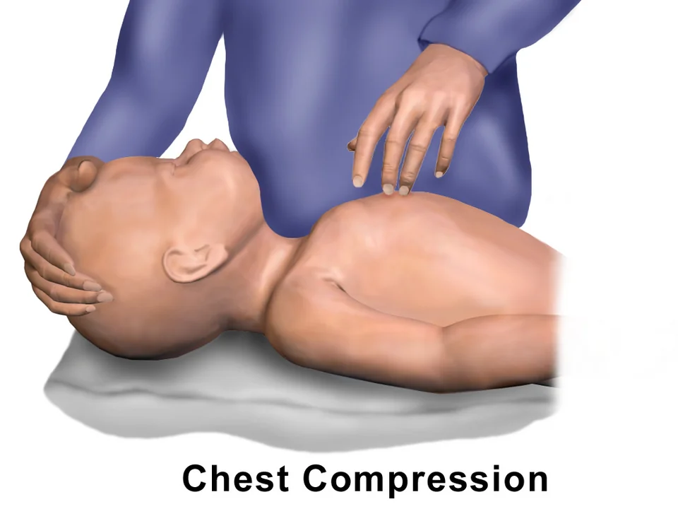
| Hand position | Two fingers on the lower half of the sternum (single rescuer), or the two-thumb encircling technique (two rescuers, preferred) | Heel of one hand, or two hands in a larger child, over the lower half of the sternum |
| Depth | At least one-third of the AP chest diameter, about 4 cm | At least one-third of the AP chest diameter, about 5 cm |
| Rate | 100-120 per minute | 100-120 per minute |
| Ratio | 15:2 (trained), 30:2 (lone lay rescuer) | 15:2 (trained), 30:2 (lone lay rescuer) |
| Airway position | Neutral | Sniffing position |
Paediatric advanced life support
Once a team and equipment are available, continue high-quality CPR with high-flow oxygen while the monitor is attached, then follow the rhythm-based pathway.
Non-shockable rhythms - asystole and pulseless electrical activity
These account for the great majority of paediatric arrests.
- Continue CPR at 15:2 with high-flow oxygen
- Obtain vascular access and give adrenaline 10 micrograms/kg (0.1 mL/kg of 1 in 10,000) IV or IO as soon as possible4
- Repeat adrenaline every 3-5 minutes, which in practice means every other 2-minute cycle
- Reassess the rhythm every 2 minutes, pausing compressions for the shortest possible time
- Identify and treat reversible causes throughout
Shockable rhythms - ventricular fibrillation and pulseless ventricular tachycardia
- Defibrillate at 4 J/kg and immediately resume CPR for 2 minutes without reassessing the rhythm or pulse
- Repeat shocks at 4 J/kg every 2 minutes
- After the third shock, give adrenaline 10 micrograms/kg and amiodarone 5 mg/kg5
- After the fifth shock, give a second dose of amiodarone 5 mg/kg
- Continue adrenaline every 3-5 minutes thereafter
- Use paediatric pads or an attenuator for children under 8 years or under 25 kg where available; adult pads are acceptable if that is all there is
Airway and ventilation
- Bag-mask ventilation is often the safest option and is entirely acceptable throughout an arrest; a poorly placed tracheal tube is far more dangerous than a well-performed bag-mask technique
- Advanced airways - supraglottic or tracheal - should be placed only by an experienced operator, with minimal interruption to compressions
- Once an advanced airway is in place, give continuous compressions with ventilations delivered asynchronously - around 25 per minute in an infant, 20 in a child of 1-8 years, 15 in a child of 8-12 years and 10 above 12 years
- Waveform capnography confirms tube placement, gives feedback on compression quality, and a sudden rise in end-tidal carbon dioxide indicates return of spontaneous circulation
- Avoid hyperventilation, which raises intrathoracic pressure and reduces venous return and coronary perfusion
Vascular access
Circulatory access is often the rate-limiting step in a small, shut-down child. Do not persist with repeated cannulation attempts.
- Peripheral intravenous access if it can be obtained rapidly
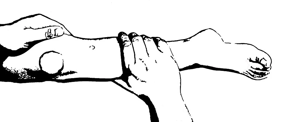
- Intraosseous access if intravenous access is not achieved within about 1 minute, or immediately in cardiac arrest. Any drug or fluid that can be given intravenously can be given intraosseously, at the same dose.
- Flush every drug generously, since drugs otherwise sit in the marrow cavity
- Take blood from the intraosseous needle at insertion for glucose, gas, electrolytes and cross-match
- Fluids in shock are given as 10 mL/kg boluses of a balanced crystalloid or 0.9% sodium chloride, reassessing after each and escalating early if more than 40-60 mL/kg is needed6
Post-resuscitation care
- Targeted temperature management - actively avoid fever, maintaining normothermia at around 36-37.5°C
- Avoid hyperoxia - titrate oxygen to saturations of 94-98% once return of spontaneous circulation is achieved
- Maintain normocapnia, guided by blood gas rather than by end-tidal values alone
- Maintain normoglycaemia and treat seizures promptly
- Support the circulation with fluids and inotropes as needed
- Transfer to paediatric intensive care via the regional retrieval service, with reassessment before and during transfer
- Debrief the team and support the family, who should be offered the option of being present during resuscitation with a dedicated member of staff to support them
Choking
Foreign body airway obstruction should be suspected where onset is sudden, in a previously well child, often while eating or playing with small objects, with no preceding illness.
| Situation | Action |
|---|---|
| Effective cough | Encourage coughing and observe continuously. Do not intervene - a coughing child is moving air better than you can. |
| Ineffective cough, conscious infant (under 1 year) | 5 back blows with the infant prone and head down along your forearm, then 5 chest thrusts - sternal, like compressions but sharper and slower. Reassess between each set. |
| Ineffective cough, conscious child (over 1 year) | 5 back blows, then 5 abdominal thrusts (Heimlich manoeuvre). Reassess between each set. |
| Unconscious | Open the airway, look in the mouth and remove only a clearly visible object - no blind finger sweeps - give 5 rescue breaths, then start CPR. Recheck the mouth for a displaced object each time before ventilating. |
Doses, weights and related emergencies
Estimating weight
Almost every paediatric emergency drug is weight-based, so an accurate weight is a resuscitation intervention in itself. Use the child's actual recent weight if it is known. Otherwise a length-based resuscitation tape is more accurate than any formula. Where neither is available, the APLS age-based formulae give a working estimate.2
| Age | Estimated weight (kg) |
|---|---|
| 1-12 months | (0.5 x age in months) + 4 |
| 1-5 years | (2 x age in years) + 8 |
| 6-12 years | (3 x age in years) + 7 |
Key emergency doses
| Drug or intervention | Dose |
|---|---|
| Adrenaline in cardiac arrest | 10 micrograms/kg IV or IO - 0.1 mL/kg of 1 in 10,000 - every 3-5 minutes |
| Defibrillation | 4 J/kg |
| Amiodarone | 5 mg/kg after the third shock, repeated after the fifth |
| Fluid bolus | 10 mL/kg balanced crystalloid or 0.9% sodium chloride, reassessing after each |
| Glucose for hypoglycaemia | 2 mL/kg of 10% glucose IV or IO |
| Adrenaline in anaphylaxis (intramuscular, 1 in 1,000) | Under 6 months 100-150 micrograms; 6 months to 6 years 150 micrograms; 6-12 years 300 micrograms; over 12 years 500 micrograms3 |
| Benzodiazepine for a seizure over 5 minutes | Buccal midazolam or rectal diazepam, repeated once after 10 minutes |
| Tracheal tube size (uncuffed, over 1 year) | Internal diameter (mm) = (age/4) + 4 |
Outcomes
Survival to hospital discharge after out-of-hospital paediatric cardiac arrest is in the region of 10%, and after in-hospital arrest around 40-45%. The gap between the two is largely explained by how long the child was hypoxic before effective resuscitation began.
Prognosis is better with a witnessed arrest, prompt bystander CPR, a short time to first intervention, a shockable rhythm, and a reversible cause such as hypothermia, poisoning or a foreign body. It is worse with prolonged downtime, asystole as the presenting rhythm, and an underlying chronic condition.
Because outcomes after arrest are poor, the most valuable thing a clinician does is upstream of the algorithm entirely: recognising the child with respiratory distress, dehydration or sepsis, escalating early, and treating them before the heart stops.7 A confident ABCDE assessment, an early senior call and a timely fluid bolus prevent more paediatric arrests than any refinement of CPR technique.
Finally, these algorithms are updated periodically. The 2021 Resuscitation Council UK guidelines lowered the paediatric fluid bolus from 20 to 10 mL/kg and revised the ventilation rates after advanced airway placement - so check the current guidance rather than relying on an older course certificate.1
References
- Resuscitation Council UK. 2021 Resuscitation Guidelines, including paediatric basic and advanced life support and newborn life support. Available here
- Advanced Life Support Group. Advanced Paediatric Life Support: A Practical Approach to Emergencies. Available here
- Resuscitation Council UK. Emergency treatment of anaphylaxis: guidelines. Available here
- BNF for Children. Adrenaline. Available here
- BNF for Children. Amiodarone hydrochloride. Available here
- NICE NG29. Intravenous fluid therapy in children and young people in hospital. 2015, updated 2020. Available here
- NICE NG51. Sepsis: recognition, diagnosis and early management. 2016, updated 2024. Available here
This article is written for revision and education. It is not clinical guidance and must not be used to make decisions about the care of a patient. Always check current NICE guidance and local protocols.