
Central Retinal Vein Occlusion
Key points
- Definition: thrombotic occlusion of the central retinal vein at or behind the lamina cribrosa, causing venous congestion, haemorrhage and macular oedema.
- Presentation: sudden or subacute painless unilateral visual loss over hours to days - less abrupt and usually less profound than arterial occlusion.
- The fundus: the blood and thunder appearance: haemorrhages in all four quadrants, dilated tortuous veins, cotton wool spots and a swollen optic disc.
- Two subtypes: non-ischaemic (about 75%, better acuity, no RAPD) and ischaemic (worse acuity, RAPD, extensive capillary non-perfusion, high risk of neovascularisation).
- The main cause of visual loss: macular oedema, treated with intravitreal anti-VEGF as first-line therapy.
- The feared complication: neovascular glaucoma, classically at around 90-100 days - the so-called 100-day glaucoma - which is why ischaemic CRVO is monitored monthly.
- Risk factors: age, hypertension, hyperlipidaemia, diabetes, smoking and glaucoma. In patients under 50, look for thrombophilia and hyperviscosity.
- Systemic workup: blood pressure, glucose, lipids, FBC and ESR in everyone; a thrombophilia and myeloma screen in the young or those with bilateral disease.
Introduction
Retinal vein occlusion is the second commonest retinal vascular disorder after diabetic retinopathy, affecting around 1 in 200 people over 40 worldwide. It divides into central retinal vein occlusion, where the trunk vein is occluded at the optic nerve head, and branch retinal vein occlusion, where a tributary vein is occluded at an arteriovenous crossing.1
Compared with arterial occlusion, it is more common, less abrupt, and considerably less bleak. Vision falls over hours to days rather than seconds, the loss is often partial, and there are effective treatments for the principal cause of visual impairment. The introduction of anti-VEGF therapy in the last fifteen years changed the outlook substantially.
It also functions as a systemic marker. A retinal vein occlusion in an adult under 50, or affecting both eyes, or recurring, should prompt a search for hyperviscosity, thrombophilia or an inflammatory cause. In an older adult it is usually a manifestation of exactly the same arteriosclerotic process that causes stroke and ischaemic heart disease, and is an opportunity to address cardiovascular risk.
Pathophysiology
The central retinal artery and central retinal vein share a common adventitial sheath as they pass through the lamina cribrosa, the sieve-like plate of collagen at the optic nerve head. A thickened, arteriosclerotic artery compresses the adjacent vein within this confined space, causing turbulence, endothelial damage and thrombosis. Virchow's triad applies precisely: stasis from compression, endothelial injury from mechanical stress, and hypercoagulability where a thrombophilia is present.
Once the vein is occluded, venous pressure rises throughout the retinal circulation. Capillaries and venules become congested and leak, producing widespread intraretinal haemorrhage and oedema. Perfusion pressure falls because the arteriovenous pressure gradient narrows, so capillaries close and areas of the retina become ischaemic. Hypoxic retina releases VEGF, which drives two consequences: increased vascular permeability causing macular oedema, and neovascularisation at the retina, disc and, most damagingly, the iris and drainage angle.
Classification
The single most useful distinction is between ischaemic and non-ischaemic occlusion, because it determines the risk of neovascular glaucoma and therefore the intensity of follow-up. Around 75% are non-ischaemic at presentation, but roughly a third of these convert to the ischaemic form within three years.
| Feature | Non-ischaemic (venous stasis retinopathy) | Ischaemic (haemorrhagic retinopathy) |
|---|---|---|
| Proportion | About 75% | About 25% |
| Visual acuity | Usually better than 6/60 | Usually 6/60 or worse, often counting fingers |
| Relative afferent pupillary defect | Absent or mild | Present and marked |
| Haemorrhages | Mild to moderate, mainly posterior | Extensive, dense, in all four quadrants and out to the periphery |
| Cotton wool spots | Few or none | Numerous |
| Capillary non-perfusion on angiography | Less than 10 disc areas | 10 disc areas or more |
| Electroretinogram | Normal or near-normal b-wave | Reduced b-wave amplitude and b:a ratio |
| Risk of neovascular glaucoma | Low (under 5%) | High - around 45-60% |
| Follow-up | 3-monthly initially | Monthly for the first 6 months, with gonioscopy at each visit |
Branch retinal vein occlusion
Occlusion of a tributary vein at an arteriovenous crossing, most often the superotemporal branch. The haemorrhages are confined to a wedge-shaped sector corresponding to the drainage territory and respect the horizontal raphe, which is diagnostically distinctive. Visual loss occurs only if the macula is involved. It is more common than central occlusion, carries a better prognosis, and its principal complications are macular oedema and neovascularisation confined to the affected sector.
Risk factors
In adults over 50
- Increasing age - over 90% of cases occur in people over 50
- Hypertension - present in around two thirds, and the single strongest modifiable factor
- Hyperlipidaemia - present in around a third
- Diabetes mellitus
- Smoking
- Glaucoma and raised intraocular pressure - an independent risk factor through compression at the lamina cribrosa. Check the intraocular pressure in both eyes in every patient with a vein occlusion.
- Obesity and obstructive sleep apnoea
- Chronic kidney disease
In patients under 50, or with bilateral or recurrent disease
- Thrombophilia - antiphospholipid syndrome, factor V Leiden, protein C or S deficiency, antithrombin deficiency, prothrombin gene mutation
- Hyperviscosity - myeloma, Waldenström macroglobulinaemia, polycythaemia vera, essential thrombocythaemia and leukaemia
- Hyperhomocysteinaemia
- Inflammatory disease - Behcet disease, sarcoidosis, systemic lupus erythematosus and retinal vasculitis
- Infection - syphilis, tuberculosis and HIV
- Combined oral contraceptive pill, pregnancy and diuretics causing dehydration
- Orbital disease - anything raising orbital or venous pressure, including thyroid eye disease and an orbital mass
Clinical features
Symptoms
- Painless unilateral visual loss developing over hours to days, sometimes noticed on waking
- Blurred or distorted central vision from macular oedema, often with metamorphopsia
- Variable severity - anything from a mild blur to counting fingers, with the ischaemic form at the worse end
- Floaters if there has been a vitreous haemorrhage from new vessels
- Pain and redness only late, if neovascular glaucoma develops
- No preceding transient obscurations, in contrast to arterial disease
Signs
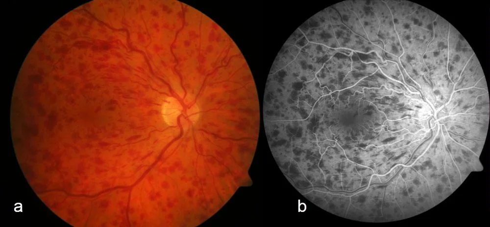
- Flame and dot-blot haemorrhages in all four quadrants - the defining feature, giving the blood and thunder fundus. In branch occlusion they are confined to one sector.
- Dilated and tortuous retinal veins, engorged and of irregular calibre
- Optic disc swelling and hyperaemia, with blurred margins
- Cotton wool spots - a marker of ischaemia, and their number correlates with the ischaemic subtype
- Macular oedema, giving a thickened, sometimes cystoid macula on examination and clearly on OCT
- Relative afferent pupillary defect - the most useful bedside discriminator of ischaemic disease
- Hard exudates in longer-standing disease
- Later - optociliary shunt vessels on the disc, neovascularisation of the disc or retina, and rubeosis iridis with new vessels visible at the pupil margin and in the angle on gonioscopy
Investigations
Ocular
- Visual acuity and pupil examination for a relative afferent pupillary defect
- Intraocular pressure in both eyes, and gonioscopy to inspect the drainage angle for new vessels
- Dilated fundus examination to define the extent of haemorrhage, cotton wool spots and neovascularisation
- Optical coherence tomography - quantifies macular oedema, guides the decision to treat, and tracks response to each injection. This is the central investigation in modern management.
- Fluorescein angiography - defines the area of capillary non-perfusion and formally classifies the occlusion as ischaemic or non-ischaemic. Often deferred until haemorrhage clears, since blood blocks fluorescence.
- Electroretinography - occasionally used to assess ischaemia where haemorrhage prevents angiography
Systemic
- Blood pressure - the highest-yield test, and abnormal in a majority
- FBC - for polycythaemia, thrombocythaemia and leukaemia
- ESR and plasma viscosity - for hyperviscosity and inflammatory disease; also to exclude giant cell arteritis where the presentation is atypical
- Fasting glucose or HbA1c, and a lipid profile
- U&Es and renal function
- In patients under 50, or with bilateral or recurrent disease - thrombophilia screen including antiphospholipid antibodies, serum protein electrophoresis and free light chains for myeloma, homocysteine, autoimmune screen, and syphilis serology
- Consider ECG and carotid assessment where there are coexisting arterial features, since the risk factors overlap with stroke
Management
There is no treatment that reopens the occluded vein. Management therefore has three aims: treat the macular oedema, detect and treat neovascularisation before it causes glaucoma, and reduce cardiovascular risk to protect the fellow eye and the patient.
Referral
- Refer to ophthalmology urgently, to be seen within one to two weeks, or sooner if acuity is poor or there is any suspicion of neovascularisation
- Same-day referral if there is a painful red eye with a high pressure, suggesting neovascular glaucoma has already developed
- Start systemic risk factor management in parallel rather than waiting for the eye clinic
Macular oedema
- Intravitreal anti-VEGF - ranibizumab, aflibercept or faricimab, given as a loading course of monthly injections then according to OCT response. This is first-line and NICE-recommended, and the CRUISE, COPERNICUS and GALILEO trials all demonstrated substantial acuity gains over sham.2,3
- Intravitreal dexamethasone implant (Ozurdex) - an alternative, particularly in pseudophakic eyes, in patients unable to attend monthly, or where anti-VEGF has failed. Causes cataract and raised intraocular pressure, so it is avoided in eyes with glaucoma.4
- Macular grid laser - has a role in branch occlusion but is not effective in central occlusion, where the Central Vein Occlusion Study showed it reduced angiographic oedema without improving acuity
- Observation may be appropriate where acuity is good and oedema is mild, since some non-ischaemic occlusions resolve spontaneously
Neovascularisation
Systemic management
- Blood pressure control to target, which is the intervention most likely to protect the fellow eye
- Statin therapy and formal cardiovascular risk assessment
- Glycaemic control and smoking cessation
- Treat coexisting glaucoma and check the intraocular pressure in the fellow eye at every visit
- Antiplatelet or anticoagulant therapy is not indicated for the vein occlusion itself - there is no evidence it improves the ocular outcome, and it may worsen retinal haemorrhage. It is prescribed only if there is a separate systemic indication.
- Haematology referral where a thrombophilia, hyperviscosity syndrome or paraproteinaemia is identified
- Review the combined oral contraceptive pill and consider an alternative
Complications and prognosis
- Macular oedema - the commonest cause of visual impairment, and the main treatment target
- Macular ischaemia - capillary dropout at the fovea, for which there is no treatment; the reason some eyes do not improve despite a dry macula on OCT
- Neovascular glaucoma - the most serious complication, often ending in a blind painful eye
- Retinal or disc neovascularisation with vitreous haemorrhage
- Tractional retinal detachment from fibrovascular proliferation
- Epiretinal membrane and chronic cystoid macular oedema
- Optociliary shunt vessels on the disc - collaterals to the choroidal circulation, which are a sign of chronic occlusion rather than a complication
- Occlusion in the fellow eye - around a 1% risk per year, since the systemic risk factors are shared
The prognosis depends principally on the subtype and on the presenting acuity. Non-ischaemic occlusion has a reasonable outlook: around half of eyes retain 6/12 or better, and some resolve substantially over months. Ischaemic occlusion is much worse, with the great majority ending at 6/60 or below and a high risk of neovascular complications.
Anti-VEGF therapy has changed the picture materially, with mean acuity gains of two to three lines in trials and a substantial proportion of treated eyes reaching 6/12.2 The corresponding burden is that treatment is prolonged, often for years, and that eyes which stop treatment frequently relapse. Discussing the likely duration honestly at the outset improves adherence and avoids the sense that the treatment has failed when a further injection is required.
Finally, it is worth returning to the systemic dimension. Retinal vein occlusion is associated with an increased risk of stroke and cardiovascular events over subsequent years, reflecting the shared arteriosclerotic substrate.5 A patient who leaves clinic with well-controlled macular oedema but an untreated blood pressure of 175/100 mmHg has had half of the necessary consultation.
References
- Royal College of Ophthalmologists. Retinal Vein Occlusion (RVO) Guidelines. Available here
- Brown DM, Campochiaro PA, Singh RP et al. Ranibizumab for macular edema following central retinal vein occlusion (CRUISE). Ophthalmology. 2010. Available here
- NICE TA283. Aflibercept for treating macular oedema secondary to central retinal vein occlusion. 2013. Available here
- NICE TA229. Dexamethasone intravitreal implant for the treatment of macular oedema secondary to retinal vein occlusion. 2011. Available here
- Wu CY, Riangwiwat T, Rattanawong P et al. Association of retinal vein occlusion with cardiovascular events and mortality: a systematic review and meta-analysis. Retina. 2019. Available here
This article is written for revision and education. It is not clinical guidance and must not be used to make decisions about the care of a patient. Always check current NICE guidance and local protocols.