
Deep Vein Thrombosis
Key points
- Deep vein thrombosis (DVT): thrombus formation within a deep vein, most often in the calf or proximal veins of the leg. With pulmonary embolism it forms the spectrum of venous thromboembolism (VTE).
- Virchow's triad: stasis, endothelial injury and hypercoagulability - the framework underlying every risk factor, from long-haul flights and surgery to malignancy and pregnancy.
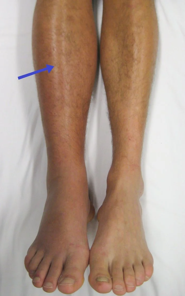
- Clinical features: unilateral limb pain, swelling, warmth, erythema and dilated superficial veins. Clinical assessment alone is unreliable, which is why a validated score plus objective testing is mandatory.
- Diagnostic pathway: the two-level Wells score directs management: 2 or more points ("likely") goes to proximal leg vein ultrasound; 1 point or less ("unlikely") goes to a D-dimer first.
- D-dimer's role: it is sensitive but not specific - useful to rule out DVT in a low-probability patient, but raised in infection, malignancy, pregnancy, trauma and old age, so a positive result never confirms the diagnosis.
- First-line treatment: a DOAC (apixaban or rivaroxaban) is now first line for most patients, started without waiting for imaging if a scan is delayed beyond 4 hours.
- Duration: at least 3 months; provoked DVT with a resolved transient risk factor usually stops there, while unprovoked DVT, active cancer or recurrence warrants consideration of long-term anticoagulation.
- Key complications: pulmonary embolism (the immediate danger) and post-thrombotic syndrome (the commonest long-term one, affecting up to a third of patients).
Introduction
Deep vein thrombosis is the formation of thrombus within the deep venous system, most commonly in the lower limb. Together with pulmonary embolism - which occurs when that thrombus embolises to the pulmonary arteries - it constitutes venous thromboembolism, one of the leading causes of preventable death in hospitalised patients.1
The clinical importance of DVT is twofold: the immediate risk that it will propagate and embolise to the lungs, and the long-term morbidity of post-thrombotic syndrome. Both are substantially reduced by prompt diagnosis and adequate anticoagulation.
Risk factors
Risk factors map onto the three components of Virchow's triad, and identifying them matters clinically because it determines whether a DVT is classed as provoked or unprovoked - which in turn drives the duration of anticoagulation.
| Component | Risk factors |
|---|---|
| Stasis | Immobility, hospital admission, long-haul travel, surgery (especially orthopaedic), lower limb fracture or plaster cast, stroke with paralysis, obesity, pregnancy (uterine compression of pelvic veins) |
| Endothelial injury | Surgery, trauma, central venous catheters, previous DVT, intravenous drug use, vasculitis |
| Hypercoagulability | Malignancy, pregnancy and the puerperium, oestrogen-containing contraception and HRT, inherited thrombophilia (factor V Leiden, prothrombin G20210A, protein C/S and antithrombin deficiency), antiphospholipid syndrome, nephrotic syndrome, myeloproliferative neoplasms, sepsis, dehydration, smoking, increasing age |
Clinical features
- Unilateral limb swelling - the most important sign; measure calf circumference 10 cm below the tibial tuberosity and compare sides, since a difference of more than 3 cm scores in the Wells criteria
- Pain and tenderness, typically along the deep venous distribution in the calf or thigh
- Warmth and erythema of the affected limb
- Dilated superficial (collateral) veins, non-varicose
- Pitting oedema, usually confined to the symptomatic leg
- Phlegmasia cerulea dolens - a rare, limb-threatening presentation with massive iliofemoral thrombosis causing a painful, swollen, cyanotic limb and compromised arterial inflow; a vascular emergency
Homans' sign (calf pain on passive dorsiflexion) is traditionally described but is neither sensitive nor specific, is uncomfortable for the patient, and is no longer recommended as a diagnostic manoeuvre. Clinical assessment alone is unreliable in either direction - which is precisely why the diagnostic pathway is built on scoring plus objective testing rather than bedside impression.
Always ask about and examine for symptoms of pulmonary embolism - pleuritic chest pain, breathlessness, haemoptysis, tachycardia - since a substantial proportion of patients with DVT have concurrent, sometimes silent, PE.
Diagnosis
The two-level DVT Wells score
The two-level Wells score stratifies patients before any test is sent. Each feature below scores +1 point, except the final item which scores -2:2
- Active cancer (treatment ongoing, within 6 months, or palliative)
- Paralysis, paresis, or recent plaster immobilisation of the legs
- Recently bedridden for 3 days or more, or major surgery within 12 weeks
- Localised tenderness along the distribution of the deep venous system
- Entire leg swollen
- Calf swelling more than 3 cm larger than the asymptomatic side
- Pitting oedema confined to the symptomatic leg
- Collateral superficial (non-varicose) veins
- Previously documented DVT
- An alternative diagnosis is at least as likely as DVT (-2 points)
| Score | Interpretation | Next step |
|---|---|---|
| 2 or more | DVT likely | Proximal leg vein ultrasound within 4 hours. If unavailable in that time, give interim anticoagulation and perform a D-dimer, with the scan within 24 hours |
| 1 or less | DVT unlikely | D-dimer (with result within 4 hours, otherwise interim anticoagulation). If positive, proceed to proximal leg vein ultrasound. If negative, DVT is excluded - consider alternative diagnoses |
Investigations
- Proximal leg vein ultrasound with Doppler - the definitive first-line imaging test. A negative scan in a Wells-likely patient with a positive D-dimer should be repeated at 6-8 days, to catch calf thrombus that has since propagated proximally
- D-dimer - a fibrin degradation product. Highly sensitive but poorly specific: it is raised in infection, inflammation, malignancy, pregnancy, recent surgery, trauma and simply with increasing age. Its value is in ruling out DVT in a low-probability patient, never in confirming it
- Baseline bloods - FBC, U&Es (renal function determines DOAC choice and dosing), LFTs and coagulation screen before starting anticoagulation
- CT pulmonary angiography if pulmonary embolism is suspected clinically
- Consider investigation for underlying malignancy in unprovoked DVT - a thorough history, examination, baseline bloods and review of age-appropriate cancer screening, rather than indiscriminate whole-body imaging
- Thrombophilia testing is not routine; consider only in unprovoked VTE with a first-degree relative affected, and remember results are unreliable during acute thrombosis or while anticoagulated
Differential diagnosis
- Cellulitis - erythema, warmth and pain, but usually with fever, a clear margin and a portal of entry; the two can coexist and are frequently confused
- Ruptured Baker's cyst - sudden calf pain and swelling, often with a history of knee arthropathy; ultrasound distinguishes it
- Calf muscle tear or haematoma - preceding trauma or sudden exertion
- Superficial thrombophlebitis - tender, palpable, erythematous cord along a superficial vein; requires ultrasound to exclude extension into the deep system
- Chronic venous insufficiency or post-thrombotic syndrome - usually bilateral or with chronic skin changes
- Lymphoedema - non-pitting, chronic, often bilateral
- Heart failure, nephrotic syndrome, liver disease - typically bilateral oedema
- Compartment syndrome and acute limb ischaemia - both surgical emergencies with distinct features
Management
Anticoagulation
- A DOAC (apixaban or rivaroxaban) is first line for most patients, offering fixed dosing without routine monitoring. Both use an initial higher-dose loading period (apixaban 10 mg twice daily for 7 days, then 5 mg twice daily; rivaroxaban 15 mg twice daily for 21 days, then 20 mg once daily) before the maintenance dose2
- If a DOAC is unsuitable, use LMWH for at least 5 days followed by dabigatran or edoxaban, or LMWH overlapped with a vitamin K antagonist (warfarin) for at least 5 days and until the INR is 2 or above on two consecutive readings, target INR 2.5
- Start interim anticoagulation immediately if diagnostic testing (scan or D-dimer result) will be delayed beyond 4 hours and there is no contraindication
- Severe renal impairment: DOACs are contraindicated or require dose adjustment - use LMWH or a vitamin K antagonist per local policy
- Antiphospholipid syndrome: use a vitamin K antagonist, not a DOAC, particularly in triple-positive disease
- Pregnancy: LMWH throughout - DOACs and warfarin are contraindicated (warfarin is teratogenic)
- Active cancer: a DOAC or LMWH, guided by bleeding risk and tumour site; caution with DOACs in luminal gastrointestinal or genitourinary cancers given higher bleeding rates
Duration of treatment
| Scenario | Duration |
|---|---|
| Provoked DVT with a resolved transient risk factor (e.g. surgery) | 3 months (3-6 months in active cancer per current practice) |
| Unprovoked DVT | At least 3 months, then assess for long-term anticoagulation, weighing recurrence risk against bleeding risk |
| Active cancer | 3-6 months minimum, then review; continue while cancer is active |
| Recurrent VTE, or antiphospholipid syndrome | Long-term/indefinite anticoagulation |
| Isolated distal (calf) DVT | Anticoagulate for 6 weeks to 3 months, or in selected low-risk patients monitor with serial ultrasound instead, by local policy |
Other measures
- Early mobilisation and adequate analgesia - bed rest is not indicated and does not reduce embolisation
- Graduated compression stockings are no longer routinely recommended for preventing post-thrombotic syndrome, though they may be used for symptomatic relief of persistent swelling
- Inferior vena cava filter - reserved for patients with a proximal DVT in whom anticoagulation is genuinely contraindicated, with a plan for removal once anticoagulation becomes possible
- Catheter-directed thrombolysis - considered in selected patients with symptomatic iliofemoral DVT of recent onset, good functional status and low bleeding risk
- Address modifiable risk factors - review oestrogen-containing contraception/HRT, encourage smoking cessation and weight management, and advise on travel prophylaxis
Complications
- Pulmonary embolism - the most serious acute complication and a leading cause of preventable hospital death
- Post-thrombotic syndrome - the commonest long-term complication, affecting up to a third of patients within 5 years. Valvular damage and venous hypertension cause chronic limb pain, swelling, hyperpigmentation, and in severe cases venous ulceration
- Recurrent VTE - substantially more likely after an unprovoked event, particularly in the first 2 years after stopping anticoagulation
- Chronic thromboembolic pulmonary hypertension - an uncommon but serious late consequence of recurrent or unresolved PE
- Bleeding from anticoagulation - the principal treatment-related harm, and the counterweight in every decision about treatment duration
- Phlegmasia cerulea dolens and venous gangrene - rare, limb-threatening
Red flags
Prognosis
With prompt diagnosis and adequate anticoagulation, the outlook after a first DVT is generally good, and the immediate risk of fatal pulmonary embolism falls sharply once treatment begins.1 The two dominant longer-term issues are recurrence and post-thrombotic syndrome.
Recurrence risk depends heavily on the provoked/unprovoked distinction: after a provoked DVT with a resolved transient trigger, recurrence after stopping treatment is relatively low, whereas after an unprovoked event it is substantially higher - which is what drives consideration of indefinite anticoagulation, balanced against cumulative bleeding risk. Post-thrombotic syndrome develops in up to a third of patients and is the main cause of long-term disability, making adequate initial anticoagulation and follow-up important beyond simply preventing embolism.
References
- Kearon C, Akl EA, Ornelas J et al. Antithrombotic Therapy for VTE Disease: CHEST Guideline. Chest. 2016. Available here
- NICE. Venous thromboembolic diseases: diagnosis, management and thrombophilia testing (NG158). 2020 (updated 2023). Available here
- Wells PS, Anderson DR, Rodger M et al. Evaluation of D-dimer in the diagnosis of suspected deep-vein thrombosis. N Engl J Med. 2003. Available here
- James Heilman, MD, CC BY-SA 3.0, via Wikimedia Commons. Available here
- NICE Clinical Knowledge Summaries (CKS). Deep vein thrombosis. 2023. Available here
This article is written for revision and education. It is not clinical guidance and must not be used to make decisions about the care of a patient. Always check current NICE guidance and local protocols.