
Endometriosis
Key points
- Endometriosis: the presence of endometrial-like tissue outside the uterus, most often on the pelvic peritoneum, ovaries (endometrioma) and uterosacral ligaments.
- Prevalence: affects roughly 1 in 10 women of reproductive age; average diagnostic delay is around 8 years from symptom onset.
- Presentation: cyclical pelvic pain, dysmenorrhoea, deep dyspareunia, dyschezia and subfertility; severity of symptoms correlates poorly with disease extent.
- Diagnosis: laparoscopy with biopsy is the gold standard; pelvic examination and transvaginal ultrasound support the diagnosis but cannot exclude it if normal.
- Medical management: NSAIDs and hormonal suppression (COCP, progestogens, GnRH analogues) as first-line, before laparoscopic diagnosis is even required in many cases.
- Surgical management: laparoscopic excision or ablation of deposits, with ovarian cystectomy for endometriomas.
- Endometrioma: a chocolate cyst - endometriosis within the ovary forming a cyst filled with old, altered blood.
- Fertility: endometriosis is a recognised cause of subfertility; referral for fertility investigation runs in parallel with symptom treatment.
Introduction
Endometriosis is a chronic, oestrogen-dependent condition in which tissue histologically similar to the endometrium is found outside the uterine cavity, most commonly on the pelvic peritoneum, ovaries, and uterosacral ligaments, and less commonly in the rectovaginal septum, bowel, bladder or, rarely, at distant sites such as the diaphragm or lungs.1
It affects approximately one in ten women of reproductive age, making it one of the most common gynaecological conditions, yet the average time from symptom onset to diagnosis in the UK is around eight years.2 This delay reflects the normalisation of period pain in wider culture, the non-specific nature of early symptoms, and the fact that examination and imaging can both be entirely normal in significant disease.
The condition behaves like ectopic endometrial tissue in miniature: deposits respond to the hormonal cycle, bleeding and shedding each month, which provokes local inflammation, fibrosis and adhesion formation - this is why the pain is classically cyclical and why chronic disease can distort pelvic anatomy over time.
Aetiology and pathophysiology
The exact cause is not fully understood, and several theories are proposed, none of which fully explains all cases.1
- Retrograde menstruation: menstrual blood containing endometrial cells flows backward through the fallopian tubes into the pelvis, where the cells implant and grow - the leading theory, though retrograde menstruation is common and most women do not develop endometriosis, implying additional immunological or genetic susceptibility
- Coelomic metaplasia: peritoneal cells undergo metaplastic transformation into endometrial-like tissue
- Lymphatic or vascular spread: explains rare distant deposits (e.g. lung, diaphragm)
- Genetic and immune factors: endometriosis clusters in families, and impaired immune clearance of ectopic endometrial cells is thought to allow deposits to establish
Ectopic deposits contain oestrogen and progesterone receptors and respond to the menstrual cycle in the same way as the endometrium, bleeding at the time of menstruation. Because this blood has no way to leave the body, it provokes a chronic inflammatory response, leading to fibrosis, adhesion formation and, over time, the classic 'chocolate cyst' when deposits form within the ovary.
Risk factors
- Early menarche
- Late menopause
- Nulliparity
- Short menstrual cycles or prolonged heavy periods
- Family history of endometriosis (first-degree relative)
- Delayed childbearing
- Mullerian anomalies obstructing outflow (increase retrograde flow)
Clinical features
The hallmark is cyclical pelvic pain, but presentation is highly variable and severity of symptoms correlates poorly with the extent of disease seen at laparoscopy - a woman with minimal peritoneal deposits can have debilitating pain, while extensive disease is occasionally found incidentally in an asymptomatic woman.3
- Chronic pelvic pain: classically cyclical, worse in the days before and during menstruation, but can become constant in longstanding disease
- Secondary dysmenorrhoea: painful periods that were not painful before, or that have become progressively worse
- Deep dyspareunia: pain with deep penetration, particularly with uterosacral ligament or pouch of Douglas involvement
- Dyschezia: pain on defecation, particularly cyclical, suggesting rectovaginal or bowel involvement
- Subfertility: endometriosis is found in around 30-50% of women investigated for infertility
- Cyclical symptoms at other sites: haematuria or dysuria (bladder involvement), or rarely cyclical haemoptysis (thoracic endometriosis)
- Chronic fatigue, which is under-recognised but common
Examination findings can be entirely normal. When present, look for tenderness or nodularity in the posterior fornix on bimanual examination, a fixed, retroverted uterus (suggesting adhesions), reduced organ mobility, and visible endometriotic lesions in the vagina or cervix on speculum examination (uncommon).
Investigations
First-line
Given diagnostic delay is a recognised problem, NICE recommends starting empirical hormonal treatment based on history and examination alone in women with suspected endometriosis, without waiting for definitive diagnosis, unless fertility is a current priority or symptoms fail to respond.2
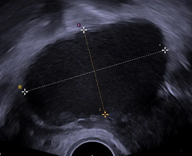
Transvaginal ultrasound
First-line imaging. It is good at identifying ovarian endometriomas but has limited sensitivity for small peritoneal deposits or adhesions, so a normal scan does not exclude the disease.
MRI pelvis
Used when deep infiltrating endometriosis is suspected (e.g. bowel or bladder involvement) or to aid surgical planning before complex excision.
Laparoscopy
The gold-standard diagnostic test, allowing direct visualisation and biopsy of deposits for histological confirmation. It also allows treatment (ablation or excision) at the same procedure. Reserved for cases where the diagnosis remains uncertain after imaging, empirical treatment has failed, or surgical management is already planned, rather than as a first-line test for every woman with suspected disease.1
Differential diagnosis
- Adenomyosis: heavy, painful periods with a bulky uterus; can coexist with endometriosis
- Pelvic inflammatory disease: fever, discharge, cervical excitation, history of STI risk
- Ovarian cyst (non-endometriotic): may present similarly on ultrasound
- Irritable bowel syndrome: overlaps significantly with dyschezia and bloating; often misdiagnosed in place of endometriosis
- Interstitial cystitis: overlapping urinary and pelvic pain symptoms
- Chronic pelvic inflammatory pain syndrome / adhesions from previous surgery
Management
Management is individualised according to symptom severity, disease extent, and whether the woman wishes to conceive, and should involve shared decision-making given the chronic, relapsing nature of the condition.2
Analgesia
NSAIDs and/or paracetamol are used first-line for pain relief, alone or alongside hormonal treatment.
Hormonal management
Hormonal treatments work by suppressing ovulation and inducing endometrial (and ectopic tissue) atrophy - they control symptoms but do not treat underlying disease and are not appropriate if the woman is trying to conceive.
| Option | Notes |
|---|---|
| Combined oral contraceptive pill | Often used continuously (skipping the pill-free interval) to suppress cyclical pain |
| Progestogens (e.g. oral, depot, or LNG-IUS) | Effective and useful where oestrogen is contraindicated |
| GnRH analogues | Induce a temporary medical menopause; effective but limited to short-term use (usually with 'add-back' HRT) due to bone density loss |
Surgical management
Laparoscopic excision or ablation of endometriotic deposits and adhesiolysis can improve pain and fertility outcomes. Ovarian endometriomas are managed with cystectomy where feasible, balancing symptom relief against the risk of reducing ovarian reserve from surgery on the ovary itself. Hysterectomy (with or without oophorectomy) is reserved for severe, refractory disease in women who have completed their family, and does not guarantee resolution of pain if endometriotic deposits remain outside the uterus.
Complications
- Subfertility
- Chronic pelvic pain and the psychological burden of a long diagnostic delay
- Ovarian endometrioma rupture, causing acute pelvic pain
- Adhesions leading to bowel obstruction in severe deep infiltrating disease (rare)
- Reduced ovarian reserve following endometrioma surgery
- A small increased risk of clear cell and endometrioid ovarian carcinoma - the relative risk is raised but the absolute lifetime risk remains low, so this warrants honest explanation rather than alarm, and does not justify routine ovarian cancer screening
Red flags
Prognosis
Endometriosis is a chronic condition that tends to improve after the menopause as oestrogen levels fall, but symptoms often recur after stopping hormonal treatment while a woman remains premenopausal, and recurrence after surgery is common. Long-term management is therefore about controlling symptoms and preserving fertility and quality of life over years rather than achieving a one-off cure, and many women need a combination of medical and surgical approaches over the course of their reproductive life.
References
- NICE NG73. Endometriosis: diagnosis and management. 2017. Available here
- NICE Clinical Knowledge Summaries (CKS). Endometriosis. Available here
- Royal College of Obstetricians and Gynaecologists. Endometriosis - patient information. Available here
This article is written for revision and education. It is not clinical guidance and must not be used to make decisions about the care of a patient. Always check current NICE guidance and local protocols.