
Uterine Fibroids (Leiomyomas)
Key points
- Fibroids (leiomyomas): benign monoclonal tumours of uterine smooth muscle, oestrogen- and progesterone-sensitive, and the most common pelvic tumour in women.
- Prevalence: found in up to 40-50% of women by age 50; more common and more severe in women of Black African and Caribbean ethnicity.
- Classification: submucosal, intramural or subserosal, by position relative to the uterine wall; submucosal fibroids cause the heaviest bleeding.
- Presentation: many are asymptomatic; when symptomatic, heavy menstrual bleeding, pelvic pressure/pain, subfertility and urinary symptoms predominate.
- Diagnosis: pelvic ultrasound is first-line; MRI is used for surgical planning or when malignancy is a concern.
- Medical management: LNG-IUS, tranexamic acid and NSAIDs for bleeding; GnRH analogues shrink fibroids short-term, usually pre-operatively.
- Surgical management: myomectomy for fertility preservation, uterine artery embolisation as a uterus-sparing alternative, and hysterectomy as definitive treatment.
- Pregnancy: fibroids can grow under oestrogen stimulation and are associated with malpresentation, red degeneration, and postpartum haemorrhage.
Introduction
Uterine fibroids (leiomyomas or myomas) are benign tumours arising from the smooth muscle of the myometrium. Each fibroid is monoclonal, arising from a single mutated myometrial cell, which is why a woman can have multiple fibroids of very different sizes growing at different rates within the same uterus.1
Fibroids are extremely common - present in up to 40-50% of women by the age of 50 - though most are small and never cause symptoms.2 They are more common, larger, and more likely to cause severe symptoms in women of Black African and Caribbean ethnicity, and incidence rises with age until the menopause, after which fibroids typically shrink as oestrogen levels fall.
Growth is driven by oestrogen and progesterone, which explains why fibroids can enlarge in pregnancy and on combined hormonal contraception, and regress after the menopause.
Classification
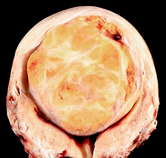
Fibroids are classified by their position relative to the uterine wall, which determines both the symptoms they cause and the treatment options available.1
| Type | Location | Typical impact |
|---|---|---|
| Submucosal | Bulges into the endometrial cavity | Heaviest menstrual bleeding; most likely to impair fertility and implantation |
| Intramural | Confined within the myometrial wall | Most common type; can cause bleeding and bulk symptoms as it enlarges |
| Subserosal | Projects outward from the outer uterine surface | Bulk/pressure symptoms (bladder, bowel); less bleeding as the endometrium is less distorted |
| Pedunculated | Subserosal or submucosal fibroid on a stalk | Can twist (torsion) causing acute pain |
Risk factors
- Black African or Caribbean ethnicity (2-3 times higher incidence)
- Increasing age until the menopause
- Obesity (peripheral aromatisation increases circulating oestrogen)
- Early menarche
- Family history of fibroids
- Nulliparity
Pregnancy and multiparity are associated with a reduced risk, and smoking has also been linked to a lower risk, though it is never advised as a means of prevention given its far greater harms.
Clinical features
Around half of fibroids are asymptomatic and found incidentally on examination or imaging. When symptomatic, the presentation reflects size, number and location.3
- Heavy menstrual bleeding: the most common symptom, especially with submucosal or large intramural fibroids
- Pelvic pain or pressure: dull, dragging discomfort from uterine enlargement
- Bulk symptoms: urinary frequency or retention from bladder compression, constipation from bowel compression, or a palpable abdominal mass in large fibroids
- Subfertility: particularly with submucosal fibroids distorting the endometrial cavity
- Dyspareunia
- Acute pain: if a pedunculated fibroid undergoes torsion, or a fibroid outgrows its blood supply (red degeneration, classically in pregnancy)
Examination may reveal a palpable, firm, irregularly enlarged, non-tender pelvic or abdominal mass arising from the pelvis - a large fibroid uterus is a classic cause of a mass that the examiner cannot get below on abdominal examination.
Differential diagnosis
- Adenomyosis: bulky, tender uterus with painful, heavy periods
- Endometrial or ovarian malignancy: especially in postmenopausal women or with rapid growth
- Ovarian cyst or mass: may be difficult to distinguish clinically from a subserosal fibroid
- Pregnancy: always exclude with a pregnancy test in a woman of reproductive age with a pelvic mass
- Leiomyosarcoma: rare malignant counterpart; suspect with rapid growth, particularly after the menopause
Investigations
Bedside and laboratory
Pregnancy test to exclude pregnancy, and full blood count to assess for iron-deficiency anaemia from chronic heavy bleeding.
Imaging
Pelvic ultrasound (transvaginal, with transabdominal for very large fibroids) is first-line, confirming the diagnosis and mapping number, size and location.2 MRI gives more detailed anatomical information and is used when planning myomectomy or uterine artery embolisation, when ultrasound findings are inconclusive, or when there is concern for leiomyosarcoma (rapid growth, atypical imaging features).
Hysteroscopy
Allows direct visualisation and, where appropriate, same-session resection of submucosal fibroids distorting the endometrial cavity.
Management
Asymptomatic fibroids require no treatment beyond routine follow-up. Management of symptomatic fibroids is guided by symptom severity, fibroid size and location, and the woman's wish to preserve fertility.2
Medical management
| Option | Notes |
|---|---|
| Levonorgestrel intrauterine system (LNG-IUS) | First-line if the uterine cavity is not significantly distorted; not always suitable with large submucosal fibroids |
| Tranexamic acid / NSAIDs | Reduce bleeding during menstruation; do not shrink the fibroid |
| GnRH analogues (e.g. goserelin) | Induce a temporary medical menopause, shrinking fibroids by reducing oestrogen; used short-term (usually ≤6 months) before surgery due to hypoestrogenic side effects (hot flushes, bone density loss) |
| Oral GnRH antagonist with hormonal add-back (e.g. relugolix combined with estradiol and norethisterone) | NICE-approved for moderate to severe fibroid symptoms;4 the add-back oestrogen offsets the hypoestrogenic side effects that limit plain GnRH analogues, allowing longer-term use |
| Ulipristal acetate | Selective progesterone receptor modulator; use has narrowed due to rare but serious hepatotoxicity, and it is reserved for specific circumstances under specialist guidance |
Surgical management
- Hysteroscopic myomectomy/resection: for submucosal fibroids, preserves the uterus and fertility
- Myomectomy (open or laparoscopic): removes fibroids while preserving the uterus; the preferred option for women wishing to conceive, though it carries a risk of uterine scarring that may necessitate caesarean section in future pregnancy
- Uterine artery embolisation (UAE): an interventional radiology technique that occludes the blood supply to the fibroid, causing it to shrink; uterus-sparing but not generally recommended if future fertility is a priority, as it can affect ovarian reserve
- Hysterectomy: definitive treatment, appropriate for women who have completed their family or where fibroids are large, multiple or causing severe symptoms unresponsive to other measures
Fibroids in pregnancy
Fibroids can enlarge during pregnancy under oestrogen stimulation, though many do not change significantly. They are associated with an increased risk of miscarriage, malpresentation, preterm labour, obstructed labour if low-lying, and postpartum haemorrhage due to impaired uterine contraction.1
Complications
- Iron-deficiency anaemia from chronic heavy menstrual bleeding
- Subfertility, particularly with submucosal fibroids
- Torsion of a pedunculated fibroid causing acute pain
- Red degeneration in pregnancy
- Urinary retention or hydronephrosis from a large fibroid compressing the ureters or bladder
- Malignant transformation to leiomyosarcoma is extremely rare and most leiomyosarcomas are thought to arise de novo rather than from a pre-existing fibroid
Red flags
Prognosis
Fibroids are benign and typically regress after the menopause as oestrogen levels decline. Most symptomatic women achieve good control of bleeding and bulk symptoms with medical or uterus-sparing surgical treatment, and fertility outcomes after myomectomy for submucosal fibroids are generally favourable. Recurrence after myomectomy is possible, as new fibroids can develop from other myometrial cells over time, which is an important counselling point for women choosing uterus-sparing procedures over hysterectomy.
References
- NICE NG88. Heavy menstrual bleeding: assessment and management. 2021. Available here
- NICE Clinical Knowledge Summaries (CKS). Fibroids. Available here
- Royal College of Obstetricians and Gynaecologists. Uterine fibroids - patient information. Available here
- NICE TA832. Relugolix-estradiol-norethisterone acetate for treating moderate to severe symptoms of uterine fibroids. 2022. Available here
This article is written for revision and education. It is not clinical guidance and must not be used to make decisions about the care of a patient. Always check current NICE guidance and local protocols.