
Adenomyosis
Key points
- Adenomyosis: the presence of endometrial glands and stroma within the myometrium, causing a diffusely enlarged, tender ('boggy') uterus.
- Typical patient: parous women in their late 30s-40s; strongly associated with previous uterine surgery (caesarean section, curettage).
- Presentation: heavy menstrual bleeding and worsening secondary dysmenorrhoea, often with a diffusely enlarged, tender uterus on examination.
- Diagnosis: transvaginal ultrasound is first-line; MRI is more sensitive and specific and used when the diagnosis is unclear or surgery is planned; histology at hysterectomy remains the definitive diagnosis.
- Distinguishing from fibroids: adenomyosis causes diffuse, symmetric enlargement with a globular uterus, whereas fibroids cause discrete, asymmetric masses.
- Medical management: LNG-IUS first-line; tranexamic acid, NSAIDs and combined hormonal contraception as alternatives.
- Surgical management: hysterectomy is the only curative treatment; uterus-sparing options have more limited and variable success.
- Association: commonly coexists with endometriosis and fibroids, and can cause subfertility and adverse pregnancy outcomes.
Introduction
Adenomyosis is a condition in which endometrial glands and stroma are found within the myometrium, the muscular wall of the uterus. This ectopic endometrial tissue responds to the menstrual cycle in the same way as the normal endometrium, bleeding and provoking inflammation within the muscle wall, which leads to smooth muscle hypertrophy and hyperplasia around the deposits - it is this hypertrophy that causes the diffusely enlarged uterus characteristic of the condition.1
It is common, though true prevalence is uncertain because historically it could only be diagnosed on histology after hysterectomy - modern imaging (transvaginal ultrasound and MRI) now allows a confident diagnosis in life. It typically affects parous women in their late 30s and 40s and is strongly associated with previous uterine instrumentation.
Adenomyosis is often discussed alongside endometriosis because both involve ectopic endometrial-like tissue, but they are distinct conditions - adenomyosis is confined to the myometrium of the uterus itself, while endometriosis involves tissue outside the uterus. The two conditions frequently coexist in the same woman.2
Aetiology and pathophysiology
The leading theory is that trauma or disruption to the junctional zone - the interface between the endometrium and myometrium - allows endometrial basal layer cells to invade directly into the myometrium.1 This explains the strong association with events that disrupt this junction:
- Previous caesarean section
- Dilatation and curettage or surgical management of miscarriage
- Endometrial biopsy or other uterine instrumentation
- Multiparity
As with endometriosis, the ectopic tissue is oestrogen-responsive, which is why symptoms are cyclical and why the condition typically regresses after the menopause.
Risk factors
- Multiparity
- Previous uterine surgery (caesarean section, curettage)
- Increasing age (peak prevalence late 30s-40s)
- Early menarche
- Short menstrual cycle length
- Coexisting endometriosis or fibroids
Clinical features
Up to a third of women with adenomyosis are asymptomatic, with the diagnosis made incidentally on imaging or at hysterectomy for another indication. When symptomatic, the two dominant features are heavy menstrual bleeding and worsening dysmenorrhoea.2
- Heavy menstrual bleeding: often the presenting symptom, sometimes with intermenstrual bleeding
- Secondary dysmenorrhoea: classically progressive, worsening over months to years, and often described as different in character from typical period pain
- Chronic pelvic pain: dull, dragging pelvic discomfort, may be present throughout the cycle rather than purely at menstruation
- Dyspareunia
- Subfertility, though the relationship with adenomyosis specifically (as opposed to coexisting endometriosis) is less well established than for endometriosis
On bimanual examination, the uterus is classically diffusely and symmetrically enlarged (usually to no more than the size of a 12-week pregnancy) and tender, often described as 'boggy'. This contrasts with fibroids, which typically produce an asymmetric, irregularly enlarged, non-tender uterus.
| Feature | Adenomyosis | Fibroids |
|---|---|---|
| Uterine enlargement | Diffuse, symmetric ('globular') | Discrete, often asymmetric masses |
| Tenderness | Often tender ('boggy') on examination | Usually non-tender |
| Pain pattern | Progressive, worsening dysmenorrhoea | Variable; bulk symptoms more prominent |
| Typical age | Late 30s-40s, parous | Any reproductive age; more common with Black African/Caribbean ethnicity |
| Imaging | Diffuse myometrial thickening, junctional zone widening | Discrete, well-circumscribed masses |
Investigations
Transvaginal ultrasound
First-line investigation. Features suggesting adenomyosis include an globally enlarged uterus, asymmetric thickening of the myometrial walls (posterior wall more commonly than anterior), a heterogeneous ('myometrial cyst') appearance, and loss of clear definition of the junctional zone.3
MRI pelvis
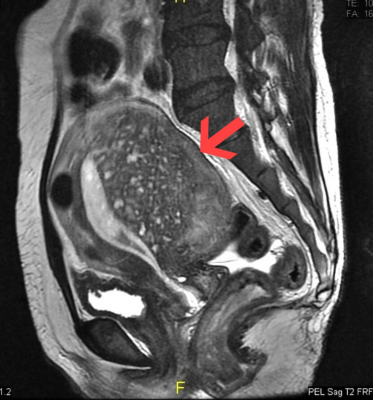
More sensitive and specific than ultrasound, particularly for distinguishing adenomyosis from fibroids and for assessing the extent of disease before planning uterus-sparing surgery. The classic finding is thickening of the junctional zone (the inner myometrial layer) to over 12 mm, along with high-signal foci within the myometrium representing ectopic endometrial tissue and cystic change.3
Histology
Definitive diagnosis is only possible on histological examination of the uterus, typically after hysterectomy, showing endometrial glands and stroma within the myometrium. In practice, imaging findings combined with a compatible clinical picture are sufficient to guide management without requiring histological confirmation in most women.
Differential diagnosis
- Uterine fibroids: discrete masses rather than diffuse enlargement (see table above)
- Endometriosis: often coexists; consider both when pain is prominent
- Endometrial hyperplasia or cancer: especially if intermenstrual or postmenopausal bleeding is present - biopsy to exclude
- Chronic pelvic inflammatory disease
Management
There is no single test that confirms cure, and management is directed at symptom control rather than eradicating the disease, since only hysterectomy is truly curative. The approach mirrors that for heavy menstrual bleeding and pain from other causes, tailored by whether fertility is a priority.2
Medical management
| Option | Notes |
|---|---|
| Levonorgestrel intrauterine system (LNG-IUS) | First-line; reduces bleeding and pain, and is generally well tolerated |
| Tranexamic acid / NSAIDs | For bleeding and pain respectively; no effect on the underlying disease |
| Combined oral contraceptive pill or cyclical progestogens | Alternative if the LNG-IUS is not suitable or acceptable |
| GnRH analogues | Induce temporary medical menopause; used short-term, e.g. pre-operatively |
Surgical management
Hysterectomy is the only definitive cure and is considered for women who have completed their family and have symptoms refractory to medical treatment. Uterus-sparing surgical options (e.g. adenomyomectomy, uterine artery embolisation, endometrial ablation) exist for women wishing to preserve the uterus or fertility, but outcomes are less predictable than for hysterectomy and recurrence of symptoms is more common.1
Adenomyosis and pregnancy
Adenomyosis has been associated with subfertility and, when pregnancy occurs, with an increased risk of miscarriage, preterm birth, pre-eclampsia and fetal growth restriction, thought to relate to impaired implantation and abnormal uterine contractility. Women with known adenomyosis who become pregnant may warrant closer antenatal surveillance, though the evidence base for specific interventions is limited.2
Complications
- Iron-deficiency anaemia from chronic heavy bleeding
- Chronic pelvic pain and reduced quality of life
- Subfertility
- Adverse pregnancy outcomes (miscarriage, preterm birth)
- Psychological impact of chronic symptoms and, where relevant, of hysterectomy ending fertility
Red flags
Prognosis
Symptoms typically improve after the menopause as oestrogen levels decline, mirroring the pattern seen in endometriosis and fibroids. Medical management provides good symptom control for many women, but relief is often incomplete, and a significant proportion of women with severe, refractory disease ultimately proceed to hysterectomy, which is curative.
References
- Royal College of Obstetricians and Gynaecologists. Adenomyosis - patient information. Available here
- NICE Clinical Knowledge Summaries (CKS). Menorrhagia - adenomyosis as a cause. Available here
- Radiopaedia. Adenomyosis. Available here
This article is written for revision and education. It is not clinical guidance and must not be used to make decisions about the care of a patient. Always check current NICE guidance and local protocols.