
Idiopathic Intracranial Hypertension: Diagnosis and Management
Key points
- Idiopathic intracranial hypertension (IIH): raised intracranial pressure of unknown cause, occurring with normal CSF composition and no structural or vascular explanation on imaging.
- Typical patient: a woman of reproductive age with obesity - this demographic accounts for the large majority of cases.
- Cardinal symptom: headache, classically worse lying flat, on waking, or with straining, often with pulsatile tinnitus and transient visual obscurations.
- Diagnosis: the modified Dandy criteria - raised opening pressure on lumbar puncture with normal CSF, normal neuroimaging (having excluded venous sinus thrombosis), and no other cause found.
- Vision is the priority: papilloedema can cause irreversible optic nerve damage - visual field testing and monitoring drive management decisions, not headache severity.
- First-line treatment: weight loss (even a modest amount is effective) and acetazolamide.
- Surgical options: optic nerve sheath fenestration or CSF shunting for progressive visual loss despite medical treatment.
- Venous sinus thrombosis must be excluded: with venography, since it can produce an identical clinical picture and is treated completely differently.
Introduction
Idiopathic intracranial hypertension (IIH), formerly known as benign (or pseudotumor cerebri), is a syndrome of raised intracranial pressure with no identifiable structural, vascular or infective cause, and normal cerebrospinal fluid composition.1 The term 'benign' has been dropped because the condition can cause permanent blindness if unrecognised or poorly managed - it is anything but benign to the optic nerve.
It occurs overwhelmingly in women of reproductive age with obesity, and its incidence is rising in step with rising rates of obesity. It is a favourite exam topic because it sits at the intersection of headache, visual loss and endocrinology, and because the diagnostic criteria and the priority given to vision over headache are both frequently tested.
Incidence in the general population is low, around 1-2 per 100,000 per year, but rises to roughly 20 per 100,000 among young women with obesity - a more than tenfold difference that explains why the demographic is such a strong part of the clinical picture. Incidence has risen in parallel with population obesity, so this is a condition that has become genuinely more common rather than simply better recognised.
Aetiology
The exact mechanism remains incompletely understood, but current thinking centres on impaired CSF resorption via the arachnoid granulations and/or impaired venous outflow (some patients have venous sinus stenosis, though whether this is cause or consequence of raised pressure is debated), combined with a metabolic contribution from obesity itself.
The role of venous sinus stenosis deserves particular comment because it is a source of genuine confusion. A proportion of patients have narrowing of the transverse sinuses on venography, and this sets up a self-reinforcing loop: raised intracranial pressure compresses the sinus, the resulting outflow obstruction raises venous pressure, and that further impairs CSF resorption and raises intracranial pressure again. Because the loop can be entered from either direction, the stenosis may be cause, consequence, or both - which is why it is not, on its own, diagnostic, and why venous stenting is reserved for carefully selected cases rather than offered routinely.
- Obesity - present in the large majority of cases and the strongest identified risk factor; recent weight gain, even without a high absolute BMI, is also a risk factor
- Female sex and reproductive age
- Drugs - tetracyclines and doxycycline, vitamin A and retinoids, growth hormone, and (less consistently) the combined oral contraceptive pill and some other hormonal agents
- Endocrine associations - polycystic ovary syndrome
Clinical features
- Headache - the commonest symptom, often daily, characteristically worse on waking, lying flat, or with coughing/straining (all of which raise venous and intracranial pressure); can also mimic migraine or tension-type headache in quality
- Pulsatile tinnitus - a whooshing sound synchronous with the pulse, a distinctive and useful symptom when present
- Transient visual obscurations - brief (seconds), often bilateral, episodes of greying or blacking out of vision, typically triggered by standing or bending forward, reflecting transient disc ischaemia
- Diplopia - from a sixth nerve palsy, a false localising sign of raised intracranial pressure rather than a lesion in the nerve's direct path
- Blurred vision and, if papilloedema is longstanding or severe, progressive peripheral visual field loss - which can occur without the patient noticing until it is advanced, since central vision is often preserved until late
- Neck and back pain, and radicular pain in some patients
Clinical examination
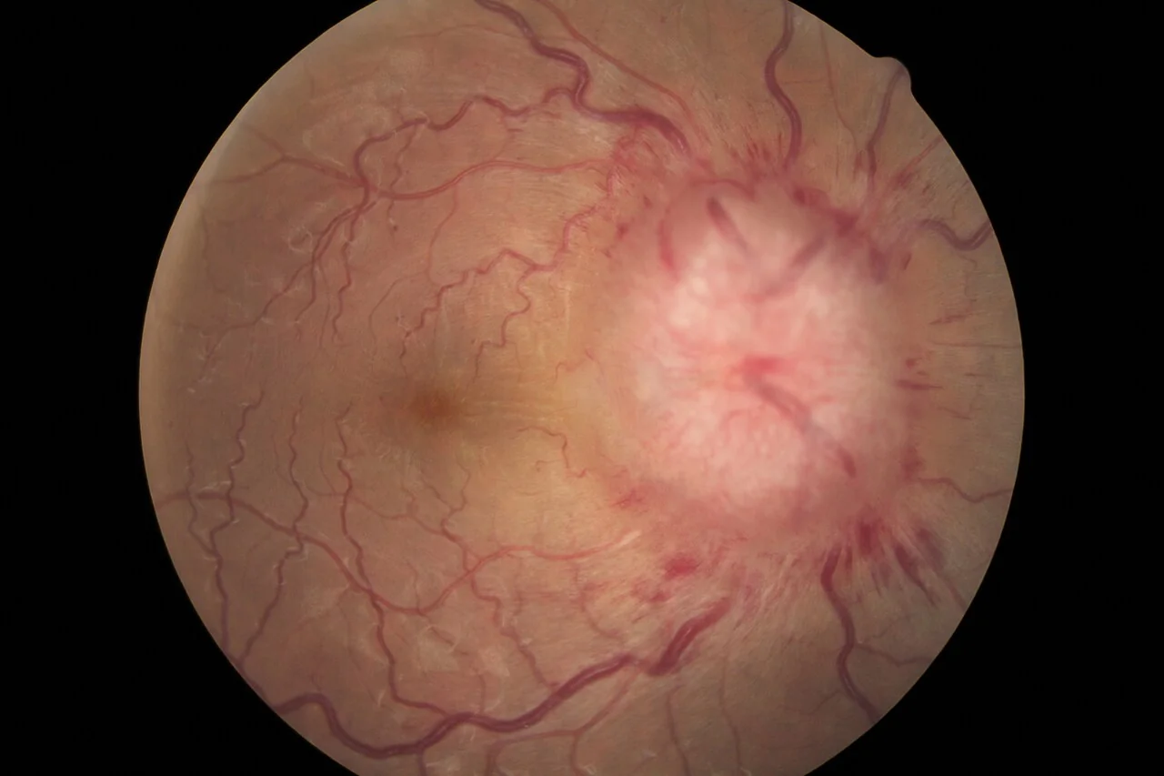
- Fundoscopy - bilateral papilloedema is the key sign; graded (for example, using the Frisén scale) and documented at every visit to track progression
- Visual acuity and formal visual field testing (Humphrey or Goldmann perimetry) - the most important objective measure to follow over time
- Cranial nerve examination - looking for a sixth nerve palsy
- Blood pressure - severe hypertension can itself cause papilloedema and must be excluded
- BMI and recent weight change
- General neurological examination - should otherwise be normal; any focal deficit should prompt reconsideration of the diagnosis
Diagnosis - the modified Dandy criteria
IIH is diagnosed using the modified Dandy criteria, which require all of the following:2
Two aspects of the lumbar puncture are worth emphasising because they are frequent sources of error. The opening pressure must be measured with the patient in the lateral decubitus position with the legs relaxed and extended - a flexed, tense or sitting patient produces a spuriously high reading that can lead to overdiagnosis. Anxiety, Valsalva and obesity itself can all elevate the measured pressure, so a borderline result in a distressed patient should be interpreted cautiously and, if necessary, repeated.
Equally, the diagnosis requires normal CSF constituents. An elevated white cell count or protein means this is not idiopathic intracranial hypertension, and should redirect the work-up towards infective, inflammatory or malignant meningeal disease.
- Symptoms and signs of raised intracranial pressure (papilloedema, or in its absence a sixth nerve palsy, plus at least three supportive neuroimaging findings)
- No localising signs on neurological examination, other than a sixth nerve palsy
- Normal neuroimaging, with no evidence of a mass lesion, hydrocephalus, or structural abnormality, and no evidence of venous sinus thrombosis
- Normal CSF composition on lumbar puncture
- Elevated CSF opening pressure (generally defined as >25 cmH2O in adults) measured in the lateral decubitus position
- No other identified cause of raised intracranial pressure
Differential diagnosis
- Cerebral venous sinus thrombosis - the most important differential, since it can present identically and must be excluded with venography before IIH is diagnosed
- Space-occupying lesion - tumour, abscess, or other mass causing raised intracranial pressure
- Hydrocephalus
- Meningitis (including chronic/malignant meningitis) - suggested by abnormal CSF composition, which by definition excludes IIH
- Drug-induced intracranial hypertension - tetracyclines, vitamin A/retinoids, growth hormone
- Severe hypertension causing papilloedema
- Migraine - can coexist with, or be mimicked by, IIH headache
Investigations
- MRI brain with MR venography - the key imaging investigation, both to exclude a structural cause and specifically to exclude venous sinus thrombosis; MRI may show supportive but non-diagnostic signs such as an empty sella, flattening of the posterior globe, or optic nerve sheath distension
- Lumbar puncture with opening pressure measurement - performed with the patient in the lateral decubitus position and legs extended (a raised pressure can be falsely produced by a flexed, tense posture), after imaging has excluded a mass lesion; CSF is sent for routine analysis, which should be normal
- Formal visual field testing at diagnosis and at every follow-up visit - the key measure that drives management decisions
- Optical coherence tomography (OCT) - increasingly used to quantify and monitor papilloedema objectively
- Screen for secondary causes - full drug history, blood pressure
Management
Management is guided principally by the degree of visual threat, not by headache severity - the overriding aim is to protect vision.
Weight management
Weight loss is the most effective disease-modifying treatment in patients with obesity, and even a modest reduction (around 5-10% of body weight) can produce meaningful improvement in papilloedema and symptoms. Referral to a weight management service or dietitian should be offered early, and bariatric surgery has good evidence in more severe or refractory obesity-associated disease.
Medical treatment
- Acetazolamide - a carbonic anhydrase inhibitor that reduces CSF production, first-line pharmacological treatment; side effects include paraesthesiae, fatigue, and taste disturbance (carbonated drinks taste flat), and it should be used cautiously in sulfonamide allergy
- Topiramate - an alternative that also promotes weight loss, though it is teratogenic and requires the same reproductive precautions as in epilepsy
- Therapeutic lumbar puncture - can provide temporary symptomatic relief by removing CSF, but the effect is short-lived (CSF reaccumulates within hours) and it is not used as a long-term strategy, though it has a role in acute severe visual threat while other treatment is arranged
Surgical treatment
Surgery is reserved for progressive visual loss despite medical treatment, or fulminant presentations with severe papilloedema threatening rapid, permanent visual loss.3
A fulminant presentation - severe visual loss developing over days rather than months - is managed differently from the usual indolent course. These patients need admission, urgent ophthalmology and neurosurgical involvement, and definitive surgical intervention within days rather than a trial of medical therapy, since the window in which vision can be preserved is very short.
- Optic nerve sheath fenestration - creates a window in the optic nerve sheath to allow CSF to decompress locally, protecting the optic nerve specifically, though it does not reliably relieve headache
- CSF shunting (ventriculoperitoneal or lumboperitoneal) - reduces intracranial pressure globally, treating both visual threat and headache, but carries the usual risks of shunt surgery including infection, blockage and overdrainage
- Venous sinus stenting - an emerging option in selected patients found to have a significant venous sinus stenosis, performed in specialist centres
Complications
- Permanent visual loss - the most feared complication, from prolonged papilloedema causing optic atrophy; can progress to complete blindness if unrecognised or inadequately treated
- Chronic, disabling headache
- Depression and anxiety, related both to chronic symptoms and to the underlying association with obesity
- Complications of treatment - side effects of acetazolamide/topiramate, or surgical complications of shunting or optic nerve sheath fenestration
- Relapse, particularly with weight regain
Red flags
Because visual field loss in IIH can progress without the patient noticing, the decision to escalate rests on objective measurement rather than reported symptoms. The features below should prompt urgent specialist review.
Prognosis
With early diagnosis, weight management and appropriate medical (or, where needed, surgical) treatment, most patients preserve useful vision and symptoms improve over time. A minority progress to significant, sometimes permanent, visual field loss - most often because papilloedema or its progression was not recognised early enough. Regular follow-up with formal visual field testing is essential for as long as papilloedema persists, and relapse can occur, particularly with weight regain.
Two practical points shape long-term follow-up. The first is that headache and papilloedema can dissociate completely: patients frequently have persistent headache after the intracranial pressure has normalised and the discs have settled, and treating that headache with escalating IIH-directed therapy is both ineffective and potentially harmful. Once papilloedema has resolved, persistent headache is usually managed as a coexisting primary headache disorder, with attention to medication overuse.
The second is pregnancy. IIH is not a contraindication to pregnancy and does not itself require caesarean delivery, but visual monitoring should intensify because weight gain can precipitate deterioration, and treatment choices change - acetazolamide is generally avoided in the first trimester and topiramate is contraindicated throughout, so management leans more heavily on monitoring and, if vision is threatened, on procedural options.
Because the condition is strongly associated with obesity and predominantly affects young women, care should be delivered in a way that supports rather than stigmatises. Weight management is the most effective intervention available, but framing it as the patient's fault is both unkind and counterproductive; referral to a structured weight management service, and where appropriate discussion of bariatric surgery as a legitimate medical treatment for this condition, is more useful than repeated advice to lose weight without support to achieve it.
References
- NICE CKS. Headache - assessment (Idiopathic intracranial hypertension). Available here
- Friedman DI, Liu GT, Digre KB. Revised diagnostic criteria for the pseudotumor cerebri syndrome in adults and children. Neurology. 2013. Available here
- Mollan SP, Davies B, Silver NC et al. Idiopathic intracranial hypertension: consensus guidelines on management. Journal of Neurology, Neurosurgery & Psychiatry. 2018. Available here
This article is written for revision and education. It is not clinical guidance and must not be used to make decisions about the care of a patient. Always check current NICE guidance and local protocols.