
Multiple Sclerosis: Diagnosis and Management
Key points
- Multiple sclerosis (MS): a chronic autoimmune disease causing demyelination and axonal damage in the central nervous system, disseminated in time and space.
- Typical patient: a woman aged 20-40, though it affects men too and can present at any adult age.
- Disease courses: relapsing-remitting (the commonest at onset), secondary progressive (relapsing-remitting that later accumulates disability between relapses), and primary progressive.
- Diagnosis: the McDonald criteria - clinical and/or MRI evidence of lesions disseminated in time and space, with alternative diagnoses excluded.
- Classic early presentation: optic neuritis - subacute unilateral visual loss with pain on eye movement.
- Acute relapse treatment: high-dose corticosteroids to shorten the relapse, which do not alter long-term disability.
- Disease-modifying therapy: reduces relapse frequency and MRI activity in relapsing disease - choice depends on disease activity and patient factors.
- Uhthoff's phenomenon: transient worsening of symptoms with heat (exercise, fever, hot baths) - a supportive but non-diagnostic clue.
Introduction
Multiple sclerosis (MS) is a chronic autoimmune disease of the central nervous system in which T-cell-mediated inflammation causes demyelination of axons and, over time, axonal loss itself.1 Because myelin can be affected anywhere in the brain, spinal cord or optic nerves, its clinical presentation is extremely varied - which is exactly why the core diagnostic concept is dissemination in time and space: separate lesions, in separate parts of the CNS, occurring at separate points in time.
MS typically presents in young adults, more commonly women, and is the commonest cause of neurological disability in young adults in the UK. It is a heavily examined topic because it combines immunology, neuroanatomy (localising lesions from symptoms) and a genuinely complex, rapidly evolving therapeutic landscape.
The UK has one of the higher prevalences worldwide, at roughly 150-200 per 100,000, with a clear latitude gradient and a female-to-male ratio of around 3 to 1 that has risen over recent decades. Onset is typically between 20 and 40, making MS a condition that characteristically strikes during education, early career and family formation - which is why its social and occupational consequences are so central to management and why early, effective treatment matters beyond the neurological outcome alone.
Aetiology
MS is thought to arise from a combination of genetic susceptibility and environmental triggers, resulting in autoreactive T-cells crossing the blood-brain barrier and initiating an inflammatory attack on myelin.
- Genetic susceptibility - strongest association is with HLA-DRB1*15:01; risk is higher with an affected first-degree relative, though MS is not straightforwardly inherited
- Epstein-Barr virus infection - now considered a near-necessary trigger, with very strong epidemiological evidence linking prior EBV infection (especially symptomatic glandular fever) to subsequent MS risk3
- Low vitamin D / reduced sunlight exposure - MS prevalence increases with distance from the equator
- Smoking - increases both the risk of developing MS and the rate of disability progression
- Obesity in adolescence
Disease course
| Course | Description | Proportion at onset |
|---|---|---|
| Relapsing-remitting (RRMS) | Discrete relapses with full or partial recovery, and stable disease between relapses | ~85% |
| Secondary progressive (SPMS) | RRMS that evolves into a phase of gradual, continuous disability accumulation, with or without superimposed relapses | Develops over time in many with RRMS |
| Primary progressive (PPMS) | Steady accumulation of disability from onset, without discrete relapses | ~10-15% |
Clinical features
Symptoms depend entirely on lesion location, and typically develop subacutely over days, then partially or fully resolve over weeks to months (in relapsing disease).
Optic neuritis
Often the first presentation of MS. Features subacute unilateral visual loss, pain on eye movement, reduced colour vision (red desaturation) out of proportion to visual acuity loss, and a relative afferent pupillary defect. The optic disc may look normal (retrobulbar neuritis) or swollen acutely; optic atrophy (a pale disc) can follow.
A useful principle when assessing any new symptom in MS is to ask whether it fits a single anatomical lesion. Demyelinating plaques are focal, so a genuine relapse produces symptoms attributable to one site in the CNS - a spinal cord lesion, an optic nerve, a brainstem plaque. Diffuse, bilateral or non-anatomical symptoms are more likely to reflect fatigue, a pseudo-relapse, mood disturbance or a coexisting condition, and treating them as relapses leads to unnecessary steroid exposure.
It is also worth remembering that MS is a disease of the central nervous system only. Symptoms that clearly localise to peripheral nerve or muscle - a glove-and-stocking sensory loss, fasciculations, or a lower motor neurone pattern of weakness - are not explained by MS and should prompt a separate diagnosis rather than being folded into the existing one.
Other common presentations
- Transverse myelitis - spinal cord involvement causing a sensory level, limb weakness, and bladder/bowel dysfunction
- Internuclear ophthalmoplegia - impaired adduction of one eye with nystagmus in the abducting eye on lateral gaze, from a lesion in the medial longitudinal fasciculus - a highly characteristic sign in a young patient
- Cerebellar syndrome - ataxia, intention tremor, dysarthria, nystagmus
- Sensory symptoms - numbness, paraesthesiae, or Lhermitte's sign (an electric shock-like sensation radiating down the spine and into the limbs on neck flexion, from cervical cord demyelination)
- Fatigue - extremely common and often disproportionately disabling relative to objective findings
- Bladder dysfunction - urgency, frequency, or retention
- Cognitive impairment - particularly processing speed and memory, often underrecognised
- Uhthoff's phenomenon - transient worsening of existing symptoms with a rise in body temperature (exercise, hot bath, fever), reflecting temperature-sensitive conduction in demyelinated axons; supportive but not diagnostic
Clinical examination
- Visual acuity, colour vision, pupillary responses and fundoscopy - looking for evidence of optic neuritis (past or present)
- Eye movements - for internuclear ophthalmoplegia and nystagmus
- Upper motor neurone signs - increased tone, hyperreflexia, extensor plantar responses, weakness in a pattern that does not fit a single peripheral nerve or root
- Cerebellar signs - dysdiadochokinesia, ataxia, intention tremor, scanning dysarthria
- Sensory examination, including testing for Lhermitte's sign
- Assessment of gait and mobility
Differential diagnosis
- Neuromyelitis optica spectrum disorder (NMOSD) - can closely mimic MS but with more severe, longitudinally extensive spinal cord lesions and optic neuritis; distinguished by aquaporin-4 antibody testing, and important because some MS treatments worsen NMOSD
- Acute disseminated encephalomyelitis (ADEM) - a monophasic, often post-infectious demyelinating illness, typically in children, that can look identical to a first MS attack radiologically
- Vitamin B12 deficiency - subacute combined degeneration of the cord can mimic MS's combined sensory and upper motor neurone signs
- Vasculitis and other autoimmune/connective tissue disease - Behçet disease, neurosarcoidosis, systemic lupus erythematosus with CNS involvement
- Migraine with brainstem aura
- Cerebrovascular disease - particularly in older patients or with vascular risk factors, since white matter lesions from small vessel disease can be mistaken for demyelination on MRI
- Functional neurological disorder
Investigations
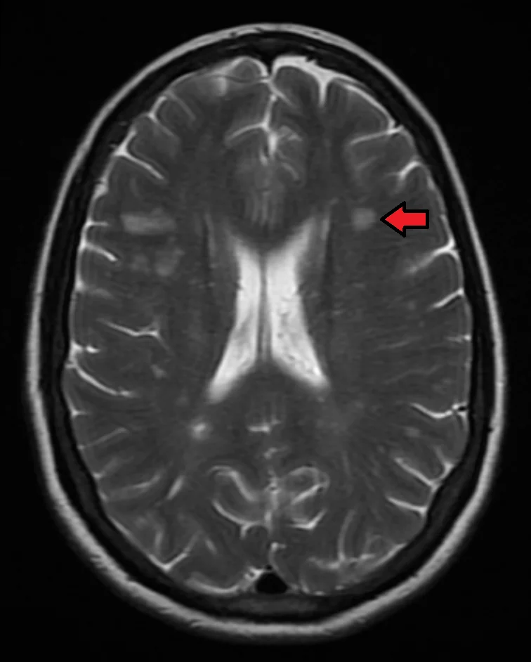
MS is diagnosed using the McDonald criteria, which require evidence of CNS lesions disseminated in space (more than one anatomical location) and disseminated in time (occurring at more than one point in time), with no better alternative explanation.2 MRI has made this possible earlier than relying on clinical relapses alone, since a single clinical event plus suitable MRI findings can now fulfil both criteria.
It is worth being clear about what a relapse actually is, because this drives treatment decisions. A relapse is new or worsening neurological symptoms lasting more than 24 hours in the absence of fever or infection, developing over days and separated from any previous episode by at least 30 days. Symptoms that recur transiently in the context of infection or heat are usually a pseudo-relapse - a temporary unmasking of existing deficits by the effect of temperature on conduction in demyelinated axons - and these require treatment of the underlying trigger, not corticosteroids.
Distinguishing the two is a frequent practical problem. A patient with known MS who presents with worsening weakness and a urinary tract infection almost always has a pseudo-relapse, and giving steroids in that situation risks worsening the infection while providing no benefit. Checking for fever and screening for infection before treating any apparent relapse is therefore routine.
- MRI brain and spinal cord with gadolinium contrast - the key investigation. Look for periventricular, juxtacortical, infratentorial and spinal cord lesions; enhancing lesions indicate active, recent inflammation while non-enhancing lesions are older, supporting dissemination in time from a single scan
- CSF analysis - oligoclonal bands present in CSF but not serum indicate intrathecal antibody synthesis and support the diagnosis, particularly useful when MRI findings are equivocal
- Visual evoked potentials - can demonstrate delayed conduction from subclinical or past optic neuritis
- Blood tests - to exclude mimics: B12, inflammatory and autoimmune markers, aquaporin-4 and MOG antibodies if NMOSD is a differential
Management
Acute relapse
A relapse causing significant functional impairment is treated with high-dose corticosteroids (for example, oral or IV methylprednisolone for 3-5 days), which shorten the relapse and speed recovery but do not alter long-term disability or the underlying disease course. Mild sensory relapses without functional impact may not need treatment.
Disease-modifying therapy
Disease-modifying therapies (DMTs) reduce relapse frequency and MRI lesion activity in relapsing forms of MS, and are broadly grouped by efficacy and risk.
| Category | Examples | Notes |
|---|---|---|
| Moderate efficacy, self-injected/oral | Interferon beta, glatiramer acetate, dimethyl fumarate, teriflunomide | Established safety profile, often first-line for milder disease activity |
| Higher efficacy, oral | Fingolimod, cladribine, siponimod | Used for more active disease or after failure of moderate-efficacy agents |
| Highest efficacy, infused | Natalizumab, alemtuzumab, ocrelizumab | Reserved for highly active disease; natalizumab carries a risk of PML in JC virus-positive patients |
Ocrelizumab is also the first disease-modifying therapy shown to slow progression in primary progressive MS4, a disease course that was historically untreatable.
Symptomatic management
- Spasticity: baclofen or gabapentin first-line; physiotherapy throughout
- Fatigue: exercise programmes, energy conservation strategies, and amantadine or modafinil in selected patients
- Bladder dysfunction: anticholinergics for urgency, intermittent self-catheterisation for retention
- Neuropathic pain and Lhermitte's sign: gabapentin, pregabalin or amitriptyline
- Depression and anxiety - actively screen for and treat, given the high prevalence in MS
- Multidisciplinary rehabilitation - physiotherapy, occupational therapy, and specialist MS nursing input are central to ongoing care, not an afterthought to drug treatment
Complications
- Progressive physical disability, particularly gait impairment, over the course of the disease
- Cognitive impairment
- Depression - both reactive and directly disease-related, with an increased suicide risk that should be actively screened for
- Recurrent urinary tract infections from bladder dysfunction
- Osteoporosis, from reduced mobility and recurrent corticosteroid use
- Complications of specific DMTs - infusion reactions, opportunistic infection, PML
Red flags
Prognosis
The course of MS is highly variable and difficult to predict in an individual. Favourable prognostic features include female sex, younger age at onset, purely sensory or optic neuritis presentation, and a low relapse rate/lesion burden in the first years. Many patients with relapsing-remitting MS eventually transition to secondary progressive disease, though earlier and more effective use of disease-modifying therapy has meaningfully delayed this transition and reduced long-term disability compared with older treatment approaches. Life expectancy is only modestly reduced overall, but quality of life and independence can be significantly affected.
Prognostic counselling should be honest about uncertainty. At diagnosis it is generally not possible to predict an individual's course, and many patients arrive with an outdated and unduly bleak picture drawn from an era before effective disease-modifying therapy. It is reasonable and accurate to say that most people with MS do not end up using a wheelchair, that life expectancy is only modestly reduced, and that treatment has changed outcomes substantially - while being clear that the course varies and cannot be forecast precisely for any one person.
References
- NICE NG220. Multiple sclerosis in adults: management. 2022. Available here
- Thompson AJ, Banwell BL, Barkhof F et al. Diagnosis of multiple sclerosis: 2017 revisions of the McDonald criteria. The Lancet Neurology. 2018. Available here
- Bjornevik K, Cortese M, Healy BC et al. Longitudinal analysis reveals high prevalence of Epstein-Barr virus associated with multiple sclerosis. Science. 2022. Available here
- MS Society UK. Disease modifying therapies. Available here
This article is written for revision and education. It is not clinical guidance and must not be used to make decisions about the care of a patient. Always check current NICE guidance and local protocols.