
Knee Pain
Key points
- Sort by age, site and mechanism: these three pieces of information narrow the differential further than any special test.
- Timing of swelling: an effusion within 2 hours is a haemarthrosis - ACL rupture, osteochondral fracture or patellar dislocation. Swelling over 6 to 24 hours suggests a meniscal tear.
- True locking: a mechanical block to full extension means a bucket-handle meniscal tear or a loose body, and needs orthopaedic referral.
- Diagnosing osteoarthritis: clinical, without imaging, in anyone 45 or over with activity-related joint pain and either no morning stiffness or stiffness lasting 30 minutes or less.
- Core OA treatment: therapeutic exercise and weight management. Topical NSAID is the first-line drug for knee osteoarthritis.
- Arthroscopy is not a treatment for osteoarthritis: or for degenerative meniscal tears - the trials show no benefit over placebo or exercise, unless there is true mechanical locking.
- Always examine the hip: hip pathology refers to the knee, and a limping child with knee pain and a normal knee has a hip problem until proven otherwise.
- Red flags: a hot swollen knee with fever is septic arthritis; night and rest pain with a mass around the knee in an adolescent raises osteosarcoma.
Introduction
The knee is the most commonly symptomatic joint in the body, and knee pain accounts for a large share of musculoskeletal presentations in primary care, sports medicine and emergency departments. The differential is wide, but it is manageable if approached systematically rather than by pattern-matching to whichever special test is remembered.
Three questions do most of the sorting.4 How old is the patient? - the causes at 15, 35 and 70 barely overlap. Where exactly does it hurt? - anterior, medial, lateral and posterior pain each have their own short list. Was there an injury, and what happened afterwards? - particularly how quickly the knee swelled.
Acute ligament and meniscal injury, and the Ottawa knee rules, are covered in more detail alongside the other soft tissue injuries. This article takes the broader view, with the emphasis on the non-traumatic causes and on osteoarthritis, which dominates in practice.
Relevant anatomy
The knee is a modified hinge with a small degree of rotation. Its bony congruity is poor, so stability depends on soft tissue: the cruciate ligaments control anteroposterior translation (the ACL preventing anterior translation of the tibia, the PCL preventing posterior), the collateral ligaments resist valgus and varus stress, and the menisci deepen the articular surface, distribute load and contribute to stability.
Two anatomical facts have direct clinical consequences. The medial meniscus is attached to the medial collateral ligament and the capsule, so it is less mobile and more often torn - and is injured together with the MCL and ACL in the classic "unhappy triad". The popliteal artery and common peroneal nerve lie immediately behind and lateral to the joint, which is why a knee dislocation is a limb-threatening injury and why LCL injuries can be accompanied by a foot drop.
The extensor mechanism runs from quadriceps through the patella and patellar tendon to the tibial tuberosity; a break anywhere along it - quadriceps tendon rupture, patellar fracture, patellar tendon rupture, or tibial tuberosity avulsion - produces the same functional deficit, an inability to perform a straight leg raise. Several bursae surround the joint: prepatellar, superficial and deep infrapatellar, and pes anserine on the medial tibia.
An approach by site
| Site | Causes |
|---|---|
| Anterior | Patellofemoral pain syndrome; patellar tendinopathy (jumper's knee); Osgood-Schlatter and Sinding-Larsen-Johansson disease in adolescents; prepatellar and infrapatellar bursitis; patellar instability or dislocation; patellofemoral osteoarthritis; fat pad impingement |
| Medial | Medial compartment osteoarthritis; medial meniscal tear; MCL injury; pes anserine bursitis; referred pain from the hip |
| Lateral | Lateral meniscal tear; LCL injury; iliotibial band syndrome; lateral compartment osteoarthritis |
| Posterior | Baker's (popliteal) cyst; hamstring or gastrocnemius injury; PCL injury; popliteal artery aneurysm; deep vein thrombosis |
| Diffuse or poorly localised | Osteoarthritis; septic arthritis; crystal arthropathy; inflammatory arthritis; effusion of any cause; referred pain from the hip or lumbar spine |
| Age | Common causes | Must not miss |
|---|---|---|
| Child and adolescent | Patellofemoral pain; Osgood-Schlatter disease; patellar instability; osteochondritis dissecans | Referred hip pathology - slipped upper femoral epiphysis, Perthes' disease, transient synovitis; septic arthritis; osteosarcoma, which characteristically arises around the knee; juvenile idiopathic arthritis; leukaemia |
| Young adult | Patellofemoral pain; ligament and meniscal injury; patellar tendinopathy; iliotibial band syndrome | Septic arthritis; inflammatory arthritis including reactive arthritis; bone tumour |
| Middle-aged | Degenerative meniscal tears; early osteoarthritis; bursitis; iliotibial band syndrome | Gout; septic arthritis; referred hip osteoarthritis |
| Older adult | Osteoarthritis; crystal arthropathy including pseudogout; Baker's cyst | Septic arthritis (which frequently occurs in an already arthritic joint); spontaneous osteonecrosis of the knee; metastatic disease |
History and examination
Key questions
- Mechanism - a twisting injury on a planted, flexed, weight-bearing foot suggests meniscal or ACL injury; a valgus blow to the lateral side suggests MCL; a blow to the front of the flexed tibia (dashboard, or falling on a bent knee) suggests PCL
- Speed of swelling - the single most useful question after trauma. Immediate swelling within 2 hours means a haemarthrosis: ACL rupture in around 70% of cases, then patellar dislocation, osteochondral fracture and intra-articular fracture. Swelling over 6 to 24 hours is a reactive effusion, typical of a meniscal tear.
- Locking - be precise about what the patient means. True locking is a mechanical block to full extension, indicating a displaced bucket-handle meniscal tear or a loose body. Patients often use "locking" to describe pain-related stiffness or catching, which is not the same thing.
- Giving way - true instability suggests ligament insufficiency (ACL) or patellar instability; giving way due to pain or quadriceps inhibition is commoner and less specific
- Pain pattern - worse going down stairs and after prolonged sitting suggests patellofemoral pain; worse with activity and at the end of the day suggests osteoarthritis; night and rest pain suggests infection, inflammation or tumour
- Systemic features - fever, weight loss, rash, urethritis or diarrhoea (reactive arthritis), psoriasis, other joints involved
Examination
- Look - gait, standing alignment (varus in medial compartment osteoarthritis, valgus in lateral), quadriceps wasting (measure thigh circumference), swelling, erythema, scars, and the position of the patella
- Feel - temperature, joint line tenderness, patellar borders, tibial tuberosity, bursae, and the popliteal fossa
- Effusion - the patellar tap detects a large effusion; the bulge or sweep test, stroking fluid from the medial gutter and watching it return, detects a small one
- Move - active and passive flexion and extension, looking for a fixed flexion deformity and for crepitus. Check the extensor mechanism by asking for a straight leg raise.
- Stress tests - valgus and varus stress at 0 and 30 degrees for the collaterals; Lachman's test at 20 to 30 degrees of flexion (the most sensitive test for the ACL), anterior drawer and pivot shift; posterior sag and posterior drawer for the PCL
- Meniscal tests - joint line tenderness (the most useful single sign), McMurray's and Thessaly tests, all of which have modest accuracy in isolation
- Patellofemoral - patellar tracking, apprehension test for instability, and patellar grind
- Always examine the hip and the lumbar spine, and assess the neurovascular status of the limb
Osteoarthritis of the knee
Osteoarthritis is by far the commonest cause of chronic knee pain, affecting around 10% of adults over 55 symptomatically. It is a whole-joint disease involving cartilage loss, subchondral bone remodelling, osteophyte formation and synovial inflammation, rather than simple "wear and tear".
Diagnosis
Clinical features are pain worse with activity and at the end of the day, short-lived stiffness after rest ("gelling"), crepitus, bony swelling from osteophytes, reduced range with a fixed flexion deformity, quadriceps wasting, varus deformity in medial compartment disease, and sometimes a mild effusion. Prolonged morning stiffness, marked warmth, or involvement of multiple small joints should prompt consideration of inflammatory arthritis instead.
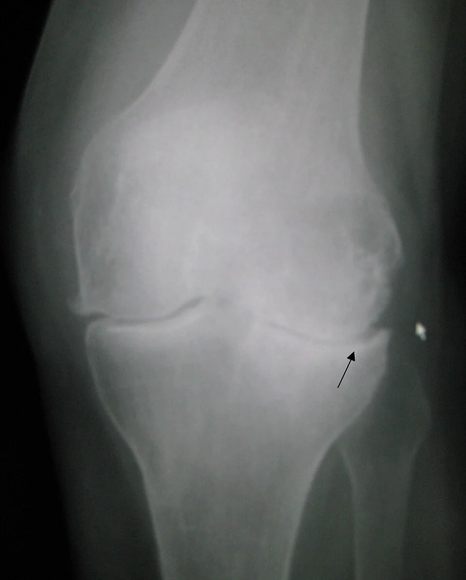
Radiographic features, where imaging is done, are conventionally remembered as LOSS: Loss of joint space, Osteophytes, Subchondral sclerosis and Subchondral cysts. Films should be weight-bearing, because joint space narrowing may be invisible on a supine view.
Management
| Category | Recommendation |
|---|---|
| Core treatment - offer to everyone | Therapeutic exercise - both local muscle strengthening and general aerobic fitness, supervised where possible, with an explanation that symptoms may worsen initially before improving. Weight loss where the person is overweight, with any amount of loss being beneficial. Information and self-management support. |
| Adjuncts | Walking aids and, for some, therapeutic footwear or insoles; manual therapy only alongside exercise. Consider TENS. |
| Topical treatment | Topical NSAID is first-line pharmacological treatment for knee osteoarthritis - effective with far lower systemic exposure than oral |
| Oral treatment | Oral NSAID at the lowest effective dose for the shortest time, with a proton pump inhibitor, having considered cardiovascular, renal and gastrointestinal risk. Do not routinely offer paracetamol alone, weak opioids, or strong opioids. |
| Injection | Intra-articular corticosteroid may be considered for short-term relief, typically lasting a few weeks, to enable engagement with exercise |
| Do not offer | Glucosamine and chondroitin; intra-articular hyaluronan injection; acupuncture; arthroscopic lavage and debridement unless there is a clear history of true mechanical locking |
| Surgery | Refer for joint replacement if symptoms substantially affect quality of life and non-surgical management has been ineffective. NICE is explicit that age, sex, smoking, comorbidity and BMI should not be barriers to referral, and that scoring tools should not be used to restrict access. |
Other important causes
Patellofemoral pain syndrome
The commonest cause of anterior knee pain in young adults, and considerably commoner in women. Pain is diffuse and poorly localised around or behind the patella, worse descending stairs, squatting, running and after prolonged sitting - the last of these being the so-called cinema sign. Examination is often unremarkable apart from tenderness of the patellar facets and sometimes abnormal tracking or quadriceps wasting; the patellar grind test is uncomfortable even in normal knees and adds little.
Management is physiotherapy - strengthening the quadriceps, particularly vastus medialis, and importantly the hip abductors and external rotators, since proximal weakness allows dynamic knee valgus - along with activity modification, load management and, in some, taping or orthoses.6 Imaging is not needed. It usually improves but frequently takes months, and a proportion have symptoms for years.
Meniscal tears
Acute traumatic tears occur in younger patients through twisting on a loaded flexed knee, with delayed swelling, joint line tenderness and sometimes locking. Degenerative tears occur in middle-aged and older patients often with no injury at all, and are extremely common as incidental MRI findings alongside osteoarthritis.
Bursitis and tendinopathy
- Prepatellar bursitis (housemaid's knee) - swelling directly over the patella, associated with kneeling; infrapatellar bursitis (clergyman's knee) sits below it. Both may become septic, and a hot, red, exquisitely tender bursa with cellulitis needs aspiration and antibiotics.
- Pes anserine bursitis - medial tibial pain a few centimetres below the joint line, common in osteoarthritis and in runners, and frequently mistaken for a medial meniscal tear
- Patellar tendinopathy (jumper's knee) - tenderness at the inferior pole of the patella in jumping athletes; treated with progressive loading rather than rest
- Iliotibial band syndrome - lateral knee pain in runners and cyclists, worse downhill, with tenderness over the lateral femoral epicondyle and a positive Noble compression test
Baker's cyst
A distension of the gastrocnemius-semimembranosus bursa communicating with the joint, almost always secondary to intra-articular pathology - osteoarthritis or a meniscal tear in adults. It presents as posteromedial fullness, most obvious on standing with the knee extended. Rupture causes sudden calf pain and swelling that closely mimics a deep vein thrombosis, sometimes with bruising tracking below the medial malleolus (the crescent sign). Ultrasound distinguishes the two, and the underlying joint problem is what needs treating.
Crystal arthropathy and inflammatory causes
- Gout - rapid onset of severe pain with a red, hot, exquisitely tender joint. The knee is the second commonest site after the first metatarsophalangeal joint. Aspiration shows negatively birefringent needle-shaped monosodium urate crystals.
- Pseudogout (calcium pyrophosphate deposition) - the knee is the commonest site, typically in older patients. Radiographs show chondrocalcinosis, and aspiration shows positively birefringent rhomboid crystals.
- Inflammatory arthritis - rheumatoid arthritis, reactive arthritis, psoriatic arthritis and the spondyloarthropathies, suggested by prolonged morning stiffness, other joint involvement, and extra-articular features
- Septic arthritis - always in the differential for a hot swollen knee, and remember that it commonly occurs in a joint already damaged by osteoarthritis or crystal disease. A raised uric acid does not exclude it, and crystals in the aspirate do not exclude co-existing infection.
Investigations
- Often none. Patellofemoral pain and typical osteoarthritis need no investigation.
- Plain radiographs - weight-bearing AP and lateral, with a skyline view for patellofemoral disease. Indicated after trauma per the Ottawa knee rules (age 55 or over, isolated patellar tenderness, tenderness at the fibular head, inability to flex to 90 degrees, or inability to weight bear for four steps both immediately and in the department), and for atypical or non-resolving pain.5
- Joint aspiration - urgently for any hot swollen joint, before antibiotics. Send for urgent Gram stain, culture, cell count and polarised light microscopy for crystals.
- MRI - for suspected internal derangement in a patient in whom the result would change management: ligament and meniscal injury in younger patients, osteochondritis dissecans, suspected tumour or osteonecrosis. It is not needed to diagnose osteoarthritis and its findings must be interpreted alongside the clinical picture.
- Bloods - FBC, CRP and ESR if infection or inflammation is suspected; urate (deferred until several weeks after an acute gout attack, when it is more reliable); rheumatoid factor, anti-CCP and HLA-B27 where indicated
- Ultrasound - for a suspected Baker's cyst, to exclude DVT, and to guide aspiration or injection
Red flags
Prognosis
Most soft tissue and overuse knee problems improve with load management and a structured exercise programme, though patellofemoral pain in particular is often more persistent than patients are led to expect - a substantial minority still have symptoms at a year, and telling them this at the outset improves adherence to rehabilitation.
Osteoarthritis progresses variably, and by no means inevitably: many patients remain stable for years, and symptoms fluctuate independently of radiographic change. Exercise and weight loss produce clinically meaningful benefit - a 5 to 10% reduction in body weight measurably improves symptoms - and their effect sizes are comparable to those of NSAIDs without the risk. Total knee replacement relieves pain effectively in the great majority, with around 80 to 90% of implants surviving 15 to 20 years, but roughly one in five patients remains dissatisfied, most often those with lower preoperative pain scores or significant psychological distress. That is worth discussing before referral.
The theme running through modern knee practice is the retreat of arthroscopy. Placebo-controlled and comparative trials have removed the indication for arthroscopic debridement in osteoarthritis and for partial meniscectomy in degenerative tears, leaving exercise therapy as the intervention with the best evidence for the commonest problems. Knowing that, and being able to say why, is what distinguishes a good answer on this topic.
References
- NICE NG226. Osteoarthritis in over 16s: diagnosis and management. 2022. Available here
- Sihvonen R, Paavola M, Malmivaara A et al. Arthroscopic partial meniscectomy versus sham surgery for a degenerative meniscal tear (FIDELITY). New England Journal of Medicine. 2013, with five-year follow-up 2020. Available here
- Katz JN, Brophy RH, Chaisson CE et al. Surgery versus physical therapy for a meniscal tear and osteoarthritis (METEOR). New England Journal of Medicine. 2013. Available here
- NICE Clinical Knowledge Summaries. Knee pain - assessment. Available here
- Stiell IG, Greenberg GH, Wells GA et al. Prospective validation of a decision rule for the use of radiography in acute knee injuries (Ottawa knee rule). JAMA. 1996. Available here
- Collins NJ, Barton CJ, van Middelkoop M et al. Consensus statement on exercise therapy and physical interventions for patellofemoral pain. British Journal of Sports Medicine. 2018. Available here
This article is written for revision and education. It is not clinical guidance and must not be used to make decisions about the care of a patient. Always check current NICE guidance and local protocols.