
Shoulder Pain
Key points
- Think by age: instability under 30, subacromial pain and frozen shoulder from 30 to 50, and cuff tears and osteoarthritis over 50.
- Active versus passive: the single most useful examination finding. Restriction of both active and passive movement means a capsular or joint problem; restricted active with full passive movement means a cuff or tendon problem.
- Frozen shoulder: global restriction with external rotation lost first and worst, strongly associated with diabetes, and self-limiting over 1 to 3 years.
- Painful arc: pain between about 60 and 120 degrees suggests subacromial pathology; pain in the last 30 degrees suggests the acromioclavicular joint.
- Cuff tear versus tendinopathy: weakness rather than pain alone. A positive drop arm test or an external rotation lag sign indicates a tear.
- Subacromial decompression: the CSAW and FIMPACT trials found it no better than placebo surgery - physiotherapy is the mainstay for subacromial pain.
- Always examine the neck: cervical radiculopathy is a common cause of shoulder pain and is missed if the neck is not moved.
- Red flags: trauma with inability to move, fever with a hot joint, a locked internally rotated shoulder after a seizure, unexplained wasting, and an apical lung mass.
Introduction
Shoulder pain has a lifetime prevalence of around 70% and is the third commonest musculoskeletal presentation in primary care after back and knee pain.1 The great majority is soft tissue in origin, and most improves with analgesia and physiotherapy - but a shoulder that is stiff, weak, hot or has followed a fall needs a different answer.
The examinable skill is one of sorting. There are perhaps seven conditions worth knowing, and they can be separated by three pieces of information: the patient's age, whether the restriction is active or also passive, and whether the problem is pain or weakness. A handful of named tests then confirm the impression.
The other half of the topic is what is not the shoulder at all. Pain referred from the cervical spine, the diaphragm, the heart and an apical lung tumour all present as shoulder pain, and they are missed by examining only the shoulder.
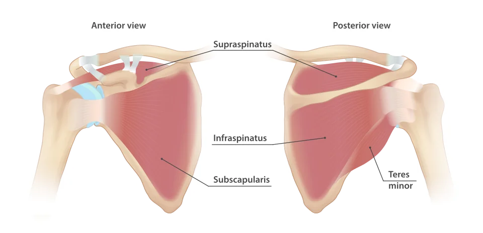
Relevant anatomy
The glenohumeral joint trades stability for range. A large humeral head articulates with a shallow glenoid - often likened to a golf ball on a tee - giving the greatest range of motion of any joint in the body, and the highest dislocation rate. Static stability comes from the glenoid labrum, the capsule and the glenohumeral ligaments; dynamic stability comes almost entirely from the rotator cuff.
| Muscle | Action | Nerve | Isolating test |
|---|---|---|---|
| Supraspinatus | Initiates the first 15 degrees of abduction, then assists deltoid | Suprascapular | Empty can (Jobe's) test - resisted abduction at 90 degrees in the scapular plane with the thumb pointing down; and the drop arm test for a full-thickness tear |
| Infraspinatus | External rotation | Suprascapular | Resisted external rotation with the elbow at the side; external rotation lag sign if there is a tear |
| Teres minor | External rotation | Axillary | Hornblower's sign - inability to hold the arm in external rotation at 90 degrees of abduction |
| Subscapularis | Internal rotation | Upper and lower subscapular | Belly press and Gerber's lift-off test |
Above the cuff, the subacromial space lies between the humeral head and the coracoacromial arch, and contains the supraspinatus tendon, the long head of biceps tendon and the subacromial bursa. Compression or irritation of these structures produces the pattern historically called impingement. Shoulder movement also depends on the acromioclavicular, sternoclavicular and scapulothoracic joints, and abnormal scapular movement (dyskinesia) is a common contributor that is easily overlooked.
Causes, by age
| Age | Most likely | Also consider |
|---|---|---|
| Under 30 | Instability and dislocation; labral tears including SLAP lesions; acromioclavicular joint injury | Fracture, septic arthritis, inflammatory arthritis |
| 30 to 50 | Subacromial pain syndrome (rotator cuff tendinopathy); adhesive capsulitis; biceps tendinopathy; calcific tendinitis | Cuff tear after trauma, acromioclavicular osteoarthritis |
| Over 50 | Rotator cuff tears, often degenerative; glenohumeral and acromioclavicular osteoarthritis | Polymyalgia rheumatica, malignancy, cervical spondylosis |
| Any age | Referred pain, septic arthritis, fracture, inflammatory arthritis | Avascular necrosis, tumour |
Subacromial pain syndrome
This is the commonest cause of shoulder pain, and the terminology has shifted deliberately. "Impingement" implied a purely mechanical cause requiring surgical decompression; subacromial pain syndrome or rotator cuff-related shoulder pain is preferred because the pathology is usually tendinopathy with secondary bursitis rather than bony compression.
- Gradual onset of pain over the lateral deltoid, worse with overhead activity and reaching behind
- Night pain, particularly when lying on the affected side - almost universal and a useful discriminator from referred pain
- Painful arc between roughly 60 and 120 degrees of abduction, with pain settling above and below that range
- Neer's and Hawkins-Kennedy tests are positive but are not specific
- Passive range is full, and power is preserved once the pain is accounted for - which distinguishes it from a tear and from a frozen shoulder
Rotator cuff tears
Tears are either acute and traumatic - typically in a younger patient after a fall onto an outstretched hand or following a first dislocation over the age of 40 - or degenerative, developing gradually in older patients, in whom asymptomatic partial tears are extremely common.
- The distinguishing feature is weakness, not just pain, and difficulty with functional tasks such as reaching a shelf or washing hair
- Drop arm test - the patient cannot lower the abducted arm smoothly from 90 degrees, or cannot hold it there; indicates a full-thickness supraspinatus tear
- External rotation lag sign - the arm drifts back into internal rotation when placed in maximal external rotation; indicates an infraspinatus tear
- Lift-off or belly press positive in subscapularis tears
- Massive chronic tears produce pseudoparalysis with the humeral head riding high on the radiograph
Adhesive capsulitis (frozen shoulder)
Inflammation and then fibrotic contracture of the glenohumeral joint capsule, particularly the rotator interval and coracohumeral ligament, producing global restriction of movement. It affects around 2 to 5% of the population, peaks between 40 and 60, and is commoner in women.6
| Phase | Duration | Features | Treatment emphasis |
|---|---|---|---|
| Freezing (painful) | 2 to 9 months | Increasing pain, often severe and worse at night, with progressive loss of movement | Analgesia and intra-articular corticosteroid injection, which works best in this phase; gentle range-of-movement exercises |
| Frozen (stiff) | 4 to 12 months | Pain settles, marked stiffness dominates, with the shoulder often surprisingly comfortable at rest | Physiotherapy and stretching; hydrodilatation |
| Thawing | 6 months to 2 years or more | Gradual return of range | Continued exercise; consider capsular release if progress stalls |
Other causes worth recognising
- Glenohumeral osteoarthritis - pain and stiffness with crepitus and restriction of both active and passive movement; radiographs show joint space narrowing, subchondral sclerosis and an inferior humeral osteophyte
- Acromioclavicular joint pathology - pain localised to the joint itself, tenderness directly over it, a high painful arc above about 140 degrees, and a positive scarf (cross-body adduction) test
- Instability - traumatic anterior instability after a dislocation, with a positive apprehension and relocation test; or atraumatic multidirectional instability in a young hypermobile patient, which is treated with physiotherapy rather than surgery
- Calcific tendinitis - abrupt, severe, often excruciating pain with a dense calcific deposit in the supraspinatus tendon on radiograph; treated with analgesia, subacromial injection or ultrasound-guided barbotage
- Long head of biceps pathology - anterior shoulder pain with positive Speed's and Yergason's tests; rupture produces the Popeye sign, a distally bunched muscle belly, which is largely a cosmetic problem in older patients
- Polymyalgia rheumatica - bilateral shoulder and hip girdle pain and stiffness over the age of 50, with morning stiffness lasting more than 45 minutes, raised ESR and CRP, and a dramatic response to low-dose prednisolone7
Referred and non-shoulder causes
- Cervical spine - C5 radiculopathy or cervical spondylosis. Suggested by neck pain, pain radiating below the elbow, paraesthesia, a full and painless passive shoulder range, and reproduction of symptoms on neck movement or Spurling's test.
- Diaphragmatic irritation - referred to the shoulder tip via C3 to C5 (Kehr's sign). Causes include subphrenic abscess, ruptured spleen, ectopic pregnancy, perforated viscus and gallbladder disease.
- Cardiac - myocardial ischaemia referred to the left shoulder and arm, particularly with exertional onset and associated breathlessness or sweating
- Pancoast tumour - an apical lung tumour causing shoulder and arm pain, with Horner's syndrome (ptosis, miosis, anhidrosis) and wasting of the small muscles of the hand from T1 involvement. Progressive, unremitting pain in a smoker demands a chest radiograph.
- Aortic dissection and pulmonary pathology including pleurisy and pneumothorax
- Neurological - brachial neuritis (Parsonage-Turner syndrome), presenting with severe shoulder pain followed days later by profound weakness and wasting; and long thoracic nerve palsy causing winging of the scapula
Examination
- Look - from front, side and behind, with both shoulders exposed. Muscle wasting (supraspinatus and infraspinatus fossae, deltoid), asymmetry, scars, swelling, a squared-off contour suggesting dislocation, and winging of the scapula on wall press.
- Feel - sternoclavicular joint, clavicle, acromioclavicular joint, acromion, greater tuberosity, bicipital groove, and the cervical spine and trapezius. Assess temperature.
- Move actively - flexion, extension, abduction, external rotation with the elbow at the side, and internal rotation assessed by how far up the back the thumb reaches. Watch for a painful arc and for abnormal scapular rhythm.
- Move passively - the crucial step. If passive range is full, the pathology is in the cuff or tendon; if it is equally restricted, it is capsular or articular.
- Power in each cuff muscle, and deltoid
- Special tests - painful arc, Jobe's empty can, drop arm, external rotation lag, lift-off, scarf test, Hawkins-Kennedy, apprehension and relocation, and Speed's test
- Examine the neck, and perform a neurovascular examination of the limb including axillary nerve sensation over the regimental badge area
- Consider the chest and abdomen where referred pain is plausible
Investigations
- Often none. A typical subacromial pain syndrome in a 45-year-old does not need imaging before a trial of physiotherapy.
- Plain radiographs - AP and an axillary or scapular Y view - for trauma, suspected dislocation, osteoarthritis, calcific tendinitis, and any atypical or non-resolving presentation. A single AP view can miss a posterior dislocation, which is why the second view is essential.
- Ultrasound - the first-line test for cuff pathology in the UK. Dynamic, quick and good for full-thickness tears, bursitis and calcific deposits; operator dependent.
- MRI or MR arthrogram - for labral and capsular pathology, suspected instability lesions, complex or recurrent cuff problems, and pre-operative planning
- Bloods - CRP and ESR if polymyalgia rheumatica, inflammatory arthritis or infection is suspected; a raised ESR in a patient over 50 with bilateral girdle pain effectively makes the diagnosis of PMR
- Chest radiograph - for suspected Pancoast tumour or referred pain
- Joint aspiration - immediately, before antibiotics, if septic arthritis is suspected
Management
General principles
- Explanation and reassurance about the natural history, which for most soft tissue shoulder pain is gradual improvement over months
- Relative rest and activity modification - avoiding the provoking overhead activity while keeping the shoulder moving. Complete rest risks stiffness.
- Analgesia - paracetamol and a short course of an NSAID, topical or oral
- Physiotherapy - the mainstay for subacromial pain, cuff tendinopathy, degenerative cuff tears and atraumatic instability. A structured, progressive loading programme, with attention to scapular control, is the best-evidenced treatment.5
- Corticosteroid injection - subacromial for subacromial pain, intra-articular for adhesive capsulitis and glenohumeral arthritis. Gives useful short-term relief, but the benefit wanes by around 3 months, so it is best used to enable rehabilitation rather than as a treatment in itself. Limit the number given.
Condition-specific treatment
| Diagnosis | First line | If that fails |
|---|---|---|
| Subacromial pain syndrome | Physiotherapy, analgesia, activity modification; subacromial injection to enable rehabilitation | Reassess the diagnosis; imaging to exclude a tear. Decompression is not routinely offered. |
| Acute traumatic cuff tear in a younger patient | Early referral - within 2 weeks - for consideration of surgical repair | Delayed repair has poorer results as the tendon retracts and the muscle undergoes fatty infiltration |
| Degenerative cuff tear | Physiotherapy; most do well without surgery | Repair in selected patients; reverse total shoulder arthroplasty for massive irreparable tears with cuff arthropathy4 |
| Adhesive capsulitis | Analgesia and early intra-articular steroid injection with physiotherapy | Hydrodilatation, manipulation under anaesthesia, or arthroscopic capsular release |
| Glenohumeral osteoarthritis | Analgesia, physiotherapy, intra-articular injection | Total shoulder arthroplasty (anatomical if the cuff is intact, reverse if not) |
| Acromioclavicular osteoarthritis | Analgesia and injection | Arthroscopic excision of the lateral clavicle |
| Traumatic anterior instability | Physiotherapy; sling and early mobilisation after a first dislocation | Arthroscopic Bankart repair, particularly in young patients and contact athletes, in whom recurrence rates without surgery are very high |
| Calcific tendinitis | Analgesia, subacromial injection | Ultrasound-guided needling and lavage (barbotage) |
| Polymyalgia rheumatica | Prednisolone 15 mg daily, with a dramatic response expected within days | Reconsider the diagnosis if the response is poor - and screen for giant cell arteritis symptoms throughout |
Red flags
Prognosis
Most soft tissue shoulder pain improves, but more slowly than patients expect. Roughly half of new episodes of shoulder pain have resolved at six months and around 60% at a year, and setting that expectation at the first consultation prevents a great deal of frustration and unnecessary imaging.
Adhesive capsulitis is usually described as self-limiting, and it is, but the timescale is one to three years and around 40% of patients have some persistent restriction of movement afterwards - though few find it functionally limiting. Diabetic patients have a longer and more resistant course. Degenerative cuff tears frequently remain manageable without surgery, whereas an acute traumatic tear in a younger patient should be referred quickly, because repair becomes progressively harder as the tendon retracts and the muscle undergoes fatty infiltration.
The recurring theme across all of these conditions is that structured exercise, not surgery or injection, does most of the work. That has become clearer as placebo-controlled trials of shoulder surgery have accumulated, and it is the reasoning examiners are looking for when they ask what to do about a painful arc in a 45-year-old.
References
- NICE Clinical Knowledge Summaries. Shoulder pain. Available here
- Beard DJ, Rees JL, Cook JA et al. Arthroscopic subacromial decompression for subacromial shoulder pain (CSAW): a multicentre, pragmatic, parallel group, placebo-controlled, three-group, randomised surgical trial. The Lancet. 2018. Available here
- Paavola M, Malmivaara A, Taimela S et al. Subacromial decompression versus diagnostic arthroscopy for shoulder impingement (FIMPACT): randomised, placebo surgery controlled clinical trial. BMJ. 2018. Available here
- British Elbow and Shoulder Society. Patient care pathways and commissioning guidance. Available here
- Littlewood C, Malliaras P, Chance-Larsen K. Therapeutic exercise for rotator cuff tendinopathy: a systematic review. International Journal of Rehabilitation Research. 2015. Available here
- NICE Clinical Knowledge Summaries. Shoulder pain - adhesive capsulitis (frozen shoulder). Available here
- Dasgupta B, Cimmino MA, Kremers HM et al. Provisional classification criteria for polymyalgia rheumatica. Arthritis and Rheumatism. 2012. Available here
This article is written for revision and education. It is not clinical guidance and must not be used to make decisions about the care of a patient. Always check current NICE guidance and local protocols.