
Bone Tumours: Metastatic and Primary
Key points
- Think metastasis first: in an adult, especially over 40, with a destructive bone lesion, metastatic disease is far more common than any primary bone tumour.
- Classic metastatic primaries: Breast, Bronchus (lung), Thyroid, Kidney (renal) and Prostate - most are lytic, but prostate is classically sclerotic and breast can be mixed.
- Osteosarcoma: the commonest primary malignant bone tumour, typically adolescents, metaphysis of the distal femur or proximal tibia - sunburst appearance and Codman triangle on X-ray.
- Ewing sarcoma: children and adolescents, diaphysis of long bones or pelvis, onion-skin periosteal reaction, t(11;22) translocation - can mimic osteomyelitis.
- Chondrosarcoma: older adults, arises from cartilage, more indolent, can follow malignant transformation of an enchondroma or osteochondroma.
- Red flag pain pattern: bone pain that is worse at night or at rest, rather than purely mechanical, points towards tumour rather than a benign musculoskeletal cause.
- Referral pathway: X-ray within 48 hours for unexplained bone pain or swelling in children/young people; very urgent specialist referral if the X-ray suggests possible bone sarcoma.
- Biopsy rule: definitive biopsy is performed at the specialist centre that will carry out surgery - a poorly placed biopsy tract can seed tumour and compromise limb-sparing surgery.
Introduction
Bone tumours span a spectrum from entirely benign incidental findings to aggressive primary sarcomas, but the single most important principle for finals - and for real practice - is one of prevalence rather than pathology. Metastatic bone disease is far more common than any primary bone tumour. In an adult, particularly one over 40, presenting with a destructive bone lesion, the correct default diagnosis is a bone metastasis from a known or occult primary cancer, not a primary bone sarcoma.
Primary malignant bone tumours are rare and are seen disproportionately in children and young adults, which is itself a useful discriminator: a lytic lesion in a 70-year-old is a metastasis until proven otherwise, while a destructive metaphyseal lesion in a 15-year-old raises concern for osteosarcoma.
This article covers metastatic bone disease, the common benign primary tumours, the three primary malignant bone tumours tested at UKMLA level - osteosarcoma, Ewing sarcoma and chondrosarcoma - and the referral pathway that gets a suspected sarcoma to a specialist centre without compromising future treatment.
Metastatic bone disease
Bone is the third most common site of metastasis after lung and liver, and skeletal metastases are far more frequently encountered in clinical practice than any primary bone tumour. Five primary cancers account for the great majority of bone metastases, often remembered with the mnemonic BLT with a kosher pickle - Breast, Lung (bronchus), Thyroid, Kidney (renal) and Prostate.
- Breast - the single commonest source of bone metastases in women, typically lytic or mixed lytic-sclerotic
- Bronchus (lung) - frequently lytic, often presents with bone pain before the primary is diagnosed
- Thyroid - particularly follicular thyroid carcinoma, lytic and can be highly vascular
- Kidney (renal cell carcinoma) - lytic, characteristically vascular and prone to brisk bleeding if biopsied or operated on
- Prostate - the classic exception: prostate metastases are sclerotic, reflecting osteoblastic new bone formation rather than pure bone destruction
Metastases favour the axial skeleton and the proximal long bones - vertebrae, pelvis, ribs, skull, and the proximal femur and humerus - because these retain red marrow and its blood supply into adult life, in contrast to the distal appendicular skeleton, which metastasis rarely reaches.
Clinical presentation
- Bone pain - typically progressive, often worse at night, and the commonest presenting symptom
- Pathological fracture - through a weakened area of bone, sometimes after trivial or no trauma
- Hypercalcaemia of malignancy - from osteoclastic bone resorption, or from tumour secretion of PTH-related peptide; presents with the classic "bones, stones, groans and psychiatric moans"
- Spinal cord or cauda equina compression - from vertebral metastases extending into the spinal canal, an oncological emergency requiring same-day MRI and urgent treatment
- Anaemia and bone marrow suppression - with extensive marrow infiltration
- Raised ALP - reflecting increased bone turnover, though this is non-specific
A metastasis may be the presenting feature of an as-yet-undiagnosed cancer, so a destructive bone lesion in an older adult should prompt a search for a primary - clinical breast and prostate examination, chest X-ray, renal and thyroid assessment, and PSA - alongside imaging of the lesion itself.
Benign primary bone tumours
Benign bone tumours are common, frequently asymptomatic, and are often picked up incidentally on imaging performed for another reason. The main task in exams is recognising the classic description of each rather than managing them in detail.
Osteochondroma
The most common benign bone tumour. It is a cartilage-capped bony outgrowth (exostosis) that arises near a growth plate and grows away from the joint, most often around the knee or proximal humerus. Most are found incidentally or present as a painless, hard lump, sometimes noticed because it catches on clothing or causes mechanical symptoms from overlying soft tissue irritation. There is a small but real risk of malignant transformation to chondrosarcoma, particularly in multiple hereditary exostoses, so a lesion that becomes painful or enlarges after skeletal maturity warrants further imaging.
Enchondroma
A benign tumour of hyaline cartilage arising within the medullary cavity of bone, most often discovered incidentally on an X-ray taken for another reason. The small tubular bones of the hands and feet are a classic site, where it appears as a well-defined lytic lesion, sometimes with characteristic stippled or popcorn calcification. It is usually asymptomatic and requires no treatment beyond surveillance; pain in an enchondroma should raise the possibility of malignant transformation to chondrosarcoma.
Giant cell tumour of bone
Typically arises in the epiphysis of a long bone in a skeletally mature patient, most classically around the knee (distal femur or proximal tibia), extending up to the articular surface. The X-ray appearance is a lytic, eccentric lesion with a characteristic "soap bubble" trabeculated appearance and no surrounding sclerosis. Although histologically benign, it is locally aggressive, can erode through the cortex, and has a significant tendency to recur after curettage, so it sits in an awkward middle ground between benign and malignant behaviour.
Osteoid osteoma
A small, benign bone-forming tumour, usually in the cortex of a long bone, that classically presents with pain worse at night and dramatically relieved by NSAIDs, reflecting prostaglandin production by the lesion. Imaging shows a small radiolucent nidus, often surrounded by dense reactive sclerosis.
Primary malignant bone tumours
Primary bone sarcomas are rare, together accounting for well under 1% of all cancers, but they are disproportionately important in exams because they occur in younger patients and have distinctive, memorable radiographic signs. The three to know are osteosarcoma, Ewing sarcoma and chondrosarcoma.
Osteosarcoma
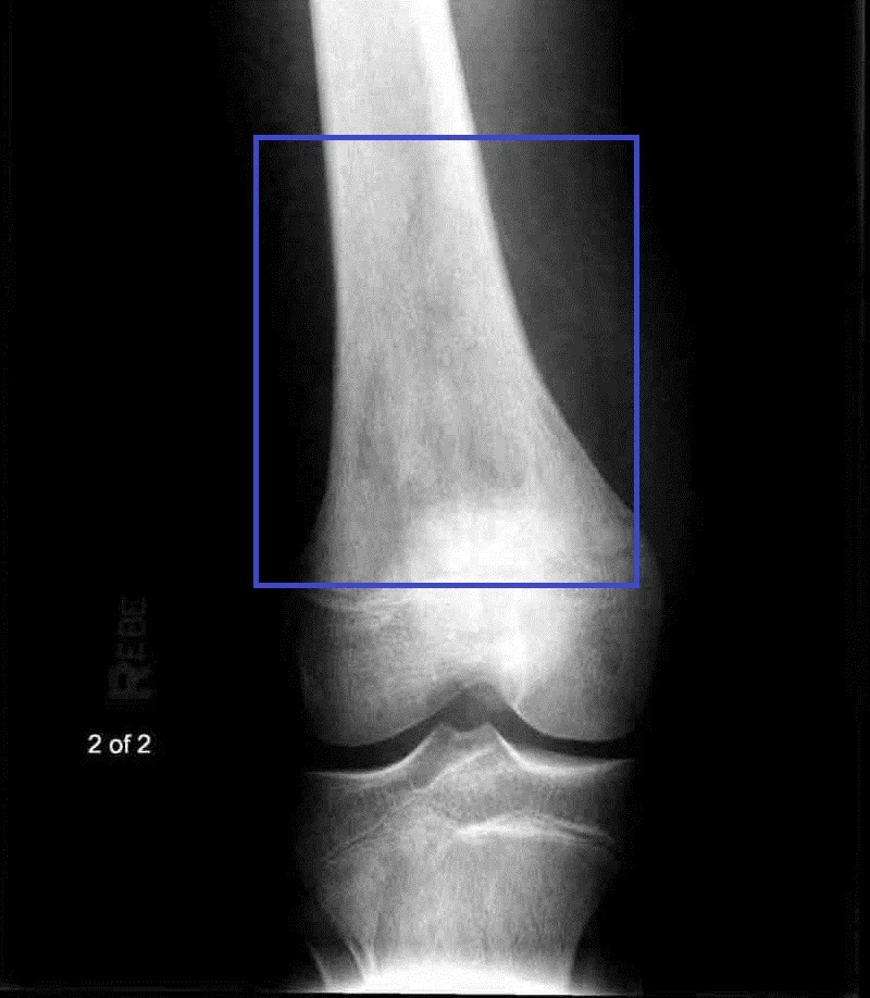
The most common primary malignant bone tumour overall. It has a bimodal age distribution, but the classic exam patient is an adolescent or young adult, with a smaller second peak in older adults, in whom it is associated with Paget's disease of bone or prior radiotherapy. It also occurs at increased frequency in hereditary retinoblastoma (RB1 mutation). It arises in the metaphysis of long bones, most classically the distal femur or proximal tibia - i.e. around the knee - reflecting the sites of fastest bone growth.
The radiograph shows an aggressive, destructive lesion with a mixed lytic and sclerotic pattern, new bone formation radiating from the cortex (the sunburst appearance), and elevation of the periosteum away from the cortex by the tumour, producing the Codman triangle.
Ewing sarcoma
Typically affects children and adolescents, slightly younger on average than the typical osteosarcoma patient. Unlike osteosarcoma, it characteristically arises in the diaphysis of long bones, or in the pelvis. The classic radiographic finding is a lamellated, "onion skin" periosteal reaction, produced by repeated cycles of the tumour breaking through reactive periosteal new bone. Ewing sarcoma is associated with a characteristic chromosomal translocation, t(11;22), fusing the EWSR1 and FLI1 genes.
It frequently presents with systemic features - fever, malaise, raised inflammatory markers, and a warm, tender, swollen area - that closely mimic osteomyelitis, a well-known exam trap that should prompt imaging before infection is assumed and antibiotics started.
Chondrosarcoma
Typically affects an older adult population, in contrast to osteosarcoma and Ewing sarcoma. It arises from cartilage and tends to be more indolent, with slower growth and a better prognosis stage for stage than the other two. It can arise de novo (primary chondrosarcoma) or from malignant transformation of a pre-existing enchondroma or osteochondroma (secondary chondrosarcoma), which is why a previously stable cartilage lesion that becomes painful or grows in adulthood needs re-imaging.
| Osteosarcoma | Ewing sarcoma | Chondrosarcoma | |
|---|---|---|---|
| Typical age | Adolescents/young adults (bimodal, second peak in older adults with Paget's disease) | Children and adolescents | Older adults |
| Typical site | Metaphysis of long bones - distal femur, proximal tibia | Diaphysis of long bones, or the pelvis | Axial skeleton and proximal long bones; often arises from a pre-existing cartilage lesion |
| Radiographic sign | Sunburst appearance, Codman triangle | Onion-skin periosteal reaction | Lytic lesion with stippled/popcorn ("rings and arcs") calcification |
| Associations | Paget's disease of bone, hereditary retinoblastoma, prior radiotherapy | t(11;22) translocation (EWSR1-FLI1); mimics osteomyelitis | Enchondroma or osteochondroma undergoing malignant transformation |
| Chemosensitivity | Chemosensitive | Chemosensitive and radiosensitive | Relatively chemo- and radio-resistant - surgery is central |
Clinical features of primary bone tumours
The features that should raise suspicion of a primary bone tumour, rather than a benign musculoskeletal problem, are consistent across osteosarcoma, Ewing sarcoma and chondrosarcoma:
- Persistent, unexplained bone pain - the single most important symptom, and the one that most often leads to delayed diagnosis if not taken seriously
- Pain worse at night or at rest, rather than purely with activity or mechanical loading - this pattern, rather than exertional pain alone, is the classic red flag
- A palpable mass or swelling, which may be tender and warm, particularly in Ewing sarcoma
- Reduced range of movement of an adjacent joint from local mass effect or pain
- Pathological fracture through the weakened area of bone, sometimes the presenting event
- Systemic features - fever, weight loss, malaise and fatigue - particularly prominent in Ewing sarcoma, less so in osteosarcoma and chondrosarcoma
Investigations
The initial investigation for suspected bone sarcoma is a plain X-ray, which in most cases is enough to raise or lower suspicion and to trigger referral. Definitive staging and tissue diagnosis then happen at a specialist centre, and the sequence matters as much as the individual tests.
- Plain X-ray - first-line; look for an aggressive periosteal reaction (sunburst, Codman triangle, onion skin), cortical destruction, and a soft tissue mass
- MRI of the whole affected bone - the key investigation for local staging, defining the extent of the tumour within the medullary cavity and any soft tissue extension, and essential for planning limb-sparing surgery
- CT chest - to look for pulmonary metastases, the commonest site of metastatic spread for both osteosarcoma and Ewing sarcoma
- Bone scan or whole-body MRI/PET-CT - to look for skip lesions or other skeletal metastases
- Bloods - ALP may be raised, particularly in osteosarcoma, reflecting osteoblastic activity; ESR and other inflammatory markers are often raised in Ewing sarcoma, contributing to the resemblance to infection; FBC and renal/liver function as a baseline before chemotherapy
- Image-guided biopsy - performed only once imaging is complete, to obtain definitive histological diagnosis and grade
Management
Primary bone sarcomas are managed by a specialist multidisciplinary team at a designated bone tumour centre, bringing together orthopaedic oncology, medical and clinical oncology, radiology and specialist pathology. This centralisation exists because these tumours are rare and because getting the surgical margins and biopsy tract right the first time has a direct effect on both local recurrence and limb salvage.
Surgery
Surgery is the mainstay of treatment for most primary bone sarcomas. The choice lies between limb-sparing surgery - wide local excision of the tumour with reconstruction using an endoprosthesis or biological graft - and amputation, decided on the basis of tumour size, location, neurovascular involvement and response to any neoadjuvant treatment. Modern limb-sparing techniques have made amputation less common than it once was, but it remains necessary where a clear margin cannot otherwise be achieved.
Chemotherapy
Osteosarcoma and Ewing sarcoma are both chemosensitive, and treatment uses combination chemotherapy given both neoadjuvantly (before surgery, to shrink the tumour and treat micrometastatic disease) and adjuvantly (after surgery). Histological response to neoadjuvant chemotherapy, assessed in the resected specimen, is an important prognostic marker. Chondrosarcoma is relatively chemo- and radio-resistant, so surgery is more central to its management and chemotherapy has a much smaller role.
Radiotherapy
Radiotherapy has a particular role in Ewing sarcoma, which is radiosensitive, and may be used instead of or alongside surgery, especially at sites where complete resection is difficult such as the pelvis. It also has a role for local control or palliation where surgery is not feasible, and for painful bone metastases generally.
Management of metastatic bone disease
This differs in emphasis: the aim is control of symptoms and prevention of skeletal complications rather than cure.
- Analgesia, following the WHO analgesic ladder, escalating as needed
- Bisphosphonates or denosumab to reduce skeletal-related events and to treat hypercalcaemia of malignancy
- Radiotherapy for pain relief, and for local control of a lesion at risk of fracturing
- Prophylactic surgical fixation of a long bone at high risk of pathological fracture, based on scoring systems that weigh site, size and radiographic appearance of the lesion
- Treatment of the underlying primary cancer, including systemic anticancer therapy appropriate to the tumour type
- Vertebroplasty/kyphoplasty or spinal surgery and urgent radiotherapy where there is spinal cord or cauda equina compression
Differential diagnosis
A destructive bone lesion has a broad differential, and age is the single most useful discriminator between them.
- Metastatic bone disease - by far the commonest cause of a destructive bone lesion in an adult, especially over 40
- Osteomyelitis - can closely mimic Ewing sarcoma in a child, with fever, local warmth, swelling and raised inflammatory markers
- Benign bone lesions - osteochondroma, enchondroma, giant cell tumour, osteoid osteoma, simple or aneurysmal bone cyst
- Stress fracture - localised pain related to activity, with a history of increased load, in a young or athletic patient
- Multiple myeloma - lytic "punched-out" lesions, particularly in the skull and axial skeleton, in an older adult with anaemia, renal impairment and a raised ESR/paraprotein
- Paget's disease of bone - can itself cause bone pain and deformity, and is a recognised risk factor for secondary osteosarcoma in older patients
Complications
Both the disease and its treatment carry a significant burden of complications.
- Pathological fracture through a weakened area of bone, from either metastatic or primary disease
- Spinal cord or cauda equina compression from vertebral metastases, or occasionally a primary tumour of the spine or pelvis
- Hypercalcaemia of malignancy, requiring IV fluids and IV bisphosphonates
- Metastatic spread, particularly to the lung, in osteosarcoma and Ewing sarcoma
- Complications of limb-sparing surgery - infection, prosthesis loosening or failure requiring revision, and in children, limb length discrepancy as the reconstructed limb no longer grows normally
- Complications of chemotherapy - myelosuppression, infection, cardiotoxicity (anthracyclines) and nephrotoxicity (cisplatin/ifosfamide)
Prognosis
Prognosis varies greatly by tumour type, histological grade, size at diagnosis and, most importantly, whether metastatic disease is present at presentation. With modern combined-modality treatment, localised osteosarcoma and localised Ewing sarcoma both carry a reasonable prospect of long-term survival, generally quoted in the region of 60-70% at five years for localised disease treated with surgery and chemotherapy.
Metastatic disease at presentation carries a much poorer outlook for both tumours, with five-year survival substantially lower, which is why prompt recognition and referral before metastatic spread has occurred makes a genuine difference to outcome. Chondrosarcoma behaves differently: low-grade lesions treated with adequate surgical excision have a good prognosis, while high-grade or dedifferentiated chondrosarcoma behaves much more aggressively and responds poorly to chemotherapy or radiotherapy.
For metastatic bone disease from a solid organ primary, prognosis is chiefly determined by the underlying cancer type and its systemic burden, but effective bone-targeted treatment - bisphosphonates, radiotherapy and prophylactic fixation - meaningfully reduces skeletal-related events and preserves quality of life even when cure is not possible.
References
- NICE NG12. Suspected cancer: recognition and referral. 2015, updated 2023. Available here
- NICE Clinical Knowledge Summaries. Bone tumours - primary and secondary. Available here
- Bone Cancer Research Trust. Understanding primary bone cancer. Available here
- Ferguson JL, Turner SP. Bone Cancer: Diagnosis and Treatment Principles. American Family Physician. 2018. Available here
- British Orthopaedic Oncology Society. Referral guidelines for suspected bone and soft tissue sarcoma. Available here
- Royal College of Radiologists. Guidance on imaging in the management of bone and soft tissue sarcoma. Available here
- Grimer RJ, Judson I, Peake D, Seddon B. Guidelines for the management of soft tissue sarcomas. Sarcoma. 2010. Available here
- NICE NG12. Metastatic bone disease and spinal metastases - suspected cancer referral criteria. Available here
This article is written for revision and education. It is not clinical guidance and must not be used to make decisions about the care of a patient. Always check current NICE guidance and local protocols.