
Oesophageal Cancer: Diagnosis, Staging and Management
Key points
- Two main types: adenocarcinoma (commonest in the UK, distal third, arising from Barrett's oesophagus) and squamous cell carcinoma (proximal two thirds).
- Adenocarcinoma risk factors: gastro-oesophageal reflux disease, Barrett's oesophagus, obesity, male sex and smoking.
- Squamous cell risk factors: smoking, alcohol, achalasia, Plummer-Vinson syndrome, and nutritional deficiency.
- Presentation: progressive dysphagia (solids before liquids), weight loss, odynophagia, and hoarseness if the recurrent laryngeal nerve is involved.
- Referral: dysphagia at any age warrants an urgent two-week-wait referral for upper GI endoscopy.
- Diagnosis: upper GI endoscopy with biopsy, then CT chest-abdomen-pelvis, PET-CT and endoscopic ultrasound for staging.
- Management: oesophagectomy with neoadjuvant chemotherapy or chemoradiotherapy for operable disease; palliative stenting and chemotherapy for advanced disease.
- Prognosis: poor, with an overall 5-year survival of around 15-20%, because most present at an advanced stage.
Introduction
Oesophageal cancer is a malignancy of the oesophageal wall, most commonly arising from the mucosal lining. It is the 14th most common cancer in the UK, with around 9,000 new cases each year, but is disproportionately lethal because symptoms appear late.1
There are two dominant histological subtypes with different risk factors, anatomical distributions and epidemiology. Adenocarcinoma is now the more common type in the UK and other Western countries, and its incidence has risen sharply over recent decades in parallel with rising rates of obesity and reflux disease. Squamous cell carcinoma predominates worldwide, particularly across the so-called oesophageal cancer belt from Iran to northern China.
Classification and risk factors
| Feature | Adenocarcinoma | Squamous cell carcinoma |
|---|---|---|
| Site | Distal third, near the gastro-oesophageal junction | Proximal and middle two thirds |
| Precursor lesion | Barrett's oesophagus (intestinal metaplasia) | Squamous dysplasia |
| Main risk factors | GORD, Barrett's oesophagus, obesity, smoking, male sex | Smoking, alcohol, achalasia, Plummer-Vinson syndrome, hot beverages, nitrosamines |
| UK incidence | Commoner, and rising | Less common, and falling |
| Global pattern | Predominates in Western countries | Predominates worldwide |
Other risk factors relevant to both types include increasing age, male sex, previous radiotherapy to the chest, and caustic (e.g. lye) injury to the oesophagus.2 Tylosis palmaris et plantaris, a rare autosomal dominant condition causing palmoplantar hyperkeratosis, carries a very high lifetime risk of squamous cell carcinoma.
Clinical features
The cardinal symptom is progressive dysphagia. Characteristically, difficulty begins with solids and progresses over weeks to months to soft foods and eventually liquids, reflecting the gradual narrowing of the lumen by an enlarging tumour. This progressive pattern contrasts with achalasia, where dysphagia affects solids and liquids from the outset.
Other features include:
- Unintentional weight loss, from both reduced intake and cancer cachexia
- Odynophagia (pain on swallowing)
- Retrosternal or epigastric discomfort, and a long history of reflux in adenocarcinoma
- Regurgitation of undigested food, and aspiration causing cough or recurrent chest infections
- Hoarseness, from recurrent laryngeal nerve involvement by tumour or nodes
- Haematemesis, melaena or iron-deficiency anaemia from tumour bleeding
- A palpable supraclavicular node (Virchow's node) or hepatomegaly in metastatic disease
By the time dysphagia develops, the tumour has usually occluded a substantial proportion of the lumen, which is why the majority of patients present with locally advanced or metastatic disease.
Differential diagnosis
Other causes of dysphagia must be considered, though malignancy should be excluded first in any new case:
- Benign peptic stricture: a long history of reflux, slowly progressive, no weight loss
- Achalasia: dysphagia to solids and liquids from the outset, regurgitation of undigested food, a dilated oesophagus with a bird's beak on barium swallow
- Eosinophilic oesophagitis: younger, atopic patients with food bolus obstruction
- Oesophageal web or ring: Plummer-Vinson syndrome (web with iron-deficiency anaemia and glossitis), Schatzki ring
- Extrinsic compression: lung cancer, mediastinal lymphadenopathy, a large left atrium, retrosternal goitre
- Neurological dysphagia: stroke, myasthenia gravis, motor neurone disease - typically causes oropharyngeal dysphagia with coughing and choking on swallowing
- Pharyngeal pouch: regurgitation of undigested food, halitosis, a gurgling neck swelling
Investigations
Referral and diagnosis
NICE recommends an urgent (two-week-wait) referral for upper GI endoscopy for dysphagia at any age, or for anyone aged 55 or over with weight loss plus upper abdominal pain, reflux or dyspepsia.3
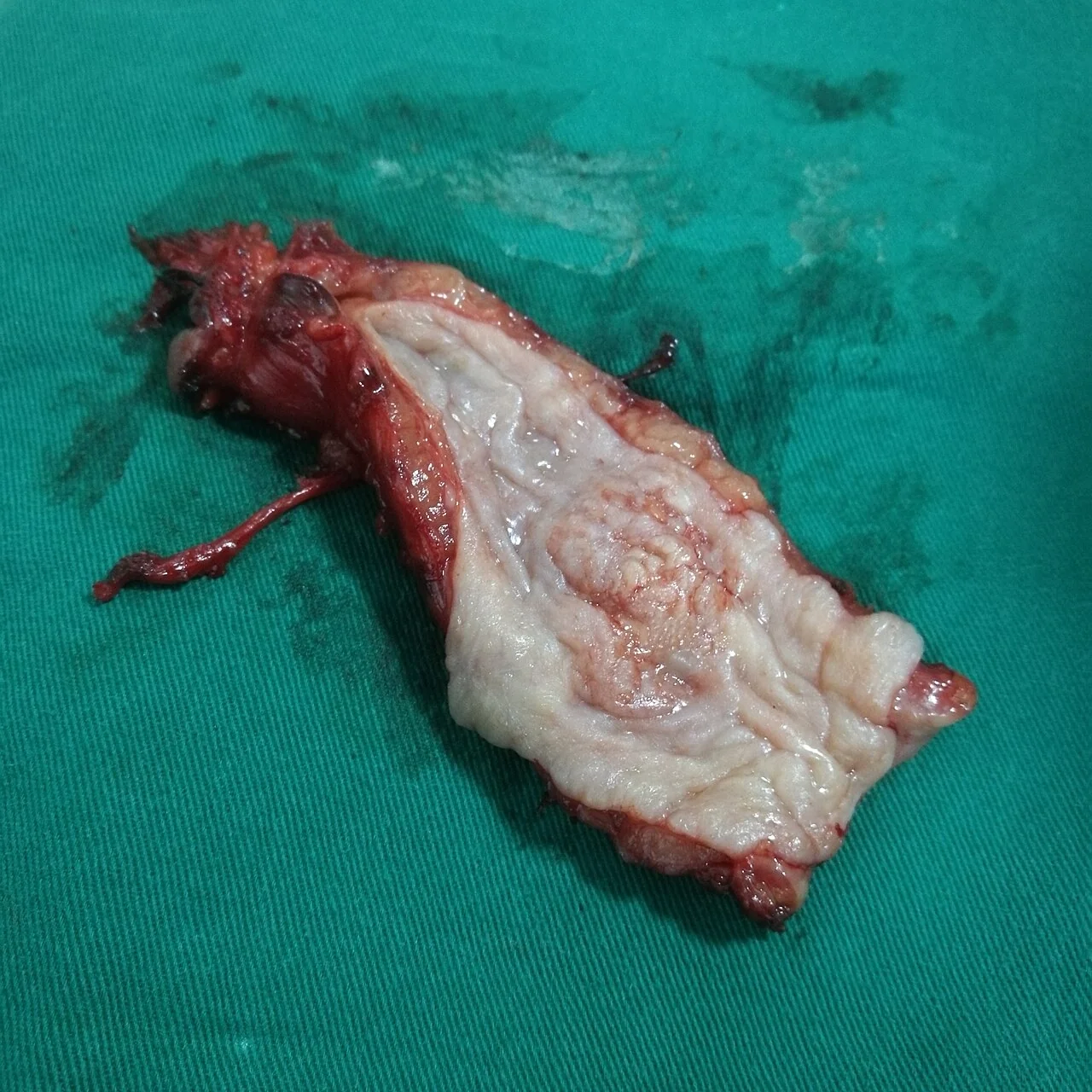
Upper GI endoscopy (OGD) with biopsy is the diagnostic investigation of choice. It allows direct visualisation of the tumour, multiple biopsies for histological diagnosis and subtype, and assessment of the tumour's distance from the incisors and its relationship to the gastro-oesophageal junction.
Staging
Accurate staging determines whether the patient is a candidate for curative treatment. It uses the TNM system and typically involves:
- CT chest, abdomen and pelvis: first-line to detect distant metastases and assess local invasion
- PET-CT: identifies occult metastatic disease and nodal involvement, and frequently changes management in apparently operable patients
- Endoscopic ultrasound (EUS): the most accurate way to assess depth of tumour invasion (T stage) and regional nodes (N stage), with fine-needle aspiration of suspicious nodes
- Staging laparoscopy: for tumours at the gastro-oesophageal junction, to detect peritoneal disease not seen on CT
- Bronchoscopy if there is concern about invasion of the trachea or bronchi in proximal tumours
Assessment of fitness for surgery is undertaken in parallel, including cardiopulmonary exercise testing, lung function and nutritional assessment, since oesophagectomy is a major undertaking.
Management
Management is decided by a specialist upper GI multidisciplinary team and depends on stage, histological subtype and patient fitness.2
Curative treatment
- Endoscopic resection: endoscopic mucosal resection or submucosal dissection for very early (T1a) tumours confined to the mucosa, often combined with radiofrequency ablation of residual Barrett's
- Oesophagectomy: the mainstay of curative treatment for operable disease, most commonly via an Ivor-Lewis (abdominal and right thoracic) approach, with reconstruction using a gastric conduit
- Neoadjuvant chemotherapy or chemoradiotherapy: given before surgery to downstage the tumour and improve survival, now standard practice for most resectable tumours
- Definitive chemoradiotherapy: an alternative to surgery, used particularly for squamous cell carcinoma, which is more radiosensitive, and for patients unfit for or declining surgery
Palliative treatment
Most patients present with disease that is not curable, so palliation of dysphagia is central. Options include self-expanding metal stent insertion to restore luminal patency, palliative radiotherapy or brachytherapy, and palliative chemotherapy or immunotherapy. Nutritional support with dietetic input and, where necessary, a feeding jejunostomy is important, alongside early involvement of the palliative care team for symptom control.
Complications
- Malnutrition and cachexia from progressive dysphagia
- Aspiration pneumonia from regurgitation or an oesophago-respiratory fistula
- Tracheo-oesophageal fistula, causing coughing on swallowing, particularly with proximal tumours
- Upper GI bleeding from tumour ulceration, occasionally catastrophic if a major vessel is eroded
- Recurrent laryngeal nerve palsy causing hoarseness and an unsafe swallow
- Metastatic disease, most commonly to liver, lung and bone
- Surgical complications: anastomotic leak, chyle leak, respiratory complications, and long-term reflux or dumping syndrome after reconstruction
Red flags
Prognosis
Oesophageal cancer has a poor prognosis, with overall 5-year survival in the UK of around 15-20%.1 This reflects late presentation: dysphagia only develops once a substantial part of the lumen is obstructed, by which point over half of patients have unresectable or metastatic disease. Stage at diagnosis is the dominant determinant of outcome - patients with early disease treated endoscopically or with surgery after neoadjuvant therapy can achieve long-term survival, while median survival in metastatic disease is under a year. This is the rationale for Barrett's oesophagus surveillance, which aims to detect neoplasia at a curable stage.
References
- Cancer Research UK. Oesophageal cancer statistics. Available here
- NICE NG83. Oesophago-gastric cancer: assessment and management in adults. 2018. Available here
- NICE NG12. Suspected cancer: recognition and referral. 2015 (updated 2023). Available here
- British Society of Gastroenterology. Guidelines on the diagnosis and management of Barrett's oesophagus. Gut. 2014. Available here
- K.Y.K.Z.K., CC BY-SA 3.0, via Wikimedia Commons. Available here
- NHS. Oesophageal cancer. 2024. Available here
- Smyth EC et al. Oesophageal cancer. Nat Rev Dis Primers. 2017. Available here
This article is written for revision and education. It is not clinical guidance and must not be used to make decisions about the care of a patient. Always check current NICE guidance and local protocols.