
Helicobacter pylori Infection: Testing and Eradication
Key points
- Helicobacter pylori: a spiral, urease-producing, Gram-negative bacterium that colonises the gastric mucus layer, infecting roughly a third of UK adults.
- Transmission: person-to-person, likely oral-oral or faecal-oral, usually acquired in childhood; more common with lower socioeconomic status and in some ethnic groups.
- Disease associations: peptic ulcer disease, chronic gastritis, gastric MALT lymphoma, and gastric adenocarcinoma.
- Presentation: most infected people are asymptomatic; when symptomatic, dyspepsia and epigastric pain predominate.
- Testing: urea breath test or stool antigen test are first-line non-invasive tests; biopsy-based tests are used when endoscopy is performed anyway.
- Test accuracy: PPIs cause false negatives and must be stopped for about 2 weeks, and antibiotics/bismuth for about 4 weeks, before testing.
- Eradication: triple therapy: a PPI plus two antibiotics for 7 days, chosen according to penicillin allergy and previous antibiotic exposure.
- Confirmation: retest with a urea breath test if symptoms persist, the ulcer was complicated, or the patient has MALT lymphoma.
Introduction
Helicobacter pylori is a spiral, flagellated, urease-producing Gram-negative bacterium that colonises the mucus layer overlying the gastric epithelium.1 It is one of the most common chronic bacterial infections worldwide, infecting an estimated 30-40% of adults in the UK and a much higher proportion in many low- and middle-income countries.
It is of major clinical importance because it is the leading identifiable cause of peptic ulcer disease and is classified by the World Health Organization as a Group 1 (definite) carcinogen for gastric cancer.2 Most infections, however, remain lifelong and entirely asymptomatic.
Microbiology and pathophysiology
H. pylori survives the acidic gastric environment by producing large amounts of urease, which hydrolyses urea to ammonia and carbon dioxide, buffering the immediate area around the bacterium. Its flagella allow it to burrow through the protective mucus layer to adhere to gastric epithelial cells, which it damages both directly, through cytotoxins such as CagA and VacA, and indirectly, through the chronic inflammatory response it provokes.1
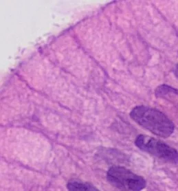
The pattern of gastritis determines the clinical outcome. Infection predominantly affecting the antrum increases gastrin release and acid secretion, favouring duodenal ulceration. Infection that spreads to involve the gastric body causes mucosal atrophy and reduced acid secretion, favouring gastric ulceration and, over decades, gastric adenocarcinoma via a sequence of atrophy, intestinal metaplasia and dysplasia. Chronic antigenic stimulation of gastric lymphoid tissue can rarely give rise to MALT lymphoma, most of which regress with eradication alone.2
Transmission and risk factors
H. pylori is transmitted person-to-person, most likely by oral-oral or faecal-oral routes, and infection is usually acquired in early childhood and, if untreated, persists for life. Risk factors for acquisition include:1
- Lower socioeconomic status and household crowding in childhood
- Birth in or migration from a region of high prevalence
- Close contact with an infected family member
- Poor access to clean water and sanitation
Clinical features
The large majority of infected people (roughly 80-90%) remain asymptomatic throughout life and are never diagnosed. When disease develops, presentation reflects the associated condition:
- Dyspepsia and epigastric pain from gastritis or peptic ulcer disease
- Bleeding, perforation, or gastric outlet obstruction as complications of peptic ulcer disease
- Weight loss, anorexia, dysphagia or an epigastric mass if gastric cancer has developed
- Unexplained iron-deficiency anaemia or, rarely, vitamin B12 deficiency or immune thrombocytopenia, both of which have recognised associations with H. pylori
Investigations
Testing is indicated for patients with dyspepsia not explained by another cause, confirmed peptic ulcer disease, a history of gastric MALT lymphoma, or before long-term NSAID or low-dose aspirin therapy in higher-risk patients.3 A critical practical point applies to every test below: PPIs cause false negatives and should be stopped for around 2 weeks, and antibiotics or bismuth-containing preparations for around 4 weeks, before testing.
| Test | Notes |
|---|---|
| Urea breath test | Non-invasive, first-line for initial diagnosis and to confirm eradication; detects active infection |
| Stool antigen test | Non-invasive, similar accuracy to the breath test; useful where breath testing is unavailable |
| Serology (IgG antibody) | Detects past exposure, cannot distinguish active from past infection, so not used to confirm eradication |
| Rapid urease test (CLO test) | Performed on a biopsy taken at endoscopy, rapid result |
| Histology | Biopsy-based, also assesses for gastritis, atrophy or malignancy |
In practice, the urea breath test or stool antigen test is used for a "test and treat" strategy in uninvestigated dyspepsia, while biopsy-based tests (rapid urease test or histology) are used opportunistically when a patient is already undergoing endoscopy.3
Management
Patients testing positive are offered eradication therapy, regardless of whether symptoms are present, given the strength of the association with peptic ulcer disease and gastric cancer.
First-line triple therapy
A proton pump inhibitor twice daily plus two antibiotics (typically amoxicillin with clarithromycin, or amoxicillin with metronidazole) for 7 days is first-line.4 In penicillin allergy, clarithromycin and metronidazole are combined instead.
Second-line and refractory infection
If first eradication fails, a different antibiotic combination is used for a second course, avoiding any antibiotic used previously wherever possible, to reduce the risk of resistance (clarithromycin resistance in particular has been rising). Persistent failure after two courses warrants specialist referral, culture and sensitivity testing where feasible, and consideration of quadruple therapy regimens.
Confirming eradication
Retesting is not routinely required after treatment of straightforward, uncomplicated dyspepsia, but is recommended when the original presentation was a complicated or bleeding peptic ulcer, symptoms persist, or the patient has MALT lymphoma, using a urea breath test at least 4 weeks after completing antibiotics and 2 weeks off any PPI.3
Complications
- Peptic ulcer disease, with its own risks of bleeding, perforation and gastric outlet obstruction
- Chronic atrophic gastritis and intestinal metaplasia, precursors of gastric cancer
- Gastric adenocarcinoma
- Gastric MALT lymphoma - a low-grade B-cell lymphoma that often regresses completely with eradication therapy alone
- Iron-deficiency anaemia and, rarely, vitamin B12 deficiency or immune thrombocytopenic purpura
Red flags
Prognosis
Eradication cures the majority of H. pylori-associated peptic ulcer disease and substantially reduces the risk of recurrence and of future ulcer complications.2 Early, low-grade gastric MALT lymphoma regresses in most patients after successful eradication alone, without chemotherapy. The impact on gastric cancer risk is greatest when eradication occurs before significant atrophic or metaplastic change has developed, which is one of the arguments for population-level screening and treatment programmes in high-incidence countries.
References
- NICE Clinical Knowledge Summaries (CKS). Helicobacter pylori infection. 2023. Available here
- IARC Working Group. Schistosomes, liver flukes and Helicobacter pylori. IARC Monographs on the Evaluation of Carcinogenic Risks to Humans, Vol. 61. 1994. Available here
- NICE NG12. Suspected cancer: recognition and referral. 2015 (updated 2023). Available here
- BNF. Helicobacter pylori eradication. Available here
- Mikael Häggström, MD, CC0, via Wikimedia Commons. Available here
- NHS. Helicobacter pylori (H. pylori) infection. 2022. Available here
- Malfertheiner P et al. Management of Helicobacter pylori infection - the Maastricht VI/Florence consensus report. Gut. 2022. Available here
This article is written for revision and education. It is not clinical guidance and must not be used to make decisions about the care of a patient. Always check current NICE guidance and local protocols.