
Clostridioides difficile Colitis: Diagnosis and Management
Key points
- C. difficile: a Gram-positive, spore-forming anaerobic bacillus that overgrows when antibiotics disrupt the normal colonic flora.
- Mechanism: toxins A (enterotoxin) and B (cytotoxin) damage the colonic mucosa, producing inflammation and the characteristic pseudomembranes.
- The 4 Cs: the highest-risk antibiotics are clindamycin, co-amoxiclav, cephalosporins and ciprofloxacin (fluoroquinolones).
- Other risk factors: age over 65, hospitalisation or care home residence, proton pump inhibitors, immunosuppression and previous C. difficile infection.
- Presentation: watery, offensive diarrhoea with crampy abdominal pain, fever and a raised white cell count, typically during or after antibiotics.
- Diagnosis: two-stage stool testing - glutamate dehydrogenase (GDH) or PCR to detect the organism, then a toxin assay to confirm active infection.
- Treatment: stop the precipitating antibiotic if possible; oral vancomycin is first-line, fidaxomicin second-line for recurrence.
- Severe disease: watch for toxic megacolon and perforation; avoid antimotility agents, and involve surgeons early in fulminant colitis.
Introduction
Clostridioides difficile (formerly Clostridium difficile) is a Gram-positive, spore-forming, anaerobic bacillus and the leading cause of healthcare-associated infectious diarrhoea in the developed world.1 It causes a spectrum of illness from mild self-limiting diarrhoea through pseudomembranous colitis to life-threatening fulminant colitis with toxic megacolon.
It is a major target of infection prevention and antimicrobial stewardship programmes, and rates in the UK fell substantially following national initiatives around antibiotic prescribing and hospital hygiene, though it remains an important cause of morbidity and mortality in older, hospitalised patients.
Pathophysiology and risk factors
The colon's normal bacterial flora provides colonisation resistance, preventing pathogenic overgrowth. Antibiotics disrupt this flora, allowing ingested C. difficile spores - which are highly resistant to heat, acid and alcohol-based hand gels - to germinate and proliferate.1
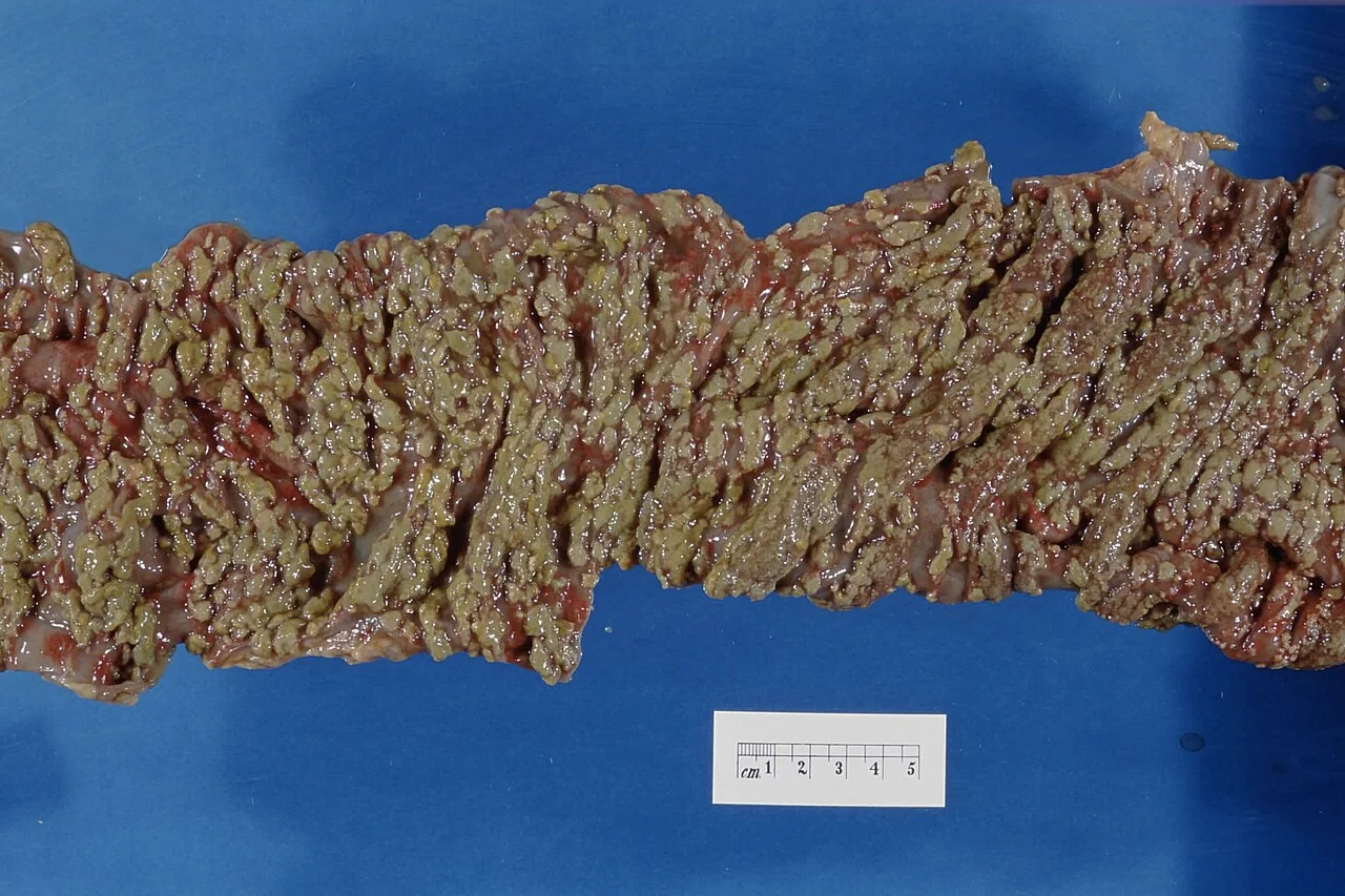
Pathogenic strains produce two exotoxins: toxin A (an enterotoxin causing fluid secretion and mucosal inflammation) and toxin B (a more potent cytotoxin causing cytoskeletal disruption and cell death). Together they produce mucosal inflammation, ulceration and the yellowish adherent plaques of fibrin, mucus and inflammatory debris known as pseudomembranes. The hypervirulent ribotype 027 (NAP1/BI) strain produces additional binary toxin and is associated with more severe disease and higher relapse rates.
Risk factors
- Antibiotic exposure - the dominant risk factor. Remember the 4 Cs: clindamycin, co-amoxiclav, cephalosporins and ciprofloxacin (and other fluoroquinolones). Risk persists for up to 3 months after a course
- Age over 65
- Hospitalisation or residence in a care home, and prolonged length of stay
- Proton pump inhibitors and other acid-suppressing drugs
- Immunosuppression, chemotherapy and serious comorbidity
- Previous C. difficile infection - the strongest predictor of a further episode
- Gastrointestinal surgery, nasogastric feeding and inflammatory bowel disease
Clinical features
The typical presentation is watery, offensive-smelling diarrhoea beginning during a course of antibiotics or within several weeks of finishing one. Associated features include:
- Crampy lower abdominal pain and tenderness
- Fever and general malaise
- Nausea and anorexia
- A markedly raised white cell count, which can be strikingly high and may precede the diarrhoea
- Dehydration and electrolyte disturbance
- Blood is usually absent or only present in small amounts, distinguishing it from many invasive bacterial causes
Severity ranges widely. Some patients have only mild diarrhoea, while others develop fulminant colitis with severe abdominal pain, distension, systemic toxicity and haemodynamic instability. Paradoxically, diarrhoea may cease as ileus and toxic megacolon develop, which can falsely reassure - worsening abdominal distension and systemic upset in a patient whose diarrhoea has suddenly stopped is an ominous sign.
Investigations
Stool testing
Testing is performed on diarrhoeal (unformed) stool only, since formed stool from an asymptomatic carrier will give a positive result without indicating disease. UK practice uses a two-stage testing algorithm.2
| Stage | Test | Interpretation |
|---|---|---|
| 1 - screening | Glutamate dehydrogenase (GDH) antigen, or nucleic acid amplification (PCR) | Highly sensitive; detects the presence of the organism but not whether it is producing toxin. A negative result effectively excludes infection |
| 2 - confirmation | Toxin A/B enzyme immunoassay | Detects active toxin production. GDH positive and toxin positive = C. difficile infection |
| Discordant | GDH positive, toxin negative | Suggests carriage or possible infection; interpret alongside the clinical picture, and treat if the patient is unwell with no alternative explanation |
Other investigations
- Full blood count: white cell count is a key severity marker and can exceed 25 × 10⁹/L in severe disease
- Urea, electrolytes and creatinine: for acute kidney injury and dehydration
- Albumin, CRP and lactate: low albumin and raised lactate indicate severe disease
- Abdominal X-ray: for colonic dilatation, mucosal thumbprinting or evidence of toxic megacolon
- CT abdomen: in severe or complicated disease, showing colonic wall thickening, the accordion sign and pericolonic stranding, and identifying perforation
- Flexible sigmoidoscopy: not routine and carries perforation risk in severe colitis, but may show pseudomembranes where the diagnosis is uncertain
Severity assessment
Severity determines the treatment setting and intensity, and should be reassessed daily.3
| Severity | Features |
|---|---|
| Mild | Fewer than 3 loose stools daily, normal white cell count |
| Moderate | 3-5 loose stools daily, raised white cell count but below 15 × 10⁹/L |
| Severe | White cell count above 15 × 10⁹/L, or acutely rising creatinine (over 50% above baseline), or temperature above 38.5°C, or evidence of severe colitis on examination or imaging. Stool frequency is an unreliable guide in severe disease |
| Life-threatening (fulminant) | Hypotension, ileus, toxic megacolon, or CT evidence of severe disease |
Management
General measures
- Stop the precipitating antibiotic wherever clinically possible, or switch to a lower-risk agent
- Review and stop proton pump inhibitors and other non-essential acid suppression
- Avoid antimotility agents such as loperamide, and opioids - these retain toxin and increase the risk of toxic megacolon
- Fluid and electrolyte replacement, with nutritional support as needed
- Isolate the patient in a side room with enteric precautions. Use soap and water for hand hygiene, as alcohol gel does not kill spores, and clean the environment with a chlorine-based disinfectant
Antibiotic treatment
| Scenario | Treatment |
|---|---|
| First episode, any severity | Oral vancomycin 125 mg four times daily for 10 days |
| No improvement, or second episode within 12 weeks | Oral fidaxomicin 200 mg twice daily for 10 days |
| Further recurrence, or failure of the above | Seek specialist advice: oral vancomycin (tapered or pulsed) with or without IV metronidazole, or fidaxomicin |
| Life-threatening infection | Oral vancomycin plus IV metronidazole, with urgent specialist and surgical review |
Vancomycin is given orally because it is not absorbed from the gut, delivering high luminal concentrations exactly where the infection is; intravenous vancomycin is ineffective for C. difficile colitis. Conversely, metronidazole is well absorbed and reaches the colon via biliary and inflammatory exudate, which is why the intravenous route is used when ileus prevents oral drug reaching the colon. Metronidazole is no longer first-line, having been shown to be less effective than vancomycin.3
Recurrent infection
Around 20-25% of patients relapse after a first episode, and the risk rises with each subsequent recurrence. Faecal microbiota transplantation (FMT) - instillation of processed stool from a screened healthy donor - restores colonisation resistance and is recommended by NICE for patients who have had two or more previous episodes, with cure rates around 90%.4 Bezlotoxumab, a monoclonal antibody against toxin B, is an alternative adjunct for reducing recurrence in selected high-risk patients.
Surgery
Urgent surgical review is needed for fulminant colitis, toxic megacolon, perforation, or deterioration despite maximal medical therapy. Subtotal colectomy with end ileostomy is the usual procedure and can be life-saving, though it carries high mortality in this frail population, so early rather than delayed involvement of the surgical team is important.
Complications
- Pseudomembranous colitis
- Toxic megacolon: colonic dilatation over 6 cm with systemic toxicity, risking perforation
- Bowel perforation and faecal peritonitis, carrying very high mortality
- Paralytic ileus, which may cause diarrhoea to cease deceptively
- Severe dehydration, acute kidney injury and electrolyte disturbance
- Hypoalbuminaemia from a protein-losing enteropathy, with oedema
- Sepsis and multi-organ failure
- Recurrent infection, affecting a quarter of patients after a first episode
- Post-infectious irritable bowel syndrome
Red flags
Prognosis
Most patients with mild to moderate infection respond well to stopping the precipitating antibiotic and a course of oral vancomycin, with symptoms improving over several days.3 Recurrence is the principal problem, affecting around a quarter of patients after a first episode and a higher proportion after each subsequent one - which is the rationale for fidaxomicin and, in repeated relapses, faecal microbiota transplantation.
Severe and fulminant disease carries substantial mortality, particularly in elderly patients with significant comorbidity, and mortality after emergency colectomy for fulminant colitis is high. Because the dominant risk factor is modifiable, prevention through antimicrobial stewardship, judicious use of proton pump inhibitors, isolation and rigorous environmental cleaning remains the most effective way to improve outcomes at a population level.
References
- Public Health England. Clostridium difficile infection: guidance on management and treatment. 2013. Available here
- Department of Health. Updated guidance on the diagnosis and reporting of Clostridium difficile. 2012. Available here
- NICE NG199. Clostridioides difficile infection: antimicrobial prescribing. 2021. Available here
- NICE IPG485. Faecal microbiota transplant for recurrent Clostridium difficile infection. 2014. Available here
- Narraburra, CC0, via Wikimedia Commons. Available here
- NICE Clinical Knowledge Summaries (CKS). Diarrhoea - antibiotic associated. 2023. Available here
- NHS. Clostridium difficile. 2023. Available here
This article is written for revision and education. It is not clinical guidance and must not be used to make decisions about the care of a patient. Always check current NICE guidance and local protocols.