
Squamous Cell Carcinoma and Bowen Disease
Key points
- Cutaneous SCC: the second commonest skin cancer, a malignant tumour of keratinocytes that, unlike basal cell carcinoma, carries a real (if generally low) risk of regional and distant metastasis.
- Bowen disease: squamous cell carcinoma in situ - malignant keratinocytes confined entirely to the epidermis - presenting as a well-demarcated, scaly or crusted erythematous plaque, with a small but real risk of progressing to invasive SCC if untreated.
- Precursor lesion: actinic keratosis is the commonest precursor to invasive SCC; only a small proportion progress, but the sheer number of actinic keratoses in the population makes them numerically important.
- Clinical features of invasive SCC: a keratotic, indurated, often tender nodule or plaque that can ulcerate, typically on chronically sun-exposed sites - the scalp, ears, lips, dorsum of the hands and forearms.
- Key risk factor beyond UV: immunosuppression carries a far stronger association with SCC than with BCC - organ transplant recipients have up to a 100-fold increased risk, and their SCCs behave more aggressively.
- Marjolin ulcer: SCC arising within a chronic wound, burn scar or area of longstanding inflammation, often after a latency of decades, and behaving more aggressively than sun-induced SCC.
- Management: Bowen disease is treated with cryotherapy, topical 5-fluorouracil, photodynamic therapy or curettage; invasive SCC needs surgical excision with a margin matched to risk, or Mohs surgery for high-risk lesions.
- Red flag: size over 2cm, depth over 4mm, poor differentiation, perineural invasion, ear or lip location, and immunosuppression are all high-risk features that meaningfully raise metastatic risk and need specialist, MDT-led management.
Introduction
Cutaneous squamous cell carcinoma (SCC) is a malignant tumour of epidermal keratinocytes and the second commonest skin cancer after basal cell carcinoma. The critical distinction from BCC is that SCC has genuine, if generally modest, metastatic potential, first to regional lymph nodes and later to distant sites, which changes the whole approach to risk-stratification and follow-up.1
Bowen disease is SCC confined entirely to the epidermis - squamous cell carcinoma in situ - and represents an intermediate step on the pathway from sun-damaged skin (actinic keratosis) to invasive disease. Recognising where a lesion sits on this spectrum, and correctly identifying the minority of SCCs with high-risk features, is the core clinical skill this topic tests.
Risk factors
- Cumulative lifetime UV exposure - the dominant risk factor, as with BCC, explaining the predilection for chronically sun-exposed sites
- Immunosuppression - a far stronger risk factor for SCC than for BCC; organ transplant recipients have up to a 100-fold increased incidence, with more numerous, more aggressive, and more rapidly metastasising tumours4
- Human papillomavirus infection - implicated particularly in periungual, genital and some immunosuppressed patients' SCCs
- Chronic wounds, burns, ulcers and scars - SCC arising within these is called a Marjolin ulcer, often after a latency of 10-30 years, and tends to behave more aggressively than typical sun-induced SCC7
- Arsenic exposure
- Smoking - particularly associated with SCC of the lip
- Chronic immunosuppressive or photosensitising drugs (voriconazole, azathioprine)
- Genetic conditions - xeroderma pigmentosum (defective DNA repair) causes multiple SCCs from a young age with minimal sun exposure
Precursor lesions and disease spectrum
SCC typically develops through a recognised progression, though not every lesion passes visibly through each stage:
- Actinic keratosis - a rough, scaly, erythematous macule or papule on chronically sun-damaged skin, representing atypical keratinocyte proliferation confined to the lower epidermis; the commonest precursor, though only a small proportion (often quoted around 1 in 1000 per lesion per year) progress to invasive SCC6
- Bowen disease (SCC in situ) - full-thickness epidermal involvement by atypical keratinocytes, but with an intact basement membrane and no dermal invasion
- Invasive SCC - malignant keratinocytes breach the basement membrane and invade the dermis, acquiring the potential for metastasis
Clinical features
Bowen disease
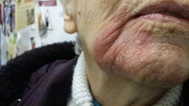
Presents as a slowly enlarging, well-demarcated, erythematous, scaly or crusted plaque, occurring predominantly in older adults (rare before age 60) and disproportionately in women, most often on the lower legs in previously or currently sun-exposed skin, though any site including mucosal surfaces can be affected. Growth is slow, over months to years, and lesions are typically asymptomatic other than mild irritation.
Invasive squamous cell carcinoma
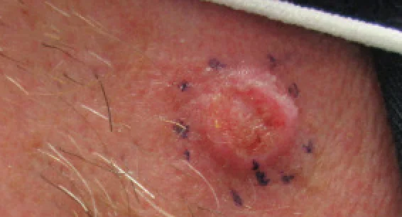
Presents as a firm, keratotic, indurated nodule or plaque, often with surface crusting, scaling or ulceration, and can be tender to touch (unlike the typically painless BCC). Sites reflect chronic sun exposure - the scalp (especially in balding men), ears, lower lip, dorsum of the hands and forearms - though it can also arise within chronic wounds or scars (Marjolin ulcer).
Keratoacanthoma
A distinctive, rapidly growing variant that develops over just a few weeks into a dome-shaped nodule with a characteristic central keratin-filled crater, closely resembling a well-differentiated SCC both clinically and histologically. Unlike typical SCC, a keratoacanthoma can spontaneously regress over several months, but because it cannot be reliably distinguished from true invasive SCC on clinical grounds alone, it is generally excised and treated as SCC rather than observed.
Clinical examination
- Size, depth and induration - larger, deeper, firmer lesions carry higher metastatic risk
- Fixation to underlying structures - suggests deeper invasion
- Regional lymph node examination - essential for any SCC with high-risk features, given the genuine metastatic potential
- Sensory testing around the lesion - perineural invasion can cause localised numbness or paraesthesia, an important and easily missed high-risk feature
- Site - the ear, lip and areas of chronic scarring or ulceration all carry a higher intrinsic risk regardless of other features
- Full skin examination for further actinic keratoses, Bowen disease or additional SCCs, particularly in a patient with a history of significant sun exposure or immunosuppression
Differential diagnosis
- Basal cell carcinoma - pearly, rolled edge with telangiectasia, generally painless, and almost never metastasises; see Basal Cell Carcinoma
- Keratoacanthoma - discussed above; managed the same way as SCC given the inability to reliably distinguish the two clinically
- Viral wart - can mimic a keratotic SCC, particularly on the hands; a lesion in an older patient that grows or changes should not be assumed to be a simple wart
- Psoriasis or eczema - considered for a scaly plaque resembling Bowen disease, particularly if multiple similar lesions are present elsewhere
- Amelanotic melanoma - can present as a non-healing, ulcerated or crusted lesion and should be considered, particularly if a presumed SCC behaves atypically or fails to respond as expected
Investigations
Biopsy confirms the diagnosis and, for invasive SCC, grades differentiation and identifies high-risk histological features that determine the intensity of further management.
- Biopsy (incisional, punch, shave, or excisional) - confirms Bowen disease versus invasive SCC and, for invasive disease, reports differentiation (well, moderate or poor), depth of invasion, and presence or absence of perineural or lymphovascular invasion
- Imaging (CT, MRI or ultrasound) - considered for high-risk SCC to assess for regional nodal disease or deep local invasion, particularly around the head and neck
- Sentinel lymph node biopsy - increasingly considered for high-risk SCC without clinically palpable lymphadenopathy, though its role is less standardised than in melanoma
- Fine needle aspiration or core biopsy of palpable lymphadenopathy - to confirm nodal metastasis if present
Management
Bowen disease
- Cryotherapy - a simple, effective first-line option for a single, well-defined lesion
- Topical 5-fluorouracil - applied over several weeks, useful for larger or multiple lesions
- Photodynamic therapy - a topical photosensitiser activated with light, giving good cosmetic results, particularly useful on the lower leg where healing after other treatments can be slow
- Curettage and cautery, or excision - for lesions where tissue diagnosis is wanted or other treatments have failed8
Invasive SCC
Management is stratified by risk, based on size, depth, differentiation, site and host immune status.1
| Risk category | Features | Management |
|---|---|---|
| Low risk | Under 2cm, well-differentiated, no perineural invasion, low-risk site, immunocompetent | Surgical excision with a 4-6mm margin |
| High risk | Over 2cm, poorly differentiated, perineural or lymphovascular invasion, ear/lip/temple location, recurrent disease, or immunosuppressed patient | Wider excision margin or Mohs micrographic surgery; consider imaging and MDT discussion |
- Mohs micrographic surgery - preferred for high-risk lesions in cosmetically or functionally sensitive sites, maximising tissue conservation while confirming complete margin clearance
- Radiotherapy - for patients unfit for surgery, as adjuvant treatment after incomplete excision or significant perineural invasion, or for regional nodal disease
- Lymph node dissection - for confirmed regional nodal metastasis, often combined with adjuvant radiotherapy
- Cemiplimab and other PD-1 checkpoint inhibitors - for locally advanced or metastatic SCC not amenable to surgery or radiotherapy, producing durable responses in a substantial proportion of patients and representing a major advance for a disease that previously had very limited systemic treatment options5
Complications
- Regional lymph node metastasis - the commonest form of spread, particularly from high-risk lesions
- Distant metastasis - uncommon overall but more likely with high-risk histological features or immunosuppression
- Local tissue destruction - invasion of underlying cartilage, bone or nerve, particularly relevant around the ear, nose and periorbital area
- Perineural spread - can extend along a nerve well beyond the visible tumour margin, causing local recurrence even after apparently complete excision, and sometimes producing progressive facial nerve palsy if the facial nerve is involved
- Progression from Bowen disease to invasive SCC if left untreated
Red flags
Prognosis
Bowen disease has an excellent prognosis when treated, since disease confined to the epidermis cannot yet metastasise; the main reason to treat it is to prevent progression to invasive disease over time.6
Low-risk invasive SCC also has a very good prognosis, with cure rates comparable to BCC after adequate excision. High-risk SCC, however, carries a meaningfully higher rate of local recurrence and regional metastasis - overall metastatic rates for cutaneous SCC are often quoted around 2-5%, but this rises substantially in the presence of the high-risk features above, particularly in immunosuppressed patients, where SCC behaves as a considerably more aggressive disease than in the general population.2,3
References
- British Association of Dermatologists. Guidelines for the management of people with cutaneous squamous cell carcinoma 2020. British Journal of Dermatology. 2021. Available here
- NICE NG12. Suspected cancer: recognition and referral. 2015, updated 2023. Available here
- Cancer Research UK. Non-melanoma skin cancer statistics. Available here
- Euvrard S, Kanitakis J, Claudy A. Skin cancers after organ transplantation. New England Journal of Medicine. 2003. Available here
- Migden MR, Rischin D, Schmults CD et al. PD-1 blockade with cemiplimab in advanced cutaneous squamous-cell carcinoma. New England Journal of Medicine. 2018. Available here
- Cockerell CJ. Histopathology of incipient intraepidermal squamous cell carcinoma ('actinic keratosis'). Journal of the American Academy of Dermatology. 2000. Available here
- Kerr-Valentic MA, Samimi K, Rohlen BH et al. Marjolin's ulcer: modern analysis of an ancient problem. Plastic and Reconstructive Surgery. 2009. Available here
- DermNet NZ. Bowen disease. Available here
This article is written for revision and education. It is not clinical guidance and must not be used to make decisions about the care of a patient. Always check current NICE guidance and local protocols.