
Osteoarthritis: Diagnosis and Management
Key points
- Osteoarthritis: a disorder of the whole joint - cartilage loss, subchondral bone remodelling and osteophyte formation - driven by an imbalance between mechanical stress and the joint's capacity to repair itself.
- Pattern: weight-bearing joints (knee, hip) and the hands - DIPs, PIPs and the first carpometacarpal joint. Unlike rheumatoid arthritis, it is often asymmetrical.
- Symptoms: pain worse with activity and relieved by rest, brief morning stiffness (under 30 minutes), and stiffness after inactivity ('gelling').
- Diagnosis: clinical, in a patient aged 45 or over with activity-related joint pain and no morning stiffness lasting more than 30 minutes - imaging is not required to diagnose.
- Radiographic features: remembered as LOSS - Loss of joint space, Osteophytes, Subchondral Sclerosis, Subchondral cysts.
- First-line management: therapeutic exercise, weight loss where relevant, and patient education - not analgesia.
- Analgesia: topical NSAIDs first-line for knee and hand OA; oral NSAIDs with gastroprotection second-line. Paracetamol and weak opioids are no longer routinely recommended.
- Definitive treatment: joint replacement for end-stage disease significantly affecting quality of life, when conservative measures have failed.
Introduction
Osteoarthritis (OA) is the commonest form of joint disease worldwide and the leading cause of pain and disability in older adults. It was once described simply as 'wear and tear', but it is now understood as an active process affecting the whole joint - cartilage, subchondral bone, synovium and the surrounding muscles and ligaments - rather than passive degeneration of cartilage alone.1
It becomes increasingly common with age, affecting most people to some degree by their seventies, though the correlation between radiographic changes and symptoms is weak - many people have osteoarthritic changes on imaging with no pain at all, and vice versa. This is a frequently tested point: do not treat the X-ray, treat the patient.
It is a favourite exam topic for the contrast it provides with rheumatoid arthritis: same small joints of the hand can be affected by both, but the pattern, timing and joints spared are different, and this distinction is exactly what exams like to probe.
Aetiology and pathophysiology
OA results from an imbalance between the mechanical stress placed on a joint and the ability of chondrocytes to repair the cartilage matrix. Early disease involves cartilage matrix breakdown and attempted repair; as this fails, cartilage thins, exposing subchondral bone, which responds with sclerosis, cyst formation and osteophyte growth at the joint margins as the body attempts to stabilise and redistribute load.
OA is broadly split into primary (idiopathic, related to age and genetic predisposition) and secondary (following joint trauma, previous inflammatory arthritis, or an underlying metabolic/structural abnormality such as haemochromatosis or joint dysplasia).
Risk factors
- Increasing age - the strongest risk factor
- Female sex, especially after the menopause
- Obesity - particularly for knee OA, both mechanical loading and metabolic/inflammatory effects of adipose tissue
- Joint trauma or previous surgery
- Occupational or repetitive joint stress
- Family history and genetic predisposition, especially for hand and hip OA
- Pre-existing joint disease - inflammatory arthritis, joint dysplasia, avascular necrosis
- Joint malalignment (varus/valgus deformity)
Clinical features
NICE recommends a clinical diagnosis, without imaging, in a patient aged 45 or over who has activity-related joint pain and either no morning joint-related stiffness or morning stiffness lasting less than 30 minutes.2
- Pain - worse with activity/loading, relieved by rest; as disease progresses, pain can occur at rest and disturb sleep
- Brief morning stiffness (under 30 minutes) and 'gelling' - stiffness after a period of inactivity that eases quickly with movement
- Reduced range of movement and crepitus on joint movement
- Bony swelling rather than the soft, boggy swelling of synovitis
- Instability or 'giving way', particularly at the knee
- Functional impact - difficulty with stairs, walking distance, fine hand tasks
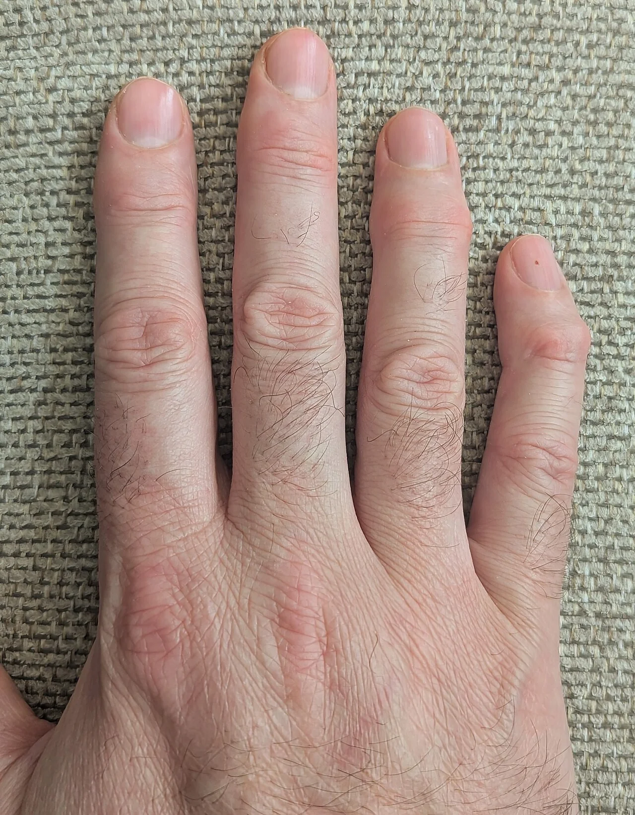
Joint distribution
| Joint | Feature |
|---|---|
| DIP joints | Heberden's nodes |
| PIP joints | Bouchard's nodes |
| First carpometacarpal joint | 'Squaring' of the thumb base |
| Knee | Medial compartment most often affected, producing varus deformity |
| Hip | Groin pain, may be referred to the knee; reduced internal rotation is often the earliest sign |
| Spine (facet joints) | Lower back and neck pain, can cause nerve root or spinal canal compromise |
Clinical examination
- Gait - antalgic gait, use of walking aids
- Look - bony swelling, joint deformity (varus knee, squared thumb base), muscle wasting from disuse
- Feel - bony, hard swelling; crepitus on movement; joint line tenderness; effusion may be present but is usually modest compared with inflammatory arthritis
- Move - reduced range of movement, pain at extremes of movement
- Special tests - hip internal rotation (often the first movement lost), Trendelenburg test for hip abductor weakness
Differential diagnosis
- Rheumatoid arthritis and other inflammatory arthropathies - symmetry, DIP sparing, prolonged morning stiffness improving with movement, raised inflammatory markers
- Crystal arthropathy (gout, pseudogout) - acute, hot, exquisitely painful joint rather than a chronic pattern
- Septic arthritis - acute, systemically unwell, single hot swollen joint - always consider and exclude if the presentation is atypical
- Referred pain - hip OA can present as knee pain; lumbar spine disease can mimic hip OA
- Bursitis / tendinopathy - localised to the bursa or tendon rather than the joint line
- Avascular necrosis - consider in a younger patient with hip pain and risk factors (steroid use, alcohol excess)
Investigations
OA is a clinical diagnosis in the typical patient described above, and NICE explicitly advises against routine blood tests or imaging to confirm it.2 Investigations are used to exclude other diagnoses when the presentation is atypical, or to plan surgery.
- Inflammatory markers (CRP/ESR) and rheumatoid factor/anti-CCP - normal in OA; used to exclude an inflammatory arthropathy when the pattern is not clear-cut
- Plain radiograph - not needed to diagnose, but useful to confirm severity, exclude other pathology, or plan joint replacement
Radiographic features - LOSS
- Loss of joint space
- Osteophytes at the joint margins
- Subchondral sclerosis
- Subchondral cysts
Management
NICE's 2022 update (NG226) restructured OA management around core, non-pharmacological treatments first, with pharmacological therapy as an adjunct rather than the mainstay.2
Core treatments (offer to everyone)
- Therapeutic exercise - local muscle strengthening and general aerobic fitness, tailored to the patient, continued long-term
- Weight loss if the patient is overweight or obese, which reduces load on weight-bearing joints and has systemic anti-inflammatory benefit
- Education about the condition and self-management strategies
Pharmacological management
Topical NSAIDs are first-line for knee and hand OA, and are preferred over oral NSAIDs where the joint is accessible, because they achieve useful local drug levels with minimal systemic absorption.
Oral NSAIDs (with a proton pump inhibitor for gastroprotection) are used second-line, or first-line where topical treatment is not suitable (for example the hip). Use the lowest effective dose for the shortest time, and consider cardiovascular and renal risk before prescribing.
Intra-articular corticosteroid injection can be considered for a flare of moderate to severe pain, particularly while awaiting other interventions, but the benefit is short-lived (weeks) and repeated injections are not recommended for regular long-term use.
- Glucosamine and chondroitin are not recommended - no consistent evidence of benefit
- Rubefacients (topical capsaicin) are not recommended for knee/hand OA
- Intra-articular hyaluronic acid is not recommended by NICE
Surgery
Referral for joint replacement should be considered when OA has a substantial impact on quality of life - pain, function, psychosocial wellbeing - and is not adequately controlled by non-surgical management, rather than being based on a fixed threshold of radiographic severity or age. Total knee and total hip replacement are highly effective at relieving pain and restoring function in end-stage disease.
Complications
Progressive pain and functional loss can significantly affect mobility, independence and mental health, with a strong association between OA and depression, poor sleep and social isolation in severe disease. Joint deformity and instability increase falls risk in older patients. Complications of joint replacement surgery include infection, venous thromboembolism, dislocation and the eventual need for revision surgery as prosthetic joints wear over time.
Red flags
Prognosis
OA is a chronic, generally slowly progressive condition, though the rate of progression varies widely between individuals and even between joints in the same person. Many people manage well long-term with exercise, weight management and simple analgesia, while others progress to needing joint replacement.
Joint replacement surgery has excellent outcomes for pain relief and function in appropriately selected patients, and is one of the most cost-effective interventions in medicine, though prosthetic longevity (typically 15-25 years) is an important consideration in younger patients.
References
- NICE NG226. Osteoarthritis in over 16s: diagnosis and management. 2022. Available here
- NICE Clinical Knowledge Summaries. Osteoarthritis. Available here
- Hunter DJ, Bierma-Zeinstra S. Osteoarthritis. The Lancet. 2019. Available here
- Arthritis Research UK / Versus Arthritis. Osteoarthritis clinical information. Available here
- National Joint Registry. Annual Report. Available here
This article is written for revision and education. It is not clinical guidance and must not be used to make decisions about the care of a patient. Always check current NICE guidance and local protocols.