
Papilloedema
Key points
- Definition: optic disc swelling caused specifically by raised intracranial pressure. It is almost always bilateral, though it may be asymmetrical.
- The key distinction: papilloedema means raised intracranial pressure. Disc swelling from any other cause - optic neuritis, ischaemia, infiltration - is called disc swelling, not papilloedema.
- Symptoms: headache worse on lying flat, coughing or straining, nausea and vomiting, pulsatile tinnitus, and transient visual obscurations lasting seconds.
- Acuity is preserved: central vision is normal until very late, which is why patients present with headache rather than visual loss. The earliest field change is an enlarged blind spot.
- Signs: blurred disc margins, loss of the physiological cup, hyperaemia, peripapillary flame haemorrhages, cotton wool spots, Paton's lines and loss of spontaneous venous pulsation.
- First step: urgent neuroimaging - CT then MRI with venography - to exclude a space-occupying lesion and cerebral venous sinus thrombosis, before lumbar puncture.
- If imaging is normal: lumbar puncture with opening pressure measurement in the lateral decubitus position. Idiopathic intracranial hypertension is a diagnosis of exclusion.
- Danger: prolonged papilloedema causes secondary optic atrophy and irreversible blindness. Falling acuity in a patient with papilloedema is a late and ominous sign.
Introduction
Papilloedema is swelling of the optic disc caused by raised intracranial pressure. The term is used precisely: disc swelling from optic neuritis, ischaemic optic neuropathy, infiltration or hypertension is called disc swelling or by its specific name, and reserving papilloedema for the raised-pressure cause keeps the diagnostic implication clear. When someone says a patient has papilloedema, they are saying the intracranial pressure is raised.
The anatomical explanation is elegant. The optic nerve is a central nervous system tract, ensheathed in meninges continuous with those of the brain, and the subarachnoid space surrounding it is continuous with the intracranial subarachnoid space. Raised intracranial pressure is therefore transmitted directly down the optic nerve sheath. The resulting pressure gradient across the lamina cribrosa obstructs axoplasmic flow within the nerve fibres. Axoplasm dams up at the disc, the axons swell, and it is this swelling - not fluid leakage, despite the name - that produces the visible appearance.
The pressure is transmitted equally to both nerves, which is why papilloedema is bilateral. Unilateral disc swelling should therefore prompt a search for a local cause, with the important exception of the Foster Kennedy syndrome described below.
Causes
Intracranial pressure rises when the volume of one of the three intracranial compartments increases without compensatory reduction in the others - the Monro-Kellie doctrine. Sorting causes by which compartment is at fault makes the list rational rather than arbitrary.
| Mechanism | Causes |
|---|---|
| Space-occupying lesion | Primary or metastatic brain tumour, abscess, intracranial haemorrhage, large infarct with oedema, tuberculoma |
| Obstruction to CSF flow | Obstructive hydrocephalus from aqueduct stenosis, colloid cyst of the third ventricle, posterior fossa tumour, Chiari malformation |
| Impaired CSF absorption | Communicating hydrocephalus after subarachnoid haemorrhage or meningitis; venous hypertension |
| Increased CSF production | Choroid plexus papilloma - rare |
| Venous outflow obstruction | Cerebral venous sinus thrombosis, superior vena cava obstruction, jugular vein thrombosis, dural arteriovenous fistula |
| Cerebral oedema | Head injury, hypoxic-ischaemic injury, hepatic encephalopathy, hyponatraemia |
| Idiopathic | Idiopathic intracranial hypertension - typically an obese young woman; a diagnosis of exclusion |
| Systemic and other | Malignant hypertension, hypercapnia, meningitis and encephalitis, craniosynostosis, and drugs including tetracyclines, retinoids, lithium, nitrofurantoin and both excess and withdrawal of corticosteroids |
Clinical features
Symptoms of raised intracranial pressure
The symptoms of the raised pressure usually dominate the presentation, and papilloedema is frequently found when the fundi are examined as part of the assessment of a headache with red flag features.4,5
- Headache - characteristically worse on lying flat, on waking, and on coughing, sneezing, straining or bending forward, all of which further raise intracranial pressure. It may improve on standing.
- Nausea and vomiting, sometimes projectile and often without preceding nausea
- Pulsatile tinnitus - a whooshing sound synchronous with the pulse, particularly characteristic of idiopathic intracranial hypertension and of venous sinus disease
- Diplopia - from a sixth nerve palsy, which is a false localising sign: the long intracranial course of the abducens nerve over the petrous ridge makes it vulnerable to stretching by any downward brain displacement, so it indicates raised pressure rather than the location of a lesion
- Neck stiffness, altered consciousness, seizures or focal neurology - suggesting a structural cause
- Cushing's triad - hypertension, bradycardia and irregular respiration - is a very late sign of impending herniation
Visual symptoms
- Transient visual obscurations - greying out or blacking out of vision in one or both eyes for a few seconds, typically on standing, bending or straining. They reflect transient ischaemia of the swollen disc head and are the hallmark visual symptom.
- Normal visual acuity - preserved until very late, because the papillomacular bundle is relatively spared until axonal loss is advanced. This is the reason papilloedema can progress silently to blindness.
- Enlarged blind spot - the earliest field defect, caused by the swollen disc displacing peripherally, and often asymptomatic
- Progressive peripheral field constriction, typically nasal inferior first, in chronic disease
- Blurring or reduced colour vision - a late and worrying development indicating optic nerve damage
- Diplopia from the sixth nerve palsy
Signs
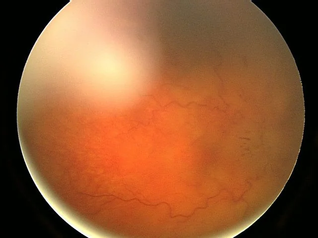
- Blurring of the disc margins, beginning nasally and superiorly and progressing around the disc. The nasal margin blurs first because the nerve fibre layer is thickest there.
- Loss of the physiological cup as swelling fills it in
- Hyperaemia of the disc with dilated surface capillaries
- Elevation of the disc above the retinal plane, best appreciated stereoscopically or by the dioptric difference needed to focus on the disc versus the retina
- Peripapillary flame haemorrhages and cotton wool spots in acute disease
- Paton's lines - concentric circumferential retinal folds radiating around the disc, caused by the swollen disc displacing the surrounding retina
- Loss of spontaneous venous pulsation - present in around 80% of normal people, and its loss suggests intracranial pressure above about 20 cmH2O. Its presence makes papilloedema unlikely, but its absence is not by itself diagnostic since a fifth of normal people never have it.
- Macular star or fan of hard exudate in severe or chronic cases
- Chronic papilloedema - the disc becomes a pale, greyish, champagne-cork shape with fewer haemorrhages, optociliary shunt vessels and eventually secondary optic atrophy with visual loss
Frisen grading
The Frisen scale grades severity from 0 to 5 and is used to track progression and treatment response.1
| Grade | Appearance |
|---|---|
| 0 | Normal disc |
| 1 | C-shaped halo of blurring sparing the temporal disc; the disc margin is subtly obscured nasally |
| 2 | Circumferential halo with elevation of the nasal border; the temporal margin is now also obscured |
| 3 | Obscuration of at least one segment of a major vessel leaving the disc; the halo has an irregular outer fringe |
| 4 | Total obscuration of a segment of a major vessel on the disc; the entire disc margin is obscured |
| 5 | Dome-shaped protrusion with obliteration of the disc margin and obscuration of all vessels on and leaving the disc |
Foster Kennedy syndrome
A frontal lobe or olfactory groove mass compresses one optic nerve directly, causing optic atrophy on that side, while raised intracranial pressure produces papilloedema in the contralateral eye. Anosmia from olfactory nerve compression completes the triad. It is rare, and much more often what is actually seen is pseudo-Foster Kennedy syndrome, in which a previous ischaemic optic neuropathy left one disc atrophic and a new episode has swollen the other. The distinction is made by the history and by imaging.
Investigations
- Urgent CT head - to exclude a mass, haemorrhage or hydrocephalus quickly, particularly out of hours
- MRI brain with contrast, and MR venography - the definitive imaging. MRV is essential because cerebral venous sinus thrombosis is a treatable cause that CT frequently misses, and it is the most important mimic of idiopathic intracranial hypertension.
- Lumbar puncture with opening pressure, measured in the lateral decubitus position with the legs relaxed, once imaging has excluded a mass. Normal is up to 20-25 cmH2O in adults; above 25 cmH2O with normal CSF constituents supports idiopathic intracranial hypertension. Send CSF for cells, protein, glucose, microscopy, culture and cytology.
- Visual acuity, colour vision and formal perimetry - Humphrey or Goldmann fields, repeated serially. Perimetry is the primary measure of optic nerve function and of treatment response, since acuity changes only late.
- Optical coherence tomography of the peripapillary retinal nerve fibre layer - quantifies swelling objectively, tracks response, and later detects the transition to atrophy
- Fundus photography - for objective comparison over time
- Blood pressure - to exclude malignant hypertension, which can produce identical disc swelling
- Bloods - FBC, clotting and thrombophilia screen if venous thrombosis is found; and consider a drug history for tetracyclines, retinoids and lithium
Management
Management has two entirely separate strands: treating the underlying cause, and protecting the optic nerves while that is happening. Both matter, and the second is frequently forgotten.
Treating the cause
- Space-occupying lesion - urgent neurosurgical referral; dexamethasone reduces peritumoural oedema, and definitive treatment is surgical or oncological
- Hydrocephalus - external ventricular drain or ventriculoperitoneal shunt
- Cerebral venous sinus thrombosis - anticoagulation, even in the presence of venous infarction with haemorrhage, and investigation for an underlying thrombophilia or malignancy
- Meningitis or encephalitis - antimicrobial treatment as appropriate
- Malignant hypertension - controlled blood pressure reduction as described in the hypertensive retinopathy article
- Drug-induced - stop the causative agent
Idiopathic intracranial hypertension
- Weight loss - the single most effective intervention. A sustained loss of around 5-10% of body weight can resolve papilloedema, and referral to a structured weight management programme or consideration of bariatric surgery is appropriate.2
- Acetazolamide - first-line drug therapy, reducing CSF production. The IIHTT trial showed benefit on visual fields when combined with a weight reduction diet.3 Doses are titrated up as tolerated; paraesthesiae, altered taste and fatigue are common.
- Topiramate - an alternative with weak carbonic anhydrase activity and the useful side effect of weight loss; also treats coexisting migraine
- Stop contributing drugs - tetracyclines, retinoids and hormonal contraception where relevant
- Therapeutic lumbar puncture - provides temporary relief and is used as a bridge, but the effect lasts hours to days and it is not a long-term strategy
- Surgery for fulminant or treatment-resistant disease - optic nerve sheath fenestration protects vision and is preferred where visual loss dominates; CSF diversion with a ventriculoperitoneal or lumboperitoneal shunt is preferred where headache dominates; venous sinus stenting is used in selected patients with a significant transverse sinus pressure gradient
Complications and prognosis
- Secondary optic atrophy - the principal ocular complication of chronic papilloedema, with permanent field loss and eventually reduced acuity
- Permanent blindness - occurring in around 10% of patients with idiopathic intracranial hypertension, and higher in fulminant disease and in Black patients and men, who tend to have more aggressive disease
- Choroidal folds, peripapillary subretinal neovascular membrane and macular exudate affecting central vision
- Persistent headache even after the pressure is controlled
- Complications of treatment - shunt blockage and infection, and the risks of optic nerve sheath fenestration including central retinal artery occlusion
- Recurrence - idiopathic intracranial hypertension frequently relapses with weight regain or in a subsequent pregnancy
Prognosis depends on the cause and on how quickly the pressure is brought under control. Papilloedema from a treatable structural cause resolves over weeks once the pressure normalises, though the disc may take two to three months to return to normal and any atrophy already present is permanent. Idiopathic intracranial hypertension has a variable course: most patients do well with weight loss and acetazolamide, but a minority have fulminant disease that blinds within weeks.
The message that should carry forward is the mismatch between how well the patient feels visually and how much damage is accruing. A patient with grade 4 papilloedema may read 6/6 and describe their vision as fine. That reassurance is false, and it is why papilloedema is followed with perimetry, why it is imaged urgently, and why any fall in acuity is treated as an emergency rather than as a gradual deterioration.
References
- Frisen L. Swelling of the optic nerve head: a staging scheme. Journal of Neurology, Neurosurgery and Psychiatry. 1982. Available here
- Mollan SP, Davies B, Silver NC et al. Idiopathic intracranial hypertension: consensus guidelines on management. Journal of Neurology, Neurosurgery and Psychiatry. 2018. Available here
- NORDIC Idiopathic Intracranial Hypertension Study Group. Effect of acetazolamide on visual function in patients with idiopathic intracranial hypertension and mild visual loss. JAMA. 2014. Available here
- NICE Clinical Knowledge Summaries. Headache - assessment. Available here
- Royal College of Ophthalmologists. Clinical guidance and resources. Available here
This article is written for revision and education. It is not clinical guidance and must not be used to make decisions about the care of a patient. Always check current NICE guidance and local protocols.