
Corneal Ulcer and Abrasion
Key points
- Abrasion: a defect of the corneal epithelium only, usually traumatic, healing within 24-72 hours without scarring.
- Ulcer: an epithelial defect with underlying stromal loss and infiltrate, usually infective, which heals with a scar and can perforate.
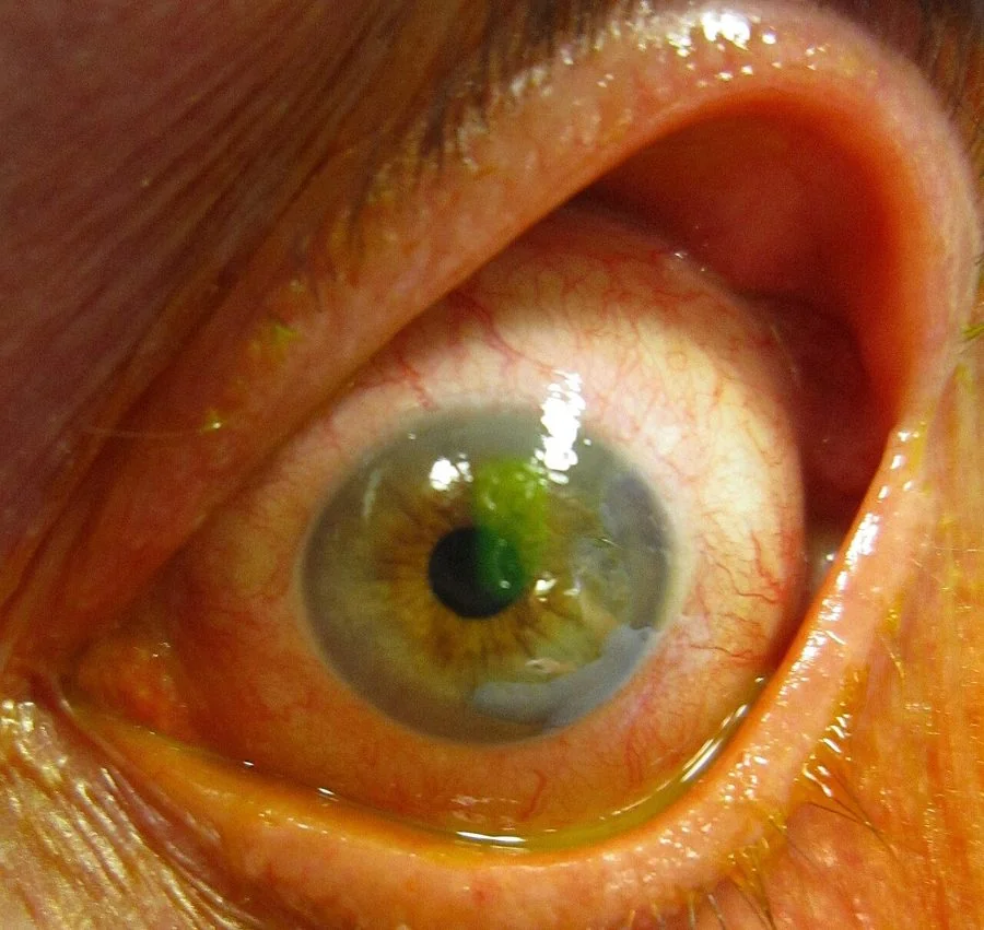
- The distinguishing sign: an ulcer has a white or grey opacity in the cornea beneath the staining area. An abrasion stains but the cornea underneath stays clear.
- Diagnosis: fluorescein 1% with a cobalt blue light, after topical anaesthetic. Always evert the upper lid to exclude a subtarsal foreign body.
- Abrasion management: topical chloramphenicol ointment, oral analgesia, no contact lenses, and review at 48 hours if not improving.
- Ulcer management: same-day ophthalmology, corneal scrape for microscopy and culture, then intensive hourly broad-spectrum topical antibiotics.
- Never: prescribe topical steroids, give topical anaesthetic for home use, or pad the eye of a contact lens wearer.
- Recurrent erosion: sudden pain on waking, weeks or months after an abrasion, from a poorly adherent epithelium. Treated with long-term ocular lubricant ointment at night.
Introduction
The cornea is five layers thick and about half a millimetre deep, yet it provides roughly two thirds of the eye's refractive power and is the most densely innervated tissue in the body. Both facts matter clinically: a small central opacity destroys vision out of all proportion to its size, and even a superficial scratch causes pain that patients describe as unbearable.1
Corneal abrasion is one of the commonest eye injuries seen in emergency departments. Corneal ulceration is far less common but is a sight-threatening emergency. The whole of the clinical skill lies in distinguishing the two at the bedside, and the distinction rests on a single observation: whether the cornea underneath the fluorescein-staining defect is clear or white.
The terminology can be confusing. Strictly, an abrasion is loss of epithelium alone, whereas an ulcer implies loss of epithelium plus underlying stroma with an inflammatory infiltrate. In UK practice, corneal ulcer is often used synonymously with microbial keratitis, since infection is by far the commonest cause of stromal loss. The related article on infective keratitis covers the microbiology and intensive treatment in more depth; this one focuses on the presentation, the bedside distinction and the management of trauma.
Anatomy and healing
Understanding the five layers explains why abrasions heal invisibly and ulcers scar.
- Epithelium - five to six cell layers, continuously renewed from limbal stem cells. It regenerates completely and without scarring, typically resurfacing a 3 mm defect within 24-48 hours.
- Bowman's layer - an acellular condensation of collagen. It does not regenerate; damage here leaves a permanent scar.
- Stroma - 90% of corneal thickness, made of precisely arranged collagen lamellae. This regular arrangement is what makes the cornea transparent, and any repair tissue laid down is disorganised, so stromal loss always scars.
- Descemet's membrane - the basement membrane of the endothelium, tough and elastic. A deep ulcer that reaches it can cause it to bulge forward as a descemetocele, which is on the point of perforating.
- Endothelium - a single layer of cells that pumps fluid out of the stroma to keep it dehydrated and clear. These cells do not divide in humans; lose enough of them and the cornea becomes permanently oedematous.
Aetiology
Corneal abrasion
- Direct trauma - fingernails, particularly a toddler's, paper edges, mascara wands, tree branches and pet claws
- Foreign bodies, including subtarsal ones which produce characteristic vertical linear scratches as the lid sweeps across the cornea
- Contact lens related - insertion or removal trauma, a torn lens, or a poorly fitting lens
- Chemical and thermal injury, including welding arc and sunbed exposure causing ultraviolet photokeratitis
- Exposure keratopathy - incomplete lid closure in facial nerve palsy, proptosis, sedation or intensive care, causing a drying inferior abrasion
- Iatrogenic - during general anaesthesia if the lids are not taped, or from instrumentation
Corneal ulcer
- Bacterial - the commonest, and overwhelmingly associated with contact lens wear. Pseudomonas aeruginosa and Staphylococcus aureus predominate.
- Herpes simplex - producing the classic branching dendritic ulcer with terminal bulbs, and reduced corneal sensation
- Varicella zoster - in herpes zoster ophthalmicus, producing pseudodendrites without terminal bulbs
- Fungal - Fusarium and Aspergillus, typically after injury with vegetable matter, with feathery infiltrate margins and satellite lesions
- Acanthamoeba - associated with tap water exposure in lens wearers, with pain grossly out of proportion to the signs and a late ring infiltrate
- Marginal keratitis - a sterile, immune-mediated peripheral infiltrate reacting to staphylococcal exotoxin in blepharitis, separated from the limbus by a clear zone
- Neurotrophic - after herpetic disease, trigeminal surgery or diabetes, where an anaesthetic cornea fails to heal
- Peripheral ulcerative keratitis - a crescentic melt at the limbus in rheumatoid arthritis or granulomatosis with polyangiitis, which can perforate and signals active systemic vasculitis
Clinical features
Both present with a painful red eye, but the tempo and the accompanying features differ.
| Feature | Corneal abrasion | Corneal ulcer |
|---|---|---|
| History | Discrete traumatic event, pain immediate | Often no trauma; gradual onset over hours to days, usually a contact lens wearer |
| Pain | Severe, sharp, foreign body sensation | Severe, deep, aching |
| Visual acuity | Normal unless the defect is central | Reduced, especially if central |
| Cornea beneath the defect | Clear | White or grey infiltrate |
| Fluorescein | Sharply demarcated linear or geographic stain | Round or irregular stain overlying the infiltrate |
| Anterior chamber | Quiet | Cells, flare, sometimes hypopyon |
| Discharge | Watery, reflex lacrimation | Mucopurulent |
| Course | Heals in 24-72 hours | Progresses without treatment; can perforate |
Symptoms common to both
- Severe pain, often described as a sensation of grit or glass in the eye
- Profuse watering and reflex lacrimation
- Blepharospasm, sometimes so marked that the lids must be prised open under topical anaesthetic
- Photophobia, from reflex ciliary spasm
- Blurred vision, especially with central lesions
- Redness, typically with ciliary flush in ulceration
Examination
- Visual acuity in each eye separately, before any drops. Use a pinhole if reduced.
- Instil topical anaesthetic - oxybuprocaine or tetracaine - which relieves surface pain and makes the rest of the examination possible. Note whether the pain resolves, which localises it to the ocular surface.
- Inspect for the pattern of redness, discharge, corneal clarity and any visible white spot. Look at the cornea against the red reflex, where an opacity shows as a dark shadow.
- Fluorescein with a moistened strip touched to the inferior fornix, examined under cobalt blue light, ideally at a slit lamp
- Evert the upper lid in every case where a foreign body is possible
- Assess corneal sensation before the anaesthetic if herpes simplex is suspected, by touching a wisp of cotton wool to the cornea from the side and comparing with the other eye
- Look for anterior chamber activity - cells, flare or a hypopyon - which upgrades the diagnosis from abrasion to ulcer
- Seidel test if perforation is suspected: apply concentrated fluorescein and look for a stream of diluted dye flowing away from the wound
Differential diagnosis
- Conjunctivitis - diffuse injection, discharge, normal acuity, no fluorescein staining of the cornea
- Anterior uveitis - deep ache, photophobia, small irregular pupil, cells in the anterior chamber but no epithelial defect
- Acute angle closure glaucoma - severe pain with vomiting, hazy oedematous cornea, fixed mid-dilated pupil, very high intraocular pressure
- Ultraviolet photokeratitis (arc eye or snow blindness) - bilateral, 6-12 hours after exposure, with diffuse punctate staining
- Recurrent corneal erosion - abrupt pain on waking with a history of previous abrasion
- Marginal keratitis - peripheral infiltrate with a clear zone separating it from the limbus, in a patient with blepharitis
- Penetrating injury - a peaked pupil, shallow anterior chamber or positive Seidel test. A high-velocity mechanism demands imaging even if the eye looks quiet.
- Endophthalmitis - profound pain and visual loss with hypopyon after intraocular surgery or injection
Investigations
- Fluorescein staining under cobalt blue light - the definitive bedside investigation and the only one needed for a straightforward abrasion
- Slit lamp examination - to assess depth, measure the infiltrate, and detect anterior chamber activity
- Corneal scrape for Gram stain, culture and sensitivity - performed by ophthalmology for any ulcer with a significant infiltrate, before starting antibiotics. Lenses, cases and solutions should be sent for culture too.
- Herpes simplex PCR on a corneal swab where a dendrite is atypical or the diagnosis is uncertain
- Confocal microscopy - used in specialist centres for suspected Acanthamoeba or fungal keratitis
- Orbital CT - if a penetrating injury or intraocular foreign body is suspected. Never MRI first with a possible metallic foreign body.
- Systemic autoimmune screen - ANCA, rheumatoid factor, CRP and ESR in peripheral ulcerative keratitis, which can be the presenting feature of systemic vasculitis
Management
Uncomplicated corneal abrasion
Most abrasions heal within 24-72 hours and can be managed without referral.2
- Remove any foreign body with a cotton bud or, if embedded, a sterile needle under topical anaesthetic by someone trained to do so. A rust ring from a metallic foreign body needs removal by ophthalmology if central.
- Topical antibiotic prophylaxis - chloramphenicol 1% ointment four times daily for 5-7 days.4 Ointment is preferred to drops because it also lubricates and reduces friction.
- Analgesia - regular oral paracetamol and an NSAID. Topical NSAIDs such as diclofenac reduce pain but may slow epithelial healing, and are not first line.
- Cycloplegia - cyclopentolate 1% relieves the ciliary spasm responsible for photophobia in larger abrasions, though it blurs near vision for a day or so
- Stop contact lens wear until the epithelium has healed and the eye has been quiet for 24 hours
- Advise no driving while vision is blurred or an eye is padded
- Review at 48 hours if symptoms are not clearly improving
Corneal ulcer
Herpes simplex dendritic ulcers are treated with topical aciclovir 3% ointment five times daily for 7-14 days, or ganciclovir 0.15% gel, with ophthalmology follow-up. Fungal and Acanthamoeba keratitis need prolonged specialist treatment measured in months, and perforation or an unresponsive ulcer may require tissue adhesive, an amniotic membrane graft or a therapeutic keratoplasty.
Recurrent corneal erosion syndrome
This deserves separate mention because it is common, frequently missed, and has a specific treatment. After an abrasion - classically from a fingernail or paper cut - the regenerated epithelium may adhere poorly to its basement membrane. During sleep the tear film thins and the lid adheres to the loose epithelium, so on opening the eyes in the morning the epithelium is torn away again.
- Presentation - sudden severe pain, watering and photophobia on waking, weeks to months after the original injury, settling over hours
- Predisposing factors - previous abrasion, epithelial basement membrane dystrophy (map-dot-fingerprint dystrophy), and diabetes
- Examination may be normal between episodes; fluorescein during an episode shows an epithelial defect or a loose flap of epithelium
- First-line treatment - ocular lubricant ointment such as simple eye ointment or a paraffin-based preparation instilled at night for at least three months, long after symptoms settle, plus hypertonic sodium chloride 5% ointment in some units
- Second line - a bandage contact lens, oral doxycycline for its anti-collagenase effect, or a short course of topical steroid under specialist supervision
- Surgical options - epithelial debridement, anterior stromal puncture or phototherapeutic keratectomy for resistant cases
Complications and prognosis
Uncomplicated abrasions heal completely within three days and leave no visual deficit, because the epithelium regenerates without scarring. The main complications are secondary infection, recurrent erosion syndrome, and traumatic uveitis in the days after a significant blunt injury.
Ulcers are a different matter. Even a well-treated bacterial ulcer heals with a stromal scar, and if that scar lies over the pupil it degrades acuity permanently. Corneal ulceration is a leading cause of monocular blindness worldwide.3 Other complications include descemetocele and perforation, secondary glaucoma, cataract, corneal neovascularisation, and endophthalmitis if the organism reaches the inside of the eye.
Outcome correlates most closely with three things: how central the ulcer is, how large the infiltrate is at presentation, and how quickly intensive treatment started. The last of these is the only one under your control, which is precisely why a red eye in a contact lens wearer is referred the same day rather than given a week of chloramphenicol and a review appointment.
References
- NICE Clinical Knowledge Summaries. Corneal superficial injury. Available here
- College of Optometrists. Clinical Management Guidelines: corneal abrasion. Available here
- Whitcher JP, Srinivasan M, Upadhyay MP. Corneal blindness: a global perspective. Bulletin of the World Health Organization. 2001. Available here
- BNF. Chloramphenicol - eye preparations. Available here
- Royal College of Ophthalmologists. Microbial keratitis guidance. Available here
This article is written for revision and education. It is not clinical guidance and must not be used to make decisions about the care of a patient. Always check current NICE guidance and local protocols.