
Infective Keratitis: Microbial and Herpetic
Key points
- Definition: infection of the cornea, producing a white stromal infiltrate with an overlying epithelial defect that stains with fluorescein.
- The single biggest risk factor: contact lens wear, which increases risk roughly tenfold, and by 5-10 times again with overnight wear.
- Presentation: a painful red eye with photophobia, watering, reduced vision, ciliary flush and a visible corneal white spot.
- Commonest organisms: Pseudomonas aeruginosa and Staphylococcus aureus. Pseudomonas can perforate a cornea in under 48 hours.
- Herpes simplex: gives a branching dendritic ulcer with terminal bulbs and reduced corneal sensation. Treat with topical aciclovir; steroids are catastrophic.
- *Acanthamoeba*: suspect when pain is grossly out of proportion to the signs in a lens wearer exposed to tap water. A ring infiltrate is a late sign.
- Management principle: scrape, then treat intensively - corneal scrape for microscopy and culture, then hourly broad-spectrum topical antibiotics day and night for 48 hours.
- Prognosis: determined by how central and how large the infiltrate is at presentation, and by how quickly treatment started. Central scarring is permanent.
Introduction
Infective keratitis is infection of the corneal stroma. It is uncommon - perhaps 40 per 100,000 contact lens wearers per year in the UK - but it is one of the genuine ophthalmic emergencies, because an organism such as Pseudomonas aeruginosa can digest a cornea from clear to perforated in under two days.1
The cornea is normally remarkably resistant to infection. It is avascular, bathed in tears containing lysozyme, lactoferrin and secretory IgA, and covered by an intact epithelium that most organisms cannot cross. Keratitis therefore almost always requires a breach in that defence: a contact lens, trauma, an anaesthetic cornea, dry eye, or topical steroid use. Identifying the breach is part of making the diagnosis.
Only two organisms are able to penetrate an intact corneal epithelium: Neisseria gonorrhoeae and Corynebacterium diphtheriae. Every other cause of microbial keratitis needs a way in. This is worth remembering because it explains the entire risk factor list and the whole of prevention.
Aetiology
Bacterial
Bacteria cause the great majority of microbial keratitis in the UK. The organism often cannot be predicted from appearance alone, which is why a scrape is taken, but there are recognisable patterns.
- Pseudomonas aeruginosa - the classic contact lens organism. It produces proteases that liquefy stroma, giving a rapidly enlarging ulcer with a soupy, greenish mucopurulent discharge and marked stromal melt.
- Staphylococcus aureus and coagulase-negative staphylococci - well-demarcated, round, dense white infiltrates, more common after trauma or in an already diseased cornea
- Streptococcus pneumoniae - historically the cause of the serpiginous creeping ulcer, often with hypopyon, seen after trauma or in association with dacryocystitis
- Moraxella - indolent, inferior, often in debilitated or alcohol-dependent patients
- Atypical mycobacteria and Nocardia - rare, indolent, typically after refractive surgery or agricultural trauma
Viral
- Herpes simplex virus type 1 - the commonest infectious cause of corneal blindness in developed countries. Primary infection is usually a childhood blepharoconjunctivitis; the virus then remains latent in the trigeminal ganglion and reactivates to cause epithelial (dendritic), stromal or endothelial (disciform) keratitis.
- Varicella zoster virus - in herpes zoster ophthalmicus, causing pseudodendrites which are raised, mucous plaques without the terminal bulbs of true dendrites, and a later stromal keratitis
Fungal
Fungal keratitis is uncommon in the UK but common in tropical agricultural settings. Filamentary fungi such as Fusarium and Aspergillus follow injury with vegetable matter - a thorn, a branch, a stalk of corn. Yeasts such as Candida affect an already compromised cornea, for instance in chronic topical steroid use or severe dry eye. The classic signs are a dry-looking infiltrate with feathery, indistinct margins, satellite lesions around the main focus, and an endothelial plaque.
Protozoal
Acanthamoeba is a free-living amoeba found in tap water, swimming pools, hot tubs and soil. Almost all UK cases occur in contact lens wearers who have rinsed lenses in tap water, showered or swum in lenses, or used homemade saline. It exists as a trophozoite and as a highly resistant cyst, which is why treatment lasts months. The defining clinical feature is pain grossly out of proportion to the signs, because the organism has a tropism for corneal nerves and causes a radial perineuritis. A ring-shaped stromal infiltrate is characteristic but appears late.2
Risk factors
| Mechanism | Examples |
|---|---|
| Contact lens wear | Extended or overnight wear, poor case hygiene, topping up rather than replacing solution, showering or swimming in lenses, rinsing in tap water, wearing lenses beyond their replacement schedule |
| Epithelial breach | Trauma, foreign body, previous abrasion, refractive or corneal surgery, sutures |
| Impaired lid or tear function | Dry eye, blepharitis, exposure keratopathy, facial nerve palsy, entropion, trichiasis, ectropion |
| Reduced corneal sensation | Previous herpetic keratitis, diabetes, trigeminal nerve surgery, long-term topical anaesthetic misuse |
| Impaired local or systemic immunity | Topical or systemic corticosteroids, diabetes, immunosuppression, HIV, vitamin A deficiency |
| Pre-existing ocular surface disease | Bullous keratopathy, chronic conjunctivitis, ocular cicatricial pemphigoid, previous graft |
Clinical features
- Pain - severe, deep and aching, worsening over hours to days. In Acanthamoeba it is out of proportion to the visible signs.
- Redness with ciliary flush - injection densest at the limbus
- Photophobia and blepharospasm
- Watering and mucopurulent discharge
- Reduced visual acuity, particularly with central lesions, from the opacity itself and from overlying oedema
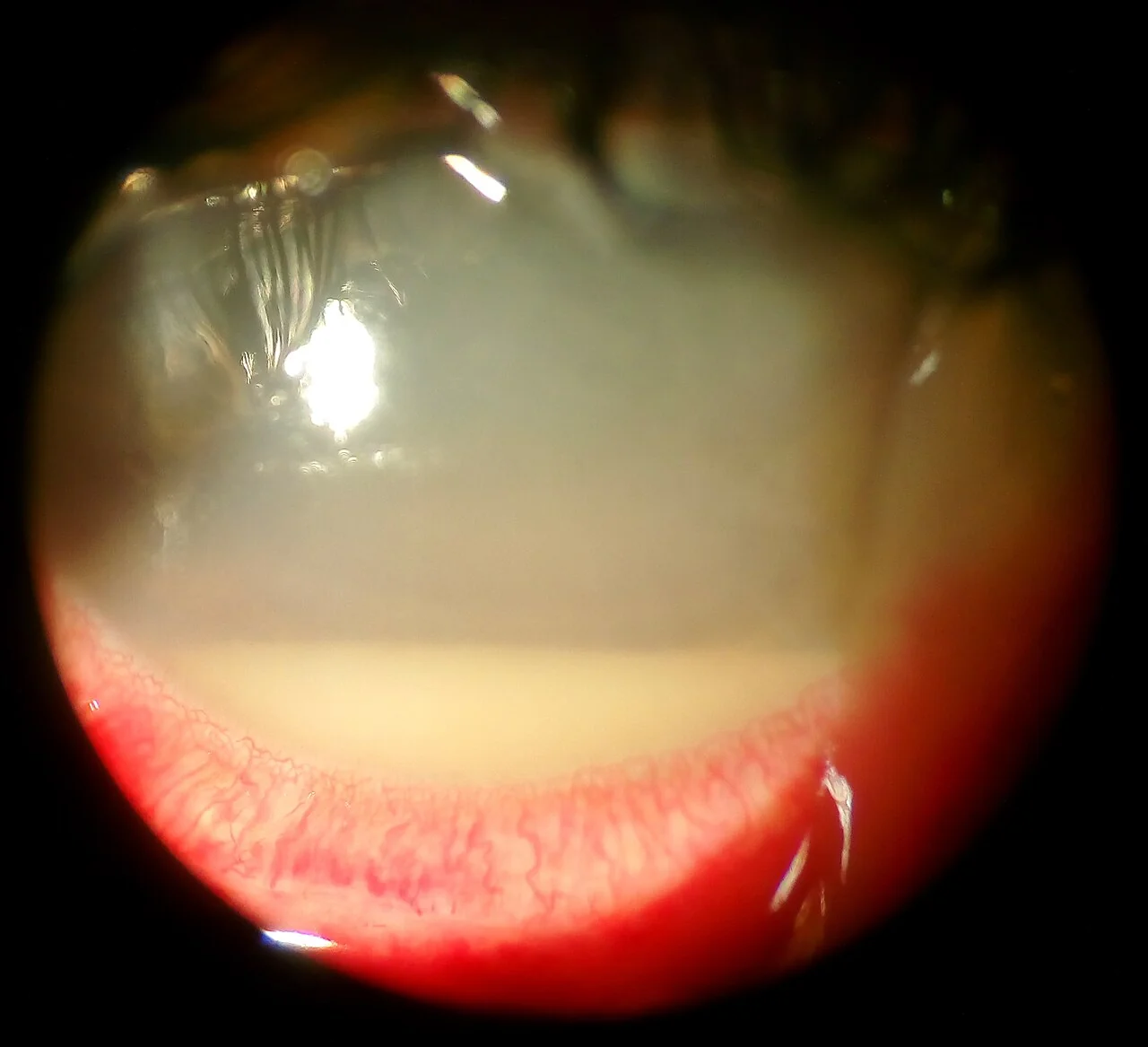
- A visible white or grey corneal spot - the infiltrate. This is the diagnostic sign and can often be seen with the naked eye against the red reflex.
- Hypopyon in severe disease - a layer of white cells in the inferior anterior chamber, which is usually sterile and reflects the intensity of inflammation rather than intraocular infection
- Lid oedema and a general appearance of an unwell eye
Herpes simplex keratitis
This deserves separate description because its management is entirely different and it is heavily examined. The epithelial form produces a dendritic ulcer: a branching, linear defect with swollen terminal bulbs at the end of each branch, which stains brilliantly with fluorescein. Untreated, or treated with topical steroid, it enlarges into a geographic ulcer with the same scalloped margins on a larger scale.
- Corneal sensation is reduced - test with a wisp of cotton wool before instilling anaesthetic, comparing the two eyes. This is the most reliable clinical discriminator.
- Almost always unilateral, and typically recurrent in the same eye
- Stromal keratitis - a later immune-mediated form with stromal opacification, neovascularisation and scarring, which does need steroid but only under specialist supervision with antiviral cover
- Disciform keratitis - a round area of stromal oedema with keratic precipitates from endothelial involvement
- Recurrence is precipitated by ultraviolet light, fever, stress, menstruation and immunosuppression
Examination
- Visual acuity in each eye before any drops, with a pinhole if reduced
- Corneal sensation with a cotton wool wisp, before topical anaesthetic, comparing sides
- Inspection for ciliary flush, discharge, lid oedema and a visible corneal opacity
- Fluorescein staining under cobalt blue light for the epithelial defect and its pattern
- Slit lamp examination - the essential investigation. Assess the size, depth, density and location of the infiltrate; the size of the epithelial defect; the presence of stromal thinning; anterior chamber cells and hypopyon.
- Measure and draw the infiltrate, since serial measurement is how response to treatment is judged
- Lid eversion for retained foreign body or a lens fragment
- Examine the fellow eye, which shares the same risk factors
Differential diagnosis
| Diagnosis | Distinguishing features |
|---|---|
| Microbial keratitis | Large, central, dense infiltrate with an overlying epithelial defect, significant pain, anterior chamber activity, contact lens wear |
| Sterile marginal keratitis | Small peripheral infiltrate with a clear zone between it and the limbus, minimal pain, associated blepharitis, minimal epithelial defect |
| Contact lens induced acute red eye (CLARE) | Peripheral sterile infiltrates on waking in an overnight lens wearer, no epithelial defect, resolves on stopping lens wear |
| Herpes simplex epithelial keratitis | Dendrite with terminal bulbs, reduced corneal sensation, recurrent, no dense stromal infiltrate |
| Corneal abrasion | Traumatic, cornea beneath the stain is clear, no infiltrate, heals in 24-72 hours |
| Anterior uveitis | Photophobia and cells, but a clear cornea with no epithelial defect |
| Peripheral ulcerative keratitis | Crescentic peripheral melt adjacent to the limbus with an epithelial defect, in known rheumatoid arthritis or vasculitis |
| Endophthalmitis | Recent intraocular surgery or injection, profound visual loss, vitritis, hypopyon with a relatively clear cornea |
Investigations
- Corneal scrape - the key investigation, taken at the slit lamp under topical anaesthetic with a sterile blade or needle, from the base and the advancing edge of the ulcer. Plated directly onto blood agar, chocolate agar, Sabouraud agar and into brain-heart infusion broth, with slides for Gram and Giemsa staining.
- Contact lens, case and solution culture - frequently yields the organism when the scrape does not, and is a good reason to ask the patient to bring them in
- Herpes simplex PCR on a corneal swab if the dendrite is atypical
- Confocal microscopy - non-invasive, and can demonstrate Acanthamoeba cysts or fungal hyphae in vivo
- Anterior chamber tap - only if endophthalmitis is suspected
- Corneal biopsy - reserved for deep infiltrates with repeatedly negative scrapes, typically suspected fungal disease
- Serial photography and measurement - the practical way of tracking response over the first 48-72 hours
Culture is negative in around 40-50% of scrapes, often because antibiotics have already been started. This is the reason the on-call team may ask you not to begin treatment before referral, and why the scrape is taken first wherever the delay is short.3
Management
Bacterial keratitis
- Corneal scrape for microscopy, culture and sensitivities
- Intensive topical antibiotics started immediately afterwards without waiting for results. A fluoroquinolone monotherapy such as levofloxacin 0.5% or ofloxacin, or dual therapy with fortified cefuroxime 5% plus gentamicin 1.5%, given hourly day and night for 48 hours, then tapered according to response.
- Cycloplegia - cyclopentolate or atropine to relieve ciliary spasm and prevent posterior synechiae
- Oral analgesia; avoid topical NSAIDs, which can mask progression and impair healing
- Daily review for the first few days, with the infiltrate measured each time. Improvement is judged by reduced pain, a smaller epithelial defect, a less dense infiltrate and a quieter anterior chamber.
- Admission for severe, central or rapidly progressive ulcers, or where hourly instillation at home is not realistic
- Topical steroid may be added cautiously by the ophthalmologist once the infection is controlled and cultures are known, to reduce scarring - never at presentation
Herpes simplex keratitis
- Epithelial (dendritic) - topical aciclovir 3% ointment five times daily for 7-14 days, or ganciclovir 0.15% gel. Gentle epithelial debridement may be used. Continue for at least three days after healing.6
- Stromal - topical steroid with antiviral cover, under specialist supervision only
- Prophylaxis - oral aciclovir 400 mg twice daily reduces recurrence by about half in those with frequent recurrences or a corneal graft4
- Avoid topical steroids in epithelial disease absolutely
Fungal and Acanthamoeba keratitis
Fungal keratitis is treated with topical natamycin 5% for filamentary fungi or topical amphotericin or voriconazole for yeasts, often with oral voriconazole, for weeks to months. Acanthamoeba requires a biguanide such as polyhexamethylene biguanide 0.02% combined with a diamidine such as propamidine, hourly initially and then tapered over six months to a year because of the cyst stage. Both are managed exclusively in specialist corneal services, and both may ultimately need therapeutic keratoplasty.
Complications and prognosis
- Corneal scarring - inevitable with stromal involvement, and permanently sight-limiting if it lies over the pupil
- Corneal thinning, descemetocele and perforation - requiring cyanoacrylate glue, amniotic membrane or emergency keratoplasty
- Secondary glaucoma from inflammatory trabecular blockage or from steroid response
- Cataract from prolonged inflammation and steroid use
- Corneal neovascularisation, which reduces clarity and makes any future graft more likely to reject
- Endophthalmitis if organisms reach the interior of the eye - rare but devastating
- Irregular astigmatism from an eccentric scar, which may need a rigid gas permeable lens to correct
The proportion of patients left with significant visual loss varies widely between series, but the determinants are consistent: the size of the infiltrate at presentation, how close it is to the visual axis, the virulence of the organism, and the delay before intensive treatment started. Only the last of these can be influenced by the person who first sees the patient.
Prevention is therefore worth as much as treatment, and is a fair OSCE counselling station. Contact lens wearers should be told never to sleep in lenses unless specifically prescribed for that, never to rinse lenses or cases in tap water, never to shower or swim in lenses, to replace the case every three months, to discard rather than top up solution, and to remove lenses and seek help immediately if an eye becomes red or painful. The last instruction is the one that most often fails to be given, and the one that most often matters.
References
- Royal College of Ophthalmologists. Microbial keratitis: clinical guidance. Available here
- Carnt N, Robaei D, Watson SL et al. The impact of topical corticosteroids used in conjunction with antiamoebic therapy on the outcome of Acanthamoeba keratitis. Ophthalmology. 2016. Available here
- Ting DSJ, Ho CS, Deshmukh R et al. Infectious keratitis: an update on epidemiology, causative microorganisms, risk factors, diagnosis and antimicrobial resistance. Eye. 2021. Available here
- Herpetic Eye Disease Study Group. Aciclovir for the prevention of recurrent herpes simplex virus eye disease. New England Journal of Medicine. 1998. Available here
- NICE Clinical Knowledge Summaries. Red eye. Available here
- BNF. Aciclovir - eye preparations. Available here
This article is written for revision and education. It is not clinical guidance and must not be used to make decisions about the care of a patient. Always check current NICE guidance and local protocols.