
Atopic Dermatitis and Eczema
Key points
- Atopic dermatitis: a chronic, relapsing, intensely itchy inflammatory skin condition arising from skin barrier dysfunction combined with a Th2-skewed immune response, usually starting in infancy.
- Mechanism: filaggrin and other barrier defects let irritants, allergens and organisms penetrate the skin and let water escape; this triggers a Th2 cytokine response that itself further impairs the barrier.
- Distribution: face and extensor surfaces in infants; flexures - antecubital and popliteal fossae, wrists, neck - in older children and adults.
- Diagnosis: clinical, using the UK Working Party criteria - an itchy skin condition plus three or more supporting features. No blood test confirms it.
- First-line management: regular emollients used as a leave-on treatment and soap substitute, plus topical corticosteroids of a potency matched to site and severity for flares.
- Second-line topical: topical calcineurin inhibitors (tacrolimus, pimecrolimus) for sensitive sites such as the face and flexures, avoiding the skin atrophy risk of prolonged steroid use.
- Escalation: phototherapy or systemic therapy - ciclosporin, methotrexate, or the biologic dupilumab - for moderate-to-severe disease uncontrolled by optimised topical treatment.
- Red flag: eczema herpeticum - widespread punched-out vesicles and erosions with systemic upset - is a dermatological emergency needing same-day assessment and aciclovir.
Introduction
Atopic dermatitis, also called atopic eczema, is a chronic, relapsing, intensely itchy inflammatory skin condition. It affects up to one in five children in the UK and around 2-10% of adults, making it one of the commonest reasons for a GP or dermatology consultation.1
The word atopic signals its place within a wider predisposition to IgE-mediated hypersensitivity that also produces asthma and allergic rhinitis. Eczema is typically the first of these to appear, often within the first year of life, and its presence increases the likelihood of the others developing later - a sequence often called the atopic march.
Course is relapsing and remitting rather than steadily progressive: patients cycle between flares and periods of relatively clear skin, and management is built around that pattern rather than around a single course of treatment. Most children improve substantially by their teenage years, but a significant minority carry the disease, or a tendency to dry and reactive skin, into adulthood.
Pathophysiology
Two processes drive atopic dermatitis, and each worsens the other: a defective skin barrier, and a Th2-skewed immune response.
Barrier dysfunction
Loss-of-function mutations in the gene encoding filaggrin, a structural protein essential for compacting the corneocytes of the stratum corneum and for generating natural moisturising factor, are found in a substantial proportion of patients with atopic dermatitis and are the strongest known genetic risk factor.3 The result is a leaky barrier: transepidermal water loss increases, the skin becomes dry, and irritants, allergens and microorganisms penetrate more easily than in normal skin.
Not everyone with atopic dermatitis carries a filaggrin mutation, and the same defect is seen in ichthyosis vulgaris, which frequently coexists. The barrier can also be impaired by acquired factors - soap and detergent use, low humidity, and the mechanical damage of scratching.
Immune dysregulation
Once the barrier is breached, keratinocytes release alarmins - thymic stromal lymphopoietin, IL-25 and IL-33 - that activate a Th2-predominant immune response. The resulting cytokines, chiefly IL-4, IL-13 and IL-31, drive itch, further suppress barrier protein expression, promote IgE class switching in B cells, and recruit eosinophils. IL-31 in particular is a key mediator of the itch, which is why IL-4/IL-13 pathway blockade with biologic therapy also markedly improves itch rather than just the visible rash.
Itch leads to scratching, which mechanically damages the barrier and releases more alarmins - the itch-scratch cycle that keeps eczema going even once an initial trigger has resolved. Colonisation with Staphylococcus aureus is near-universal in active eczema; its exotoxins act as superantigens that amplify the cutaneous inflammation, which is one reason flares can settle with antiseptic measures alone.
Risk factors
The strongest predictor of atopic dermatitis is a personal or family history of atopic disease, reflecting the substantial genetic contribution to both barrier function and immune skew.
- Family history of atopy - eczema, asthma or allergic rhinitis in a first-degree relative
- Filaggrin null mutations, more common in people of Northern European ancestry
- Personal history of asthma or allergic rhinitis
- Early onset - most cases present before 12 months and the great majority by age 5
- Urban living and higher socioeconomic status, consistent with the hygiene hypothesis
- Small family size, reducing early exposure to childhood infections
- Hard water exposure, an association seen in UK cohort studies, though causation is unproven
- Early cessation of breastfeeding, though the evidence for a protective effect of breastfeeding is inconsistent
Clinical features
The cardinal symptom is itch, which is usually present before any visible rash and typically worsens at night. Diagnosis is clinical, using the UK Working Party diagnostic criteria: an itchy skin condition in the last 12 months, plus three or more of the following.4
- Visible flexural dermatitis (or dermatitis of the cheeks, forehead and outer limbs in children under 4)
- History of flexural involvement
- Personal history of asthma or hay fever (or atopy in a first-degree relative if the child is under 4)
- History of generally dry skin in the last year
- Onset before age 2 (not used as a criterion if the child is under 4)
Distribution changes characteristically with age, which is a favourite feature to test in exams.
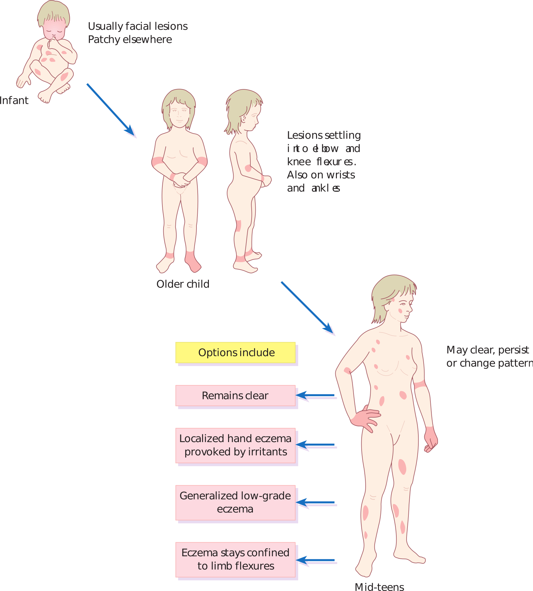
| Age group | Typical distribution |
|---|---|
| Infants | Face, scalp and extensor surfaces of the limbs; the napkin area is often relatively spared |
| Older children | Flexural - antecubital and popliteal fossae, wrists and ankles, and around the neck |
| Adults | Flexures as above, plus hands, eyelids and nipples; may become more lichenified with a chronic pattern of scratching |
In an acute flare, expect erythema, papules, vesicles and weeping or exudation. Chronic, repeatedly scratched skin instead shows lichenification (thickened skin with exaggerated skin markings), excoriation and post-inflammatory pigment change, which can be marked in darker skin tones.
Associated findings include Dennie-Morgan folds (an extra fold of skin below the lower eyelid), keratosis pilaris, hyperlinear palms, pityriasis alba (hypopigmented, faintly scaly patches, especially on the face), and a personal or family history of ichthyosis vulgaris.
Assessing severity
The Patient-Oriented Eczema Measure (POEM), a simple seven-item questionnaire completed by the patient or carer, is used in primary care to monitor severity and response to treatment over time. In secondary care and in trials, the SCORAD and EASI indices combine an objective assessment of extent and morphology with symptom scores. NICE's severity categories - clear, mild, moderate and severe - map directly onto the stepwise treatment ladder described below.
Clinical examination
Examination establishes the distribution and morphology, looks for signs of complications, and screens for other atopic disease.
- Distribution and morphology - flexural versus extensor, acute weeping change versus chronic lichenification
- Signs of secondary bacterial infection - increased weeping, yellow crusting, pustules, or rapidly worsening erythema
- Signs suggesting eczema herpeticum - clustered punched-out vesicles or erosions of similar size, particularly on a background of active eczema, with associated fever or malaise
- Growth parameters in children - height and weight plotted on a growth chart, as severe uncontrolled eczema and, rarely, chronic potent steroid overuse can both affect growth
- Other atopic features - wheeze, allergic shiners, nasal crease, associated conjunctivitis
Differential diagnosis
Several other rashes can mimic atopic dermatitis, and distinguishing them changes management.
- Seborrhoeic dermatitis - greasy scale affecting the scalp, eyebrows and nasolabial folds, and in infants the classic yellow, crusted 'cradle cap'
- Contact dermatitis (irritant or allergic) - a distribution corresponding to contact with a specific product or material is the key clue
- Psoriasis - well-demarcated, salmon-pink plaques with silvery scale, typically extensor and less itchy
- Scabies - intense itch, worse at night, with burrows in the finger webs and a household contact history
- Tinea corporis - an asymmetrical, annular, expanding plaque with a scaly, active edge
- Discoid (nummular) eczema - well-defined coin-shaped plaques, which can occur as a variant or overlapping pattern
- Wiskott-Aldrich syndrome and hyper-IgE (Job) syndrome - rare primary immunodeficiencies that present with severe, early, atypical eczema plus recurrent infections, and should be considered in an infant with unusually severe or treatment-resistant disease
Investigations
Atopic dermatitis is a clinical diagnosis, and investigations are reserved for suspected complications or triggers rather than for confirming the diagnosis itself.
- Skin swab - if secondary bacterial infection is suspected, to guide antibiotic choice
- Viral swab for HSV PCR - if eczema herpeticum is suspected, though treatment should not be delayed while awaiting the result
- Specific IgE testing or skin prick testing - considered in young children with moderate-to-severe eczema and a clear history suggesting a food trigger (for example immediate reaction after egg or milk), not performed routinely or on every patient
- Patch testing - if allergic contact dermatitis is suspected as a superimposed or complicating factor, particularly in eczema that is resistant to treatment or has an unusual distribution
- Total IgE and eosinophil count - often raised, but neither is sensitive or specific enough to help with diagnosis and neither is requested routinely
Management
Treatment is stepwise, matched to severity, and built around patient (or parent) education, since this is a condition managed largely by the patient at home between flares.2
Emollients - the foundation
Emollients are used by everyone, all the time, not just during flares. They should be applied liberally and frequently - often several times a day, and in quantities of 250-500g a week in an adult with moderate disease - both as a leave-on treatment and as a soap substitute for washing.5 Ointments are generally more effective than creams because they contain less water and more occlusive lipid, though they are greasier and adherence can suffer as a result; the best emollient is the one the patient will actually use.
- Apply in the direction of hair growth to reduce the risk of folliculitis
- Use a pump dispenser or clean spoon/spatula rather than dipping fingers into a shared pot, to reduce bacterial contamination
- Avoid aqueous cream as a leave-on emollient - it contains sodium lauryl sulfate, which can itself irritate and worsen barrier function, though it remains acceptable as a short-contact wash product
- Leave at least 30 minutes between applying emollient and a topical corticosteroid so the two are not simply diluted together
Topical corticosteroids for flares
Topical corticosteroids treat the inflammatory component during a flare. Potency is matched to the site and to severity: use the least potent preparation that controls the flare, for the shortest effective period, then step back down to emollients alone.6
| Potency | Example | Typical use |
|---|---|---|
| Mild | Hydrocortisone 1% | Face, eyelids, flexures, and children |
| Moderate | Clobetasone butyrate 0.05% | Trunk and limbs, mild-moderate flares |
| Potent | Betamethasone valerate 0.1% | Trunk and limbs, moderate-severe flares; short courses only on the face |
| Very potent | Clobetasol propionate 0.05% | Palms, soles, lichenified plaques resistant to potent steroid; specialist-initiated in most cases |
For patients who flare frequently at the same sites, weekend (twice-weekly) maintenance therapy with a low-to-moderate potency steroid on previously affected but currently clear skin reduces relapse. Continue treatment for about 48 hours after the flare has visibly settled rather than stopping the moment it looks better.
Second-line topical therapy
Topical calcineurin inhibitors - tacrolimus ointment and pimecrolimus cream - are used where topical corticosteroids are poorly tolerated, insufficiently effective, or needed long-term on sensitive sites such as the face, eyelids and flexures, since they do not cause skin atrophy. They commonly cause a transient burning or stinging sensation on application, which usually settles within the first few days and can undermine adherence if not warned about in advance. They are licensed from age 2.
Adjuncts during a flare
- Wet wrap therapy - a layer of emollient (with or without diluted topical steroid) under a wrapping of tubular bandage, used for widespread or resistant flares, particularly in children
- Sedating antihistamines (for example chlorphenamine) for short-term use where itch is disturbing sleep; non-sedating antihistamines are not recommended for itch control in eczema outside a coexisting allergic trigger such as hay fever
- Antiseptic measures - antimicrobial emollients or diluted bleach baths, used by some specialists for recurrent bacterial infection, reflecting the role of Staphylococcus aureus colonisation
- Oral antibiotics (typically flucloxacillin) for clinically infected eczema, and aciclovir for eczema herpeticum, discussed under Red flags below
Escalation to secondary care
Referral to dermatology is appropriate when the diagnosis is uncertain, when eczema fails to respond to optimised primary care treatment, when infections recur, when contact allergy is suspected, or when the impact on quality of life or growth is significant. Secondary care options include:
- Phototherapy - narrowband UVB for widespread disease in adults
- Ciclosporin - typically the first-line systemic agent, acting quickly but requiring blood pressure and renal function monitoring
- Methotrexate and azathioprine - alternative steroid-sparing systemic agents; check thiopurine methyltransferase (TPMT) activity before starting azathioprine
- Dupilumab - a monoclonal antibody blocking the IL-4 receptor alpha subunit, approved by NICE for moderate-to-severe atopic dermatitis inadequately controlled by other systemic treatments or where they are contraindicated8
- JAK inhibitors (for example upadacitinib, baricitinib) - newer oral options for moderate-to-severe disease, used under specialist supervision because of their wider side-effect and monitoring profile
Complications
Most complications arise from breakdown of the skin barrier and from the treatments used to restore it.
- Secondary bacterial infection, usually Staphylococcus aureus and occasionally streptococcal, presenting as weeping, crusting or pustules on a background of active eczema
- Eczema herpeticum - disseminated HSV infection, discussed below
- Eczema coxsackium - a similar but generally milder disseminated viral superinfection with coxsackievirus
- Skin atrophy, striae and telangiectasia from prolonged, unsupervised use of potent topical steroids, particularly on thin skin such as the face and flexures
- Growth impact - rare, but severe uncontrolled eczema or extensive potent steroid use can affect a child's growth, which is one reason growth is monitored
- Sleep disturbance and psychosocial impact - itch-related sleep loss affects the whole family, and visible, chronic skin disease in children and adults alike is associated with anxiety and low mood
- Progression along the atopic march - a substantial proportion of children with atopic dermatitis go on to develop asthma or allergic rhinitis9
Red flags
Prognosis
The natural history is generally favourable: around 60% of children with atopic dermatitis have substantially clearer skin by their early teenage years, and many achieve long remission.9 A minority have disease that persists into or first appears in adulthood, often with a more chronic, lichenified pattern concentrated on the hands and flexures.
Even where the skin itself settles, the atopic tendency does not necessarily disappear - patients who had eczema in childhood carry a higher lifetime risk of asthma and allergic rhinitis, and often retain a more reactive, easily irritated skin barrier. With consistent emollient use and appropriately targeted anti-inflammatory treatment during flares, the great majority of patients achieve good control and a normal quality of life; the exceptions are largely those with severe, refractory disease, for whom the systemic and biologic options above now offer a substantially better outlook than a decade ago.
References
- NICE Clinical Knowledge Summaries. Eczema - atopic. Available here
- NICE CG57. Atopic eczema in under 12s: diagnosis and management. 2007, updated 2023. Available here
- Palmer CN, Irvine AD, Terron-Kwiatkowski A et al. Common loss-of-function variants of the epidermal barrier protein filaggrin are a major predisposing factor for atopic dermatitis. Nature Genetics. 2006. Available here
- Williams HC, Burney PG, Pembroke AC, Hay RJ. The U.K. Working Party's diagnostic criteria for atopic dermatitis. British Journal of Dermatology. 1994. Available here
- BNF. Emollients and barrier preparations. Available here
- BNF. Corticosteroids - topical. Available here
- British Association of Dermatologists. Eczema herpeticum patient information leaflet. Available here
- NICE TA534. Dupilumab for treating moderate to severe atopic dermatitis. 2018. Available here
- Spergel JM, Paller AS. Atopic dermatitis and the atopic march. Journal of Allergy and Clinical Immunology. 2003. Available here
This article is written for revision and education. It is not clinical guidance and must not be used to make decisions about the care of a patient. Always check current NICE guidance and local protocols.