
Malignant Bowel Obstruction
Key points
- Definition: obstruction of the bowel beyond the ligament of Treitz caused by intra-abdominal malignancy, in a patient with incurable disease.
- Who: commonest in ovarian and colorectal cancer, then gastric, pancreatic and peritoneal disease from any primary.
- Often multi-level: peritoneal disease typically obstructs at several points at once, which is why surgery frequently fails to relieve it.
- Mechanism: extrinsic compression, intraluminal tumour, or a functional obstruction from malignant infiltration of nerve plexuses - and often more than one.
- Decision: surgery, endoscopic stenting or medical management, made by the MDT on the basis of performance status, disease burden and the patient's wishes.
- The vicious cycle: distension causes secretion, which causes more distension - which is why the aim of drug treatment is to reduce secretions.
- Core drugs: a subcutaneous syringe driver with an analgesic, an antiemetic (usually cyclizine or levomepromazine) and an antisecretory drug (hyoscine butylbromide, then octreotide).
- Not always nil by mouth: in medically managed obstruction patients are usually allowed to eat and drink small amounts for pleasure once symptoms are controlled.
Introduction
Malignant bowel obstruction is obstruction of the small or large bowel caused by advanced intra-abdominal cancer, in a patient whose disease is not curable. It complicates around 5 to 15% of intra-abdominal cancers overall, and up to 40% of advanced ovarian cancer - the tumour that most characteristically produces it.1
It is a difficult clinical problem for two reasons. First, the classic surgical reflex - drip, suck and operate - is often the wrong answer, because the obstruction is at several levels and operating on a patient with widespread peritoneal disease produces high mortality and frequently fails to restore transit. Second, the decision requires an honest assessment of prognosis, which is uncomfortable to make and to discuss.
The examinable skill is therefore not the diagnosis, which is usually straightforward, but the triage: which patients benefit from surgery or a stent, which are better served by medical management, and what that medical management actually consists of.
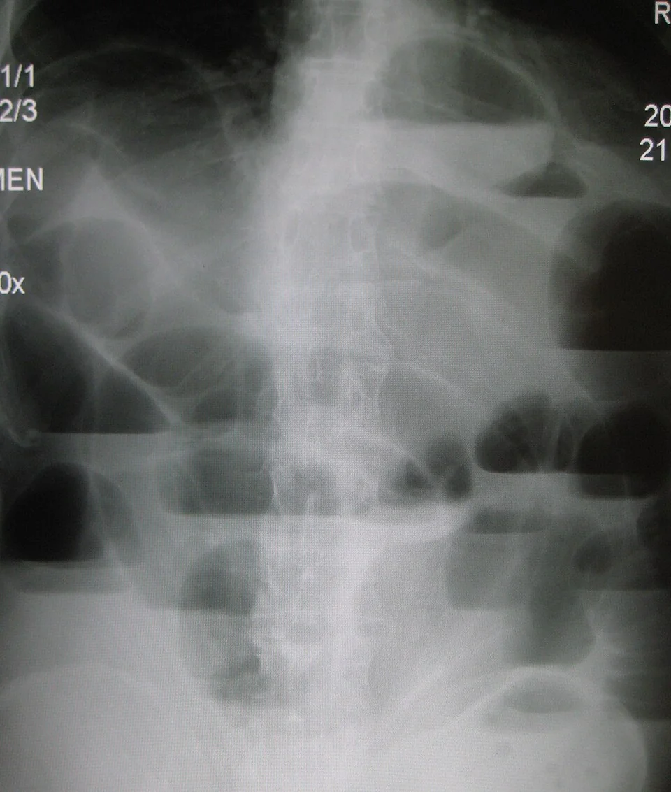
Aetiology and pathophysiology
Which cancers
- Ovarian cancer - the classic cause, through transcoelomic spread producing widespread peritoneal deposits and multi-level small bowel obstruction
- Colorectal cancer - either an obstructing primary or recurrence at the anastomosis or in the pelvis
- Gastric and pancreatic cancer - often causing gastric outlet obstruction rather than small bowel obstruction
- Peritoneal metastases from any primary - breast, lung, melanoma and cancer of unknown primary
- Non-malignant causes in a patient with cancer - adhesions from previous surgery, radiation enteritis, a hernia, faecal impaction or constipation from opioids. These account for around a third of episodes and are important because they are more readily treated.
Mechanisms
| Mechanism | Description |
|---|---|
| Extrinsic compression | A tumour mass or peritoneal deposits compress the bowel from outside - the commonest pattern in ovarian cancer |
| Intraluminal occlusion | Tumour growing into the lumen, typical of a primary colorectal cancer |
| Intramural infiltration | Tumour spreading along the bowel wall, producing a rigid, non-peristalsing segment - linitis plastica-like disease |
| Functional (pseudo-obstruction) | Malignant infiltration of the mesenteric nerve plexuses, or a paraneoplastic or drug-induced ileus, with no mechanical blockage |
| Adhesional or radiation | Non-malignant, but occurring in a patient with cancer; potentially reversible |
The vicious cycle
Understanding the physiology is what makes the drug treatment make sense. Obstruction causes proximal distension. Distension stretches the bowel wall, which stimulates further secretion of fluid into the lumen and impairs its absorption, so the bowel distends more. The stretched, oedematous wall releases inflammatory mediators and prostaglandins, which drive further secretion and generate colic and pain.
Two therapeutic implications follow. Reducing secretions breaks the cycle, which is why antisecretory drugs such as hyoscine butylbromide and octreotide are so effective in medical management - they reduce the volume the patient vomits and often relieve the distension without any drainage. And corticosteroids may reduce peritumoural oedema enough to restore a lumen, which is why a trial of dexamethasone is often given.
Clinical features
Malignant obstruction usually develops gradually over days to weeks, often with a preceding history of intermittent symptoms, in contrast to the abrupt onset of adhesional or strangulating obstruction.
- Vomiting - the character indicates the level. Early, large-volume, undigested vomiting suggests a high (gastric outlet or proximal small bowel) obstruction; late, feculent vomiting suggests a distal small bowel or colonic level.
- Nausea - often continuous, and frequently the most distressing symptom
- Abdominal pain - two distinct types that need different drugs. Colic is intermittent and cramping; continuous background pain arises from tumour, distension and peritoneal irritation.
- Distension - marked in distal obstruction, minimal in a high obstruction
- Absolute constipation - no flatus or faeces in complete obstruction, though overflow diarrhoea can occur with partial obstruction and is easily misread
- Dehydration and prerenal failure from vomiting and third-space losses
Examination
- General - dehydration, cachexia, jaundice, lymphadenopathy including a left supraclavicular node
- Abdomen - distension, visible peristalsis, surgical scars, palpable masses, hepatomegaly, ascites and shifting dullness
- Bowel sounds - characteristically high-pitched and tinkling in mechanical obstruction, absent in ileus. This distinction is less reliable than textbooks imply.
- Hernial orifices - always examine them; an obstructed hernia is a treatable cause
- Rectal examination - an empty rectum supports mechanical obstruction; a rectal or pelvic mass may be palpable. Impacted faeces suggests a very different problem.
Investigations
- Bloods - FBC, U&Es (looking for the hypokalaemic hypochloraemic picture of persistent vomiting and for acute kidney injury), LFTs, corrected calcium since hypercalcaemia causes an ileus, CRP, magnesium and a venous gas for lactate
- Erect chest radiograph - to look for free air under the diaphragm if perforation is suspected
- Abdominal radiograph - confirms obstruction and gives a crude level, but does not establish the cause or the number of levels
- CT of the abdomen and pelvis with contrast - the key investigation. It shows the site, the number of levels, the transition point, whether there is a single resectable lesion, the burden of peritoneal disease, ascites, and complications such as ischaemia or perforation. It is what the surgical decision rests on.
- Water-soluble contrast study - both diagnostic and occasionally therapeutic in adhesional obstruction
- Endoscopy - where stenting is being considered, and to obtain histology if the diagnosis is not established
It is entirely appropriate to omit investigations in a patient who is clearly dying, in whom no result would change management. But CT should not be withheld from someone who might benefit from a stent or from surgery simply because they have advanced cancer - that decision needs the information.
Management
Initial resuscitation
Whatever the eventual plan, the first steps are the same: correct dehydration with intravenous fluid, correct potassium and magnesium, provide adequate analgesia and antiemesis, and pass a nasogastric tube on free drainage if there is large-volume vomiting. The nasogastric tube is a temporary measure to give relief and buy time for a decision - it is not itself a treatment, and it is unpleasant enough that it should be removed once a longer-term plan is in place.
Deciding on the approach
The decision is made by a multidisciplinary team including surgeons, oncologists and palliative care, and the patient's own priorities carry considerable weight - the trade-off between the chance of eating again and the risk of dying in hospital after an operation is a personal one.2
| Favours surgery or stenting | Against surgery |
|---|---|
| A single, identifiable transition point on CT | Multiple levels of obstruction, or diffuse peritoneal disease |
| Good performance status (0 to 2) and reasonable nutrition | Poor performance status (3 to 4), cachexia, low albumin |
| Expected survival of more than 2 to 3 months | Expected survival of weeks |
| No or minimal ascites | Large-volume ascites - a strong predictor of surgical failure |
| Disease responsive to further systemic treatment | Disease progressing through multiple lines of therapy |
| No previous radiotherapy to the field, and few previous laparotomies | Previous abdominal or pelvic radiotherapy, multiple previous operations |
| Signs of ischaemia, strangulation or perforation - which force the issue | Patient's clearly expressed wish to avoid surgery |
- Surgery - resection, bypass or a defunctioning stoma. It offers the best chance of restoring transit where obstruction is at a single level, but carries operative mortality of the order of 10 to 30% in this population, with high rates of re-obstruction and prolonged hospital stay.
- Endoscopic stenting - a self-expanding metal stent is very effective for a single obstructing lesion in the oesophagus, gastric outlet, duodenum or left colon, and avoids a laparotomy and a stoma. It is not an option for multi-level small bowel obstruction.
- Venting gastrostomy - a percutaneous gastrostomy placed for drainage rather than feeding. It relieves vomiting without a nasogastric tube and allows the patient to eat and drink for pleasure, and is a good option in refractory high obstruction.
- Medical management - the appropriate route for most patients with multi-level obstruction from peritoneal disease
Medical management
The aim is to control nausea, vomiting and pain well enough that the patient is comfortable, usually without a nasogastric tube and often able to take small amounts orally. Drugs are given by continuous subcutaneous infusion through a syringe driver, since oral absorption is unreliable.3,4
| Purpose | Drug | Notes |
|---|---|---|
| Background pain | Morphine or, in renal impairment, oxycodone or alfentanil | Titrate from the previous oral opioid dose; a syringe driver is the usual route |
| Colic | Hyoscine butylbromide (Buscopan) 60 to 120 mg | Antispasmodic and antisecretory. Stop prokinetics if there is colic. |
| Nausea and vomiting - complete obstruction | Cyclizine 100 to 150 mg, or levomepromazine 6.25 to 25 mg | Cyclizine is antihistaminic and anticholinergic; levomepromazine is broad-spectrum and sedating at higher doses |
| Nausea and vomiting - partial obstruction with no colic | Metoclopramide 30 to 60 mg | A prokinetic. Effective in functional or partial obstruction, but contraindicated in complete obstruction or where there is colic, as it worsens pain and risks perforation. |
| Reducing secretions | Octreotide 300 to 600 micrograms | A somatostatin analogue. More effective than hyoscine butylbromide at reducing gastrointestinal secretion, and often dramatically reduces vomiting volume. Expensive, so usually second-line. |
| Reducing peritumoural oedema | Dexamethasone 6 to 16 mg | May relieve obstruction altogether. Given as a time-limited trial of about 5 days, then stopped if there is no benefit. |
| Refractory nausea | Haloperidol 1.5 to 5 mg | Useful where a chemical or metabolic component is suspected, and can be combined with cyclizine |
Nutrition, hydration and eating
- Allow oral intake for comfort and pleasure once symptoms are controlled - small amounts of fluid and low-residue food. Blanket nil-by-mouth orders in a patient not heading for theatre serve no purpose and cause distress.
- Mouth care is essential and does more for thirst than intravenous fluid
- Clinically assisted hydration - subcutaneous or intravenous fluid may relieve thirst and reduce nausea and delirium, but does not prolong life and can worsen secretions and oedema. The decision is individual and should be reviewed regularly.
- Parenteral nutrition is rarely appropriate; it is considered only where the patient has a good performance status, an expected survival of months, and obstruction is the limiting problem rather than the disease itself
- Bowel care below the obstruction - a stool softener may still be needed, but stimulant laxatives are avoided in complete obstruction
Complications
Bowel ischaemia, perforation and faecal peritonitis are the feared complications, and they change the situation entirely: a patient who was being managed medically and develops peritonism, tachycardia, fever and a rising lactate needs an urgent surgical opinion even if their prognosis is poor, because perforation is a distressing way to die and a stoma may be the kinder option.
- Aspiration pneumonia from large-volume vomiting, particularly in a drowsy patient
- Dehydration, acute kidney injury and electrolyte disturbance, which cause delirium and opioid accumulation
- Malnutrition and progressive cachexia
- Enterocutaneous or enterovaginal fistula, particularly after pelvic radiotherapy
- Stent complications - migration, occlusion by tumour ingrowth, and perforation. Colonic stenting is generally avoided in patients on bevacizumab because of the perforation risk.
- Surgical complications - anastomotic leak, wound dehiscence, prolonged ileus, stoma problems, and death
Red flags
Prognosis
Malignant bowel obstruction generally signals the terminal phase of intra-abdominal cancer. Median survival after an episode is in the region of 3 to 6 months where it can be relieved, and often only weeks in patients with multi-level obstruction from diffuse peritoneal disease who are managed medically.1,5
Adverse features are poor performance status, low albumin, large-volume ascites, multiple levels of obstruction, previous abdominal radiotherapy, and disease that has progressed through several lines of systemic treatment. Patients with a single obstructing lesion who are otherwise well may do considerably better, particularly if stented successfully.
For that reason, an episode of malignant obstruction is one of the clearest indications to have an explicit conversation about the future: what treatment would and would not be wanted, whether readmission is wanted if it recurs, preferred place of care, and a DNACPR decision. Symptoms can usually be controlled well at home or in a hospice with a syringe driver, provided the anticipatory medications and community support are set up before discharge rather than after the next crisis.
References
- Ripamonti C, Easson AM, Gerdes H. Management of malignant bowel obstruction. European Journal of Cancer. 2008. Available here
- Cousins SE, Tempest E, Feuer DJ. Surgery for the resolution of symptoms in malignant bowel obstruction in advanced gynaecological and gastrointestinal cancer. Cochrane Database of Systematic Reviews. 2016. Available here
- Scottish Palliative Care Guidelines. Bowel obstruction. Available here
- Palliative Care Formulary and BNF. Hyoscine butylbromide, octreotide, levomepromazine. Available here
- NICE Clinical Knowledge Summaries. Palliative care - nausea and vomiting. Available here
This article is written for revision and education. It is not clinical guidance and must not be used to make decisions about the care of a patient. Always check current NICE guidance and local protocols.