
Complications of Labour
Key points
- Failure to progress: analysed as the three Ps - the powers, the passenger and the passages; in a parous woman, never assume it is simply the powers.
- First stage delay: less than 2 cm of cervical dilatation in 4 hours, managed by amniotomy then oxytocin augmentation.
- Second stage delay: diagnosed after 2 hours of active pushing in a nulliparous woman and 1 hour in a parous woman.
- Cord prolapse: an immediate emergency; relieve pressure on the cord, avoid handling it, and deliver by the quickest safe route.
- Shoulder dystocia: manage with the HELPERR sequence, starting with McRoberts and suprapubic pressure, which resolve the great majority.
- Fetal blood sampling: pH below 7.20 or lactate above 4.9 mmol/L is abnormal and indicates the need for delivery.
- Uterine rupture: suspect with severe continuous pain, fetal bradycardia, loss of contractions and a rising presenting part in a scarred uterus.
- Caesarean categories: category 1 within 30 minutes for immediate threat to life, category 2 within 75 minutes, categories 3 and 4 are non-urgent and elective.
Introduction
Most labours proceed without incident, but the labour ward exists for those that do not. The complications covered here fall into two groups: those in which labour fails to progress, which usually allow time for assessment and a considered decision, and those that require action within minutes because the fetus or mother is acutely compromised.
Recognising which group a situation belongs to is the central skill. Delay in the first stage is managed methodically over hours; a cord prolapse, a shoulder dystocia or a uterine rupture is managed in seconds, from memory, with the whole team called at once. This is why obstetric emergencies are drilled with mandatory multiprofessional simulation training, and why the RCOG guidelines specify sequences rather than principles.1
Failure to progress
Progress in labour depends on the powers, the passenger and the passages. A structured assessment covers all three rather than reaching immediately for oxytocin.
| Factor | Problems | Assessment |
|---|---|---|
| Powers | Hypotonic or incoordinate contractions; commonest cause in a nulliparous woman | Palpate frequency, duration and strength; aim for 4-5 contractions in 10 minutes lasting 45-60 seconds |
| Passenger | Malposition (usually occipito-posterior), malpresentation (brow, face, breech), macrosomia, fetal anomaly such as hydrocephalus | Abdominal palpation for lie, presentation and fifths palpable; vaginal examination for position, caput and moulding |
| Passages | Contracted or unfavourable pelvis, cephalopelvic disproportion, soft tissue obstruction such as a cervical fibroid, full bladder | Assess station and descent, look for excessive moulding, ensure the bladder is empty |
Managing delay in the first stage
- Confirm delay: less than 2 cm dilatation in 4 hours, or slowing of progress in a parous woman
- Provide support, analgesia, hydration and encourage mobility and position change
- Ensure the bladder is empty
- Offer amniotomy if the membranes are intact, and reassess in 2 hours
- If progress remains inadequate, offer oxytocin augmentation with continuous cardiotocography, and reassess after 4 hours
- If dilatation increases by less than 2 cm after 4 hours of oxytocin, obstetric review is required and caesarean section is usually indicated
Delay in the second stage
Delay is diagnosed after 2 hours of active pushing in a nulliparous woman and 1 hour in a parous woman, and obstetric review is required. The options are continued pushing with support, oxytocin augmentation if contractions are inadequate and the membranes are ruptured, instrumental vaginal birth, or caesarean section. The decision rests on the station and position of the head, the presence of caput and moulding, and the fetal condition.
Malposition and malpresentation
Position describes the orientation of the fetal occiput relative to the maternal pelvis; presentation describes which part of the fetus occupies the lower pole of the uterus.
| Type | Features | Management |
|---|---|---|
| Occipito-posterior position | Around 10-20% at the onset of labour; back pain, early urge to push, slow progress, a deflexed head presenting a larger diameter | Most rotate spontaneously; otherwise rotational instrumental birth or caesarean section |
| Occipito-transverse (deep transverse arrest) | Head fails to rotate at the pelvic floor and arrests in the transverse position | Rotational ventouse, Kielland's forceps or manual rotation by an experienced operator, or caesarean section |
| Breech | 3-4% at term; frank (extended), complete (flexed) or footling | Offer external cephalic version from 36 weeks in a nullipara and 37 weeks in a multipara; otherwise planned caesarean or, with an experienced operator, vaginal breech birth |
| Brow presentation | The head is midway between flexion and extension, presenting the mentovertical diameter of around 13 cm | Vaginal birth is impossible unless it converts; caesarean section |
| Face presentation | Fully extended head; mento-anterior can deliver vaginally, mento-posterior cannot | Mento-anterior may be allowed to labour; mento-posterior requires caesarean section |
| Transverse or oblique lie | Shoulder presents; associated with polyhydramnios, multiparity, placenta praevia and uterine anomaly | Caesarean section; high risk of cord prolapse when membranes rupture |
Umbilical cord prolapse
Cord prolapse is descent of the umbilical cord through the cervix alongside or past the presenting part, after rupture of the membranes. It occurs in around 1 in 500 births and causes fetal hypoxia by two mechanisms: mechanical compression of the cord between the presenting part and the pelvis, and vasospasm of the cord vessels on exposure to cold air.
Risk factors are anything that prevents the presenting part from filling the pelvis: prematurity, low birth weight, breech and other malpresentation, transverse or unstable lie, polyhydramnios, multiple pregnancy, and a second twin. A substantial proportion are iatrogenic, following artificial rupture of membranes with a high presenting part.
Shoulder dystocia
Shoulder dystocia is a vaginal cephalic delivery that requires additional manoeuvres to deliver the fetus after the head has delivered and gentle traction has failed. It complicates around 0.6% of vaginal births. The anterior shoulder becomes impacted behind the pubic symphysis, and the fetus cannot breathe or receive placental gas exchange, so it is a genuine time-critical emergency.
Risk factors include macrosomia, maternal diabetes, maternal obesity, previous shoulder dystocia, induction of labour, prolonged labour, and instrumental delivery. Crucially, most cases occur in babies of normal weight with no risk factors at all, which is why it cannot be reliably predicted and why every birth attendant must know the drill.
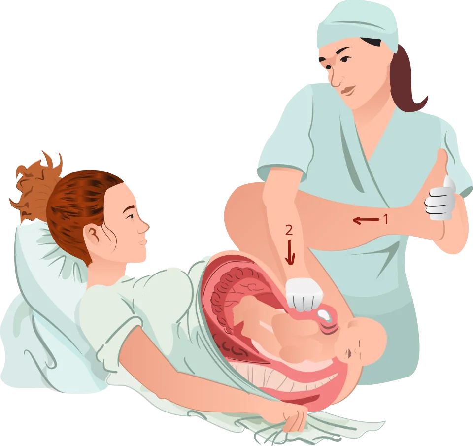
| Step | Action |
|---|---|
| H - Help | Call for senior obstetric, anaesthetic, midwifery and neonatal help; note the time |
| E - Evaluate for episiotomy | An episiotomy does not free the bony obstruction but may create room for internal manoeuvres |
| L - Legs (McRoberts) | Hyperflex and abduct the maternal hips; resolves around 40-50% of cases alone |
| P - Suprapubic pressure | Apply pressure behind the fetal anterior shoulder to adduct it and push it into the oblique diameter; with McRoberts this resolves around 90% |
| E - Enter (internal rotational manoeuvres) | Rubin II presses the posterior aspect of the anterior shoulder; the Wood's screw and reverse Wood's screw rotate the fetus 180 degrees |
| R - Remove the posterior arm | Sweep the posterior arm across the chest and deliver it, reducing the shoulder diameter |
| R - Roll the patient | The all-fours position increases the true pelvic diameters and may free the shoulder |
Complications for the baby are brachial plexus injury, most often Erb's palsy affecting C5 and C6, which resolves in the majority; fracture of the humerus or clavicle, which heals well; and hypoxic-ischaemic encephalopathy or death if delivery is delayed. Maternal complications are postpartum haemorrhage, occurring in around 11%, and third- or fourth-degree perineal tears. Full documentation, including the time of head and body delivery, the manoeuvres used and which shoulder was anterior, is a medicolegal necessity.
Fetal compromise in labour
A pathological cardiotocograph indicates that the fetus may be hypoxic, but it is a poor positive predictor: most fetuses with an abnormal trace are not acidotic. The response is therefore graded, beginning with intrauterine resuscitation and correcting reversible causes, before proceeding to fetal blood sampling or delivery.
- Change maternal position to left lateral, relieving aortocaval compression
- Correct hypotension with intravenous fluid, particularly after an epidural top-up
- Stop or reduce oxytocin if there is tachysystole; consider tocolysis with terbutaline
- Exclude a precipitating event: vaginal examination for cord prolapse, and consider abruption or uterine rupture
- Do not give routine maternal oxygen, which has no evidence of benefit and may be harmful
- Consider fetal blood sampling where the trace is pathological, the cervix is at least 3-4 cm dilated and delivery is not immediately indicated
| Result | pH | Lactate | Action |
|---|---|---|---|
| Normal | 7.25 or above | 4.1 mmol/L or below | Repeat in 1 hour if the trace remains abnormal |
| Borderline | 7.21-7.24 | 4.2-4.8 mmol/L | Repeat in 30 minutes |
| Abnormal | 7.20 or below | 4.9 mmol/L or above | Urgent obstetric review and expedite birth |
Fetal blood sampling is contraindicated in maternal sepsis, suspected fetal bleeding disorders, prematurity below 34 weeks, and where there is an acute bradycardia requiring immediate delivery. A sudden prolonged deceleration lasting more than 3 minutes is managed by preparing for immediate birth while intrauterine resuscitation is under way, not by attempting to sample.5
Uterine rupture and inversion
Uterine rupture
Uterine rupture is a full-thickness breach of the uterine wall, and is usually but not always associated with a previous caesarean scar. The risk of scar rupture in a planned vaginal birth after one previous lower segment caesarean is approximately 0.5%, rising with induction and further with oxytocin augmentation or prostaglandin use. Other risk factors are previous myomectomy or uterine surgery, high parity, obstructed labour and injudicious oxytocin.
- Fetal bradycardia or an abnormal cardiotocograph - the commonest and earliest sign
- Severe constant abdominal pain, which may persist between contractions and break through an effective epidural
- Cessation of contractions and loss of uterine tone
- Scar tenderness, vaginal bleeding and haematuria
- Loss of station: the presenting part rises out of the pelvis
- Easily palpable fetal parts abdominally, and maternal cardiovascular collapse
Management is immediate laparotomy after resuscitation, with repair of the defect or, where the tear is extensive, hysterectomy. Perinatal mortality is high and depends almost entirely on the interval between rupture and delivery.
Acute uterine inversion
Uterine inversion is turning of the uterus inside out, usually following excessive cord traction on an uncontracted uterus or a fundal placenta. It presents with severe lower abdominal pain, haemorrhage and profound shock that is often out of proportion to the blood loss, because of vagal stimulation from traction on the peritoneum and ligaments. Management is immediate manual replacement by pushing the fundus back through the cervix (the Johnson manoeuvre), before the cervical ring contracts; the placenta should be left attached until the uterus is replaced. If manual replacement fails, hydrostatic reduction with warm saline or surgery is needed. Uterotonics are withheld until replacement is complete, then given to maintain it.6
Amniotic fluid embolism
Amniotic fluid embolism is rare, at roughly 2 per 100,000 births in the UK, but carries high mortality. It is now understood as an anaphylactoid reaction to fetal antigens entering the maternal circulation rather than a mechanical embolic phenomenon. It occurs typically during labour, at delivery or immediately postpartum.
The presentation is a biphasic collapse: sudden hypoxia, dyspnoea, cyanosis and cardiovascular collapse with acute right heart failure, often preceded by a sense of impending doom, followed within an hour by left ventricular failure and severe disseminated intravascular coagulation with torrential haemorrhage. Seizures occur in a substantial minority.
It is a diagnosis of exclusion made clinically, and management is entirely supportive: immediate resuscitation with high-flow oxygen and intubation, circulatory support, massive transfusion protocol with early blood products for the coagulopathy, and delivery of the fetus if the arrest occurs antenatally, including perimortem caesarean section within four minutes of cardiac arrest.7
Assisted birth
| Feature | Ventouse | Forceps |
|---|---|---|
| Success rate | Lower; more likely to fail and require forceps or caesarean | Higher |
| Maternal trauma | Less perineal and vaginal trauma, fewer third- and fourth-degree tears | More maternal trauma; anal sphincter injury more common |
| Neonatal trauma | Cephalhaematoma, chignon, retinal haemorrhage; subgaleal haemorrhage is rare but dangerous | Facial bruising and facial nerve palsy; skull fracture rare |
| Requirements | Gestation of 34 weeks or more; requires maternal effort | Can be used at earlier gestations and with less maternal effort |
The prerequisites for any instrumental birth are conventionally remembered as a checklist: fully dilated cervix, ruptured membranes, cephalic presentation, defined position, engaged head with no more than one-fifth palpable abdominally, adequate analgesia, empty bladder, adequate contractions, consent, and a clear plan of where to go if it fails. Attempts should be abandoned if there is no progressive descent with each pull, or after three contractions with a correctly applied instrument.
| Category | Indication | Target decision-to-delivery interval |
|---|---|---|
| 1 | Immediate threat to the life of the woman or fetus - cord prolapse, uterine rupture, prolonged bradycardia | 30 minutes |
| 2 | Maternal or fetal compromise that is not immediately life-threatening | 75 minutes |
| 3 | No compromise, but early delivery needed | No fixed target |
| 4 | Elective, at a time to suit the woman and the service | Planned, usually from 39 weeks |
Retained placenta and perineal trauma
The placenta is retained if it has not delivered within 30 minutes with active management or 60 minutes with physiological management. Management is to empty the bladder, give an oxytocin infusion if not already running, and attempt controlled cord traction once more; if this fails, manual removal in theatre under regional or general anaesthesia is required, with prophylactic antibiotics. Do not give oxytocin into the umbilical vein, which is ineffective. Consider placenta accreta if the placenta will not separate, and stop rather than persisting.8
Obstetric anal sphincter injury, comprising third- and fourth-degree tears, affects around 3% of vaginal births and up to 6% of first vaginal births. Risk factors are nulliparity, birth weight above 4 kg, occipito-posterior position, prolonged second stage, shoulder dystocia and forceps delivery. Every woman should have a systematic perineal and rectal examination after birth, because tears are otherwise missed. Repair is performed in theatre by a trained clinician, with broad-spectrum antibiotics, laxatives for 10 days, physiotherapy and follow-up in a perineal clinic. Around 60-80% are asymptomatic at 12 months, but a significant minority have long-term faecal urgency or incontinence.
Red flags
References
- NICE CG190. Intrapartum care for healthy women and babies. 2014 (updated 2023). Available here
- RCOG Green-top Guideline No. 20a. External cephalic version and reducing the incidence of term breech presentation. Available here
- RCOG Green-top Guideline No. 50. Umbilical cord prolapse. Available here
- RCOG Green-top Guideline No. 42. Shoulder dystocia. Available here
- NICE NG229. Fetal monitoring in labour. 2022. Available here
- RCOG Green-top Guideline No. 52. Prevention and management of postpartum haemorrhage. Available here
- UKOSS. Amniotic fluid embolism surveillance. Available here
- NICE NG192. Caesarean birth. 2021 (updated 2024). Available here
This article is written for revision and education. It is not clinical guidance and must not be used to make decisions about the care of a patient. Always check current NICE guidance and local protocols.