
Bursitis
Key points
- Bursa: a fluid-filled sac that reduces friction between a tendon or the skin and underlying bone. Bursitis is inflammation of this sac, not of the joint itself.
- Common sites: olecranon (student's elbow), prepatellar (housemaid's knee), infrapatellar (clergyman's knee), trochanteric, and subacromial.
- Key discriminator: active and passive range of motion of the underlying joint is usually preserved in simple bursitis, unlike septic arthritis where movement is globally painful and restricted.
- Septic bursitis: usually from direct inoculation through an overlying skin break, most commonly with Staphylococcus aureus. Suspect it with fever, marked or spreading erythema, and disproportionate pain.
- Key investigation: aspiration when septic bursitis or crystal arthropathy is suspected - send fluid for Gram stain, culture and sensitivity, and microscopy for crystals.
- Non-septic management: activity modification, ice, NSAIDs, aspiration if tense, and corticosteroid injection for persistent inflammatory bursitis once infection is excluded.
- Septic management: empirical antibiotics against Staph aureus (for example flucloxacillin), aspiration or drainage, and bursectomy for recurrent or resistant cases.
- Exam trap: a hot, swollen bursa with a fever is septic bursitis until proven otherwise - do not manage it as simple inflammatory bursitis and wait and see.
Introduction
A bursa is a small, flattened, fluid-filled sac lined by synovium. Its job is purely mechanical: it sits between a tendon or the skin and an underlying bony prominence, letting the two surfaces glide over one another with minimal friction during movement. There are around 150 bursae in the body, most of them at sites of repeated mechanical stress over a joint.
Bursitis is inflammation of a bursa, producing swelling and pain over that bony prominence.1 It matters clinically for two reasons. First, it can closely mimic septic arthritis and cellulitis, both of which need urgent treatment, so distinguishing between them is a core clinical skill. Second, a bursa can itself become infected, and septic bursitis is a genuine emergency that is easy to under-call if it is assumed to be 'just' inflammatory bursitis.
Superficial bursae, such as the olecranon and prepatellar bursae, lie directly under the skin and are the ones most often affected by trauma and infection. Deeper bursae, such as the subacromial and trochanteric bursae, are not directly inoculable from the skin and are almost always inflammatory rather than septic.
Common sites
The site of bursitis usually points straight to the mechanism, and the eponyms below are a favourite in written exams.
| Site | Eponym | Typical cause |
|---|---|---|
| Olecranon bursa | Student's elbow | Leaning on the elbows, or direct trauma; a superficial bursa, so the commonest site for septic bursitis |
| Prepatellar bursa | Housemaid's knee | Prolonged kneeling upright, e.g. carpet fitters, cleaners |
| Infrapatellar bursa | Clergyman's knee | Kneeling with the trunk more upright and weight further forward than in prepatellar bursitis |
| Trochanteric bursa | Greater trochanteric pain syndrome | Repetitive friction of the iliotibial band over the greater trochanter; overlaps with, and often coexists with, gluteal tendinopathy |
| Subacromial/subdeltoid bursa | - | Associated with rotator cuff pathology and subacromial impingement rather than direct pressure |
Olecranon and prepatellar bursitis are the two sites most often seen acutely and most often septic, because both bursae are superficial and vulnerable to a break in the overlying skin. Trochanteric and subacromial bursitis, by contrast, present as chronic regional pain syndromes rather than acute swellings, since both bursae lie deep to muscle and are not directly exposed to the skin.
Trochanteric bursitis classically affects middle-aged and older women with lateral hip pain that is worse lying on the affected side and with prolonged walking or stair climbing. It is now usually considered part of a broader greater trochanteric pain syndrome, in which bursal inflammation coexists with, and is often secondary to, gluteus medius and minimus tendinopathy, so isolated bursitis is probably less common than the name suggests.6
Aetiology
Bursitis has four broad mechanisms, which often overlap in an individual patient.
- Repetitive friction, pressure or overuse - the commonest cause, from occupational or recreational activities that repeatedly load a bursa (kneeling, leaning on the elbows, running)
- Direct trauma - a fall or blow onto the bursa, which can also introduce infection if the skin is broken
- Inflammatory arthropathy - rheumatoid arthritis, gout and pseudogout can all cause bursitis, either from the same systemic inflammatory process or from crystal deposition directly into the bursal fluid
- Infection - almost always by direct inoculation through an overlying skin break (an abrasion, laceration or insect bite) rather than by haematogenous spread; Staphylococcus aureus accounts for the large majority of cases5
Occupation is a useful clue in the history: anyone whose job or hobby involves sustained kneeling, leaning on the elbows, or repetitive shoulder overhead activity is at higher risk of the corresponding bursitis. Diabetes, alcohol excess, immunosuppression, chronic kidney disease and corticosteroid use all raise the risk that any bursitis - especially one arising from trauma with a skin breach - becomes septic.
Clinical features
The typical picture is a well-defined, fluctuant swelling directly over a bony prominence, with overlying tenderness and warmth. Onset can be acute, following a single episode of trauma or a break in the skin, or gradual, following weeks of repetitive friction. The overlying skin may be normal, mildly erythematous, or thickened if the bursitis is chronic and recurrent.
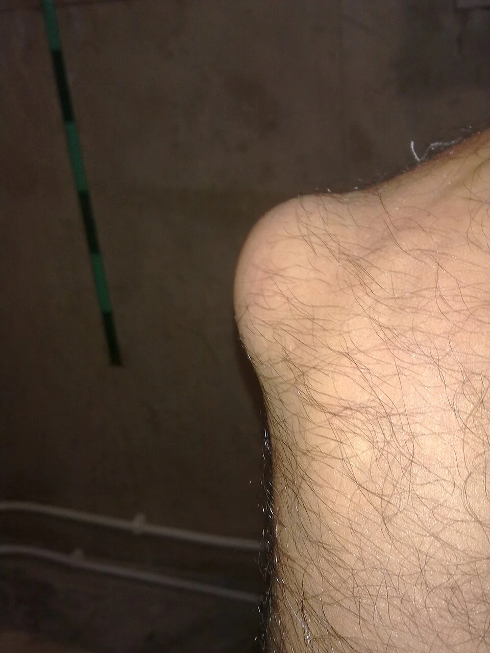
Active and passive range of motion of the underlying joint is usually preserved, or only mildly reduced at the extremes of movement where the swollen bursa itself is mechanically compressed. This is because the inflammation is confined to the bursa, outside the joint capsule, rather than involving the joint space itself.
Systemic features - fever, malaise, rigors - are absent in simple inflammatory or traumatic bursitis. Their presence, alongside spreading erythema or disproportionate pain, should immediately raise concern for septic bursitis or an alternative diagnosis such as septic arthritis or cellulitis.
Septic versus non-septic bursitis
This distinction is the single most important judgement to make in a patient with bursitis, because it determines whether the patient needs antibiotics and aspiration or simple conservative measures. Septic bursitis accounts for a substantial minority of cases presenting acutely, particularly at the olecranon and prepatellar bursae, so it must be actively considered rather than assumed away.4
Non-septic bursitis is either traumatic (from a single injury or repetitive friction, without infection) or inflammatory (secondary to rheumatoid arthritis, gout or pseudogout). Septic bursitis follows direct inoculation of the bursa, almost always through an overlying skin break, with Staphylococcus aureus the dominant organism; streptococci account for most of the remainder.
| Feature | Non-septic (traumatic/inflammatory) | Septic |
|---|---|---|
| Onset | Gradual, or acute after a single non-penetrating injury | Often acute, frequently after a penetrating skin injury or abrasion over the bursa |
| Erythema | Absent or mild, confined to the bursa | Marked, may extend beyond the margins of the bursa into surrounding tissue |
| Warmth | Mild | Pronounced |
| Pain | Proportionate to swelling | Disproportionate to swelling |
| Fever/systemic upset | Absent | Often present |
| Skin breach nearby | Uncommon | Common - actively look for one |
| Range of motion of underlying joint | Preserved or mildly reduced at extremes | Preserved or mildly reduced at extremes (this alone does not exclude sepsis of the bursa) |
| Risk factors | Occupational friction, gout, rheumatoid arthritis | Diabetes, immunosuppression, alcohol excess, corticosteroid use, skin breach |
Note that preserved range of motion helps separate bursitis (septic or not) from septic arthritis of the joint itself, but it does not, on its own, tell you whether the bursa is infected - for that, look at the systemic features, the degree and spread of erythema, and any history of a skin breach, and have a low threshold to aspirate.
Aspiration is the key investigation because clinical assessment alone cannot reliably exclude infection, and the bursal fluid gives a direct answer: a Gram stain and culture confirm or exclude sepsis and identify the organism, while microscopy for crystals under polarised light distinguishes gout (negatively birefringent needle-shaped crystals) and pseudogout (positively birefringent rhomboid crystals) from infection when the picture is inflammatory rather than septic.
Investigations
Bursitis is usually a clinical diagnosis, made on the appearance and location of the swelling together with the history. Investigations are targeted at confirming or excluding infection and crystal arthropathy, rather than at diagnosing bursitis itself.
- Aspiration - indicated whenever septic bursitis or crystal arthropathy is suspected, or to relieve a tense, symptomatic effusion. Send fluid for Gram stain, culture and sensitivity, and polarised light microscopy for crystals.
- FBC, CRP - a raised white cell count and CRP support infection but neither is sensitive or specific enough to rule sepsis in or out alone
- Blood cultures - if the patient is systemically unwell or febrile
- Ultrasound - confirms the presence and extent of bursal fluid, helps distinguish bursitis from a solid mass or a joint effusion, and can guide aspiration, particularly at deeper sites such as the trochanteric or subacromial bursa
- Plain X-ray - not routinely needed, but occasionally useful to look for an underlying bony spur, foreign body, or to exclude an associated fracture after trauma
Management
Non-septic bursitis
- Activity or pressure modification - avoiding the precipitating friction or pressure (kneepads, elbow pads, activity modification)
- Ice and simple analgesia
- NSAIDs, oral or topical, for inflammatory or traumatic bursitis once infection has been excluded
- Aspiration if the bursa is tense and symptomatic, which also provides diagnostic fluid
- Corticosteroid injection into the bursa for persistent inflammatory bursitis once infection has been confidently excluded - effective, but carries a small risk of introducing infection and of skin atrophy or depigmentation, and should not be given if there is any suspicion of sepsis
- Treatment of any underlying cause, such as urate-lowering therapy after recurrent gouty bursitis or optimising rheumatoid disease control
Septic bursitis
- Empirical antibiotics targeted at Staphylococcus aureus - flucloxacillin is the typical first-line choice per local policy, with a glycopeptide such as vancomycin considered where MRSA is a risk3
- Antibiotics are then rationalised according to culture and sensitivity results from the aspirate
- Aspiration or formal drainage of the bursa, both diagnostic and therapeutic, and often repeated if fluid reaccumulates
- Surgical bursectomy for cases that are recurrent, fail to respond to antibiotics and drainage, or become chronically discharging
- Admission and intravenous antibiotics for anyone systemically unwell, immunocompromised, or not responding to oral treatment
Mild septic bursitis in an otherwise well patient can sometimes be managed with oral antibiotics and outpatient review, but the threshold to admit should be low in anyone systemically unwell, diabetic, immunosuppressed, or where the diagnosis is uncertain between septic bursitis and septic arthritis.
Differential diagnosis
- Septic arthritis - global painful restriction of active and passive movement through the joint, rather than swelling confined to a bursa2
- Cellulitis - diffuse skin and soft tissue erythema and warmth without a discrete fluctuant swelling over a bony prominence
- Gout or pseudogout - can cause bursitis directly, or present as an acute monoarthritis mimicking it; joint aspiration with crystal microscopy distinguishes the two
- Rheumatoid nodule - a firm, non-fluctuant subcutaneous nodule, classically also at the olecranon, in a patient with established rheumatoid arthritis
- Tumour or ganglion - a chronic, slowly enlarging, non-inflamed swelling without the acute pain and warmth of bursitis
Complications
- Chronic or recurrent bursitis, with thickened, fibrotic bursal walls from repeated episodes
- Abscess formation within an inadequately treated septic bursa
- Spread of infection to the adjacent joint (septic arthritis) or into the surrounding soft tissue as cellulitis
- Septicaemia if septic bursitis is left untreated, particularly in immunocompromised or diabetic patients
- Sinus formation or chronic discharge after incomplete drainage, sometimes requiring bursectomy
Red flags
References
- NICE Clinical Knowledge Summaries. Bursitis. Available here
- NICE Clinical Knowledge Summaries. Septic arthritis. Available here
- BNF. Flucloxacillin - indications and dose. Available here
- Baumbach SF, Lobo CM, Badyine I, Mutschler W, Kanz KG. Prepatellar and olecranon bursitis: literature review and current recommendations. Musculoskeletal Surgery. 2014. Available here
- Zimmermann B 3rd, Mikolich DJ, Ho G Jr. Septic bursitis. Seminars in Arthritis and Rheumatism. 1995. Available here
- Mallow M, Nazarian LN. Greater trochanteric pain syndrome diagnosis and treatment. Physical Medicine and Rehabilitation Clinics of North America. 2014. Available here
- Solomon L, Warwick D, Nayagam S. Apley and Solomon's System of Orthopaedics and Trauma. 10th edition. Available here
- Orthobullets. Olecranon Bursitis. Available here
This article is written for revision and education. It is not clinical guidance and must not be used to make decisions about the care of a patient. Always check current NICE guidance and local protocols.